 ムービー
ムービー コントローラー


+ データを開く

- 基本情報
基本情報
| 登録情報 | データベース: PDB / ID: 3a60 | ||||||
|---|---|---|---|---|---|---|---|
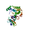
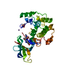
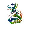
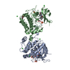
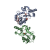
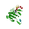
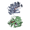
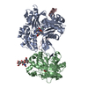
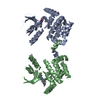
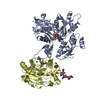
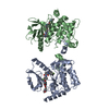
| タイトル | Crystal structure of unphosphorylated p70S6K1 (Form I) | ||||||
 要素 要素 | Ribosomal protein S6 kinase beta-1 リボソーム リボソーム | ||||||
 キーワード キーワード | TRANSFERASE (転移酵素) / Kinase (キナーゼ) / Kinase domain (キナーゼ) / Inactive / Active / Ribosomal S6 kinase (リボソームタンパク質S6キナーゼ) / Activation (活性化) / Alternative initiation / ATP-binding / Cell junction (細胞結合) / Cytoplasm (細胞質) / Nucleotide-binding / Phosphoprotein / Polymorphism / Serine/threonine-protein kinase / Synapse (シナプス) / Synaptosome | ||||||
| 機能・相同性 |  機能・相同性情報 機能・相同性情報long-chain fatty acid import into cell / response to electrical stimulus involved in regulation of muscle adaptation / skeletal muscle atrophy / positive regulation of skeletal muscle tissue growth / regulation of glucose import / ribosomal protein S6 kinase activity / response to L-leucine / cellular response to nutrient / phosphatidylinositol-mediated signaling / response to glucagon ...long-chain fatty acid import into cell / response to electrical stimulus involved in regulation of muscle adaptation / skeletal muscle atrophy / positive regulation of skeletal muscle tissue growth / regulation of glucose import / ribosomal protein S6 kinase activity / response to L-leucine / cellular response to nutrient / phosphatidylinositol-mediated signaling / response to glucagon / response to testosterone / positive regulation of smooth muscle cell migration / MTOR / mTORC1-mediated signalling / germ cell development / positive regulation of translational initiation / skeletal muscle contraction / long-term memory / behavioral fear response / response to tumor necrosis factor / response to glucose / response to mechanical stimulus / negative regulation of insulin receptor signaling pathway / positive regulation of TORC1 signaling / protein serine/threonine/tyrosine kinase activity / cellular response to dexamethasone stimulus / positive regulation of mitotic cell cycle / response to nutrient levels / positive regulation of translation / protein phosphatase 2A binding / phosphatidylinositol 3-kinase/protein kinase B signal transduction / PDZ domain binding / negative regulation of extrinsic apoptotic signaling pathway / peptide binding / positive regulation of smooth muscle cell proliferation / G1/S transition of mitotic cell cycle / modulation of chemical synaptic transmission / response to toxic substance / cellular response to growth factor stimulus / cellular response to type II interferon / cellular response to insulin stimulus / 遊走 / postsynapse / peptidyl-serine phosphorylation / response to ethanol / mitochondrial outer membrane / response to lipopolysaccharide / non-specific serine/threonine protein kinase / neuron projection / protein kinase activity / response to xenobiotic stimulus / protein serine kinase activity / protein serine/threonine kinase activity / glutamatergic synapse / apoptotic process / negative regulation of apoptotic process / perinuclear region of cytoplasm / 細胞膜 / シグナル伝達 / ミトコンドリア / 核質 / ATP binding / identical protein binding / 細胞核 / 細胞質基質 / 細胞質類似検索 - 分子機能 | ||||||
| 生物種 |  Homo sapiens (ヒト) Homo sapiens (ヒト) | ||||||
| 手法 | X線回折 / シンクロトロン / 分子置換 / 解像度: 2.8 Å | ||||||
 データ登録者 データ登録者 | Sunami, T. / Byrne, N. / Diehl, R.E. / Funabashi, K. / Hall, D.L. / Ikuta, M. / Patel, S.B. / Shipman, J.M. / Smith, R.F. / Takahashi, I. ...Sunami, T. / Byrne, N. / Diehl, R.E. / Funabashi, K. / Hall, D.L. / Ikuta, M. / Patel, S.B. / Shipman, J.M. / Smith, R.F. / Takahashi, I. / Zugay-Murphy, J. / Iwasawa, Y. / Lumb, K.J. / Munshi, S.K. / Sharma, S. | ||||||
 引用 引用 | ジャーナル: J.Biol.Chem. / 年: 2010 タイトル: Structural basis of human p70 ribosomal S6 kinase-1 regulation by activation loop phosphorylation. 著者: Sunami, T. / Byrne, N. / Diehl, R.E. / Funabashi, K. / Hall, D.L. / Ikuta, M. / Patel, S.B. / Shipman, J.M. / Smith, R.F. / Takahashi, I. / Zugay-Murphy, J. / Iwasawa, Y. / Lumb, K.J. / Munshi, S.K. / Sharma, S. | ||||||
| 履歴 |
|
- 構造の表示
構造の表示
| 構造ビューア | 分子: MolmilJmol/JSmol |
|---|
- ダウンロードとリンク
-ダウンロード
| PDBx/mmCIF形式 | 3a60.cif.gz | 118.6 KB | 表示 | PDBx/mmCIF形式 |
|---|---|---|---|---|
| PDB形式 | pdb3a60.ent.gz | 91.2 KB | 表示 | PDB形式 |
| PDBx/mmJSON形式 | 3a60.json.gz | ツリー表示 | PDBx/mmJSON形式 | |
| その他 |  その他のダウンロード その他のダウンロード |
-検証レポート
| アーカイブディレクトリ | https://data.pdbj.org/pub/pdb/validation_reports/a6/3a60ftp://data.pdbj.org/pub/pdb/validation_reports/a6/3a60 | HTTPS FTP |
|---|
-関連構造データ

-リンク
 PDBj
PDBj


- 集合体
集合体
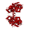
| 登録構造単位 | 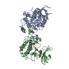
| ||||||||
|---|---|---|---|---|---|---|---|---|---|
| 1 |
| ||||||||
| 単位格子 |
|
-要素
| #1: タンパク質 | リボソーム / Ribosomal protein S6 kinase I / S6K1 / S6K / 70 kDa ribosomal protein S6 kinase 1 / p70 S6 kinase ...Ribosomal protein S6 kinase I / S6K1 / S6K / 70 kDa ribosomal protein S6 kinase 1 / p70 S6 kinase alpha / p70(S6K)-alpha / p70-S6K / P70S6K / p70-alpha 分子量: 36936.641 Da / 分子数: 2 / 断片: UNP residues 75-399 / 由来タイプ: 組換発現 / 由来: (組換発現) Homo sapiens (ヒト) / プラスミド: pVL1392 / 細胞株 (発現宿主): Sf21発現宿主:   Spodoptera frugiperda (ツマジロクサヨトウ) Spodoptera frugiperda (ツマジロクサヨトウ)参照: UniProt: P23443, non-specific serine/threonine protein kinase#2: 化合物 | スタウロスポリン  分子量: 466.531 Da / 分子数: 2 / 由来タイプ: 合成 / 式: C28H26N4O3 分子量: 466.531 Da / 分子数: 2 / 由来タイプ: 合成 / 式: C28H26N4O3コメント: 抗がん剤, 抗真菌剤, 抗生剤, alkaloid*YM #3: 水 | ChemComp-HOH / | 水 分子量: 18.015 Da / 分子数: 6 / 由来タイプ: 天然 / 式: H2O 分子量: 18.015 Da / 分子数: 6 / 由来タイプ: 天然 / 式: H2O |
|---|
-実験情報
-実験
| 実験 | 手法: X線回折 / 使用した結晶の数: 1 |
|---|
- 試料調製
試料調製
| 結晶 | マシュー密度: 2.9 Å3/Da / 溶媒含有率: 57.62 % |
|---|---|
| 結晶化 | 温度: 293 K / 手法: 蒸気拡散法, ハンギングドロップ法 / pH: 5.5 詳細: 0.1M Bis-Tris, pH 5.5, 0.2M lithium sulfate, 22.5% PEG 3350, VAPOR DIFFUSION, HANGING DROP, temperature 293.0K |
-データ収集
| 回折 | 平均測定温度: 100 K |
|---|---|
| 放射光源 | 由来: シンクロトロン / サイト: APS  / ビームライン: 17-ID / 波長: 1 Å / ビームライン: 17-ID / 波長: 1 Å |
| 検出器 | タイプ: MAR CCD 165 mm / 検出器: CCD / 日付: 2008年3月19日 |
| 放射 | プロトコル: SINGLE WAVELENGTH / 単色(M)・ラウエ(L): M / 散乱光タイプ: x-ray |
| 放射波長 | 波長: 1 Å / 相対比: 1 |
| 反射 | 解像度: 2.8→19.81 Å / Num. obs: 20565 / % possible obs: 97.9 % / 冗長度: 3.6 % / Biso Wilson estimate: 70.1 Å2 / Rsym value: 0.049 / Net I/σ(I): 14.3 |
| 反射 シェル | 解像度: 2.8→2.9 Å / 冗長度: 2.4 % / Mean I/σ(I) obs: 2.85 / Rsym value: 0.282 / % possible all: 83.8 |
- 解析
解析
| ソフトウェア |
| ||||||||||||||||||||||||||||||||||||||||||||||||||||||||||||||||||||||||||||||||
|---|---|---|---|---|---|---|---|---|---|---|---|---|---|---|---|---|---|---|---|---|---|---|---|---|---|---|---|---|---|---|---|---|---|---|---|---|---|---|---|---|---|---|---|---|---|---|---|---|---|---|---|---|---|---|---|---|---|---|---|---|---|---|---|---|---|---|---|---|---|---|---|---|---|---|---|---|---|---|---|---|---|
| 精密化 | 構造決定の手法: 分子置換 開始モデル: p70S6K1 homology model based on the published RSK1 structure; PDB ENTRY 2Z7R 解像度: 2.8→19.81 Å / Rfactor Rfree error: 0.006 / Data cutoff high absF: 1025624.23 / Data cutoff low absF: 0 / Isotropic thermal model: RESTRAINED / 交差検証法: THROUGHOUT / σ(F): 3
| ||||||||||||||||||||||||||||||||||||||||||||||||||||||||||||||||||||||||||||||||
| 溶媒の処理 | 溶媒モデル: FLAT MODEL / Bsol: 24.6967 Å2 / ksol: 0.325502 e/Å3 | ||||||||||||||||||||||||||||||||||||||||||||||||||||||||||||||||||||||||||||||||
| 原子変位パラメータ | Biso mean: 51.9 Å2
| ||||||||||||||||||||||||||||||||||||||||||||||||||||||||||||||||||||||||||||||||
| Refine analyze |
| ||||||||||||||||||||||||||||||||||||||||||||||||||||||||||||||||||||||||||||||||
| 精密化ステップ | サイクル: LAST / 解像度: 2.8→19.81 Å
| ||||||||||||||||||||||||||||||||||||||||||||||||||||||||||||||||||||||||||||||||
| 拘束条件 |
| ||||||||||||||||||||||||||||||||||||||||||||||||||||||||||||||||||||||||||||||||
| LS精密化 シェル | 解像度: 2.8→2.97 Å / Rfactor Rfree error: 0.027 / Total num. of bins used: 6
| ||||||||||||||||||||||||||||||||||||||||||||||||||||||||||||||||||||||||||||||||
| Xplor file |
|
