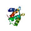
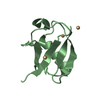
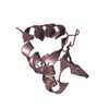
ジャーナル: Structure / 年: 2015 タイトル: The modular structure of the inner-membrane ring component PrgK facilitates assembly of the type III secretion system basal body. 著者: Julien R C Bergeron / Liam J Worrall / Soumya De / Nikolaos G Sgourakis / Adrienne H Cheung / Emilie Lameignere / Mark Okon / Gregory A Wasney / David Baker / Lawrence P McIntosh / Natalie C J Strynadka / 要旨: The type III secretion system (T3SS) is a large macromolecular assembly found at the surface of many pathogenic Gram-negative bacteria. Its role is to inject toxic "effector" proteins into the cells ...The type III secretion system (T3SS) is a large macromolecular assembly found at the surface of many pathogenic Gram-negative bacteria. Its role is to inject toxic "effector" proteins into the cells of infected organisms. The molecular details of the assembly of this large, multimembrane-spanning complex remain poorly understood. Here, we report structural, biochemical, and functional analyses of PrgK, an inner-membrane component of the prototypical Salmonella typhimurium T3SS. We have obtained the atomic structures of the two ring building globular domains and show that the C-terminal transmembrane helix is not essential for assembly and secretion. We also demonstrate that structural rearrangement of the two PrgK globular domains, driven by an interconnecting linker region, may promote oligomerization into ring structures. Finally, we used electron microscopy-guided symmetry modeling to propose a structural model for the intimately associated PrgH-PrgK ring interaction within the assembled basal body.
NOE constraints total: 1912 / NOE intraresidue total count: 147 / NOE long range total count: 272 / NOE medium range total count: 296 / NOE sequential total count: 1197 / Protein phi angle constraints total count: 50 / Protein psi angle constraints total count: 51
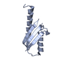
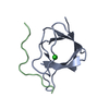
代表構造
選択基準: lowest energy
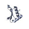
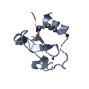
NMRアンサンブル
コンフォーマー選択の基準: target function / 計算したコンフォーマーの数: 200 / 登録したコンフォーマーの数: 20
 ムービー
ムービー コントローラー


 データを開く
 基本情報
基本情報 要素
要素 キーワード
キーワード Secretion systems (分泌) / macromolecular assemblies
 機能・相同性情報
 データ登録者
 引用
引用

 構造の表示
構造の表示 ダウンロードとリンク
 その他のダウンロード


 PDBj
PDBj 集合体
集合体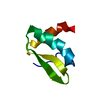
 試料調製
試料調製 解析
解析