ムービー
ムービー コントローラー


+ データを開く

- 基本情報
基本情報
| 登録情報 | データベース: PDB / ID: 1xyr | ||||||
|---|---|---|---|---|---|---|---|
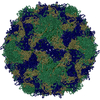
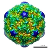
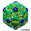
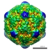
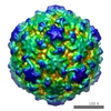
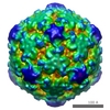
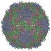
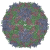
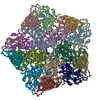
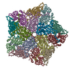
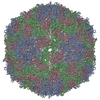
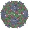
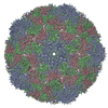
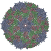
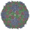
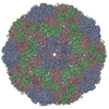
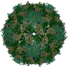
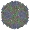
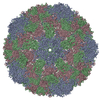
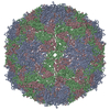
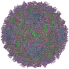
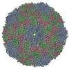
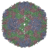
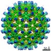
| タイトル | Poliovirus 135S cell entry intermediate | ||||||
 要素 要素 | (Genome polyprotein, Coat protein ...) x 7 | ||||||
 キーワード キーワード |  VIRUS (ウイルス) / BETA BARREL (Βバレル) / VIRAL CAPSID (カプシド) / CELL ENTRY INTERMEDIATE / Icosahedral virus VIRUS (ウイルス) / BETA BARREL (Βバレル) / VIRAL CAPSID (カプシド) / CELL ENTRY INTERMEDIATE / Icosahedral virus | ||||||
| 機能・相同性 |  機能・相同性情報 機能・相同性情報symbiont-mediated suppression of host translation initiation / symbiont-mediated suppression of host cytoplasmic pattern recognition receptor signaling pathway via inhibition of MDA-5 activity / symbiont-mediated suppression of host cytoplasmic pattern recognition receptor signaling pathway via inhibition of RIG-I activity / ピコルナイン2A / symbiont-mediated suppression of host mRNA export from nucleus / symbiont-mediated suppression of host cytoplasmic pattern recognition receptor signaling pathway via inhibition of MAVS activity / symbiont genome entry into host cell via pore formation in plasma membrane / picornain 3C / ribonucleoside triphosphate phosphatase activity / T=pseudo3 icosahedral viral capsid ...symbiont-mediated suppression of host translation initiation / symbiont-mediated suppression of host cytoplasmic pattern recognition receptor signaling pathway via inhibition of MDA-5 activity / symbiont-mediated suppression of host cytoplasmic pattern recognition receptor signaling pathway via inhibition of RIG-I activity / ピコルナイン2A / symbiont-mediated suppression of host mRNA export from nucleus / symbiont-mediated suppression of host cytoplasmic pattern recognition receptor signaling pathway via inhibition of MAVS activity / symbiont genome entry into host cell via pore formation in plasma membrane / picornain 3C / ribonucleoside triphosphate phosphatase activity / T=pseudo3 icosahedral viral capsid / host cell cytoplasmic vesicle membrane / endocytosis involved in viral entry into host cell / : / nucleoside-triphosphate phosphatase / protein complex oligomerization / monoatomic ion channel activity / RNA helicase activity / induction by virus of host autophagy / RNA依存性RNAポリメラーゼ / symbiont-mediated suppression of host gene expression / viral RNA genome replication / cysteine-type endopeptidase activity / RNA-dependent RNA polymerase activity / DNA-templated transcription / host cell nucleus / structural molecule activity / virion attachment to host cell / タンパク質分解 / RNA binding / ATP binding / 生体膜 / metal ion binding類似検索 - 分子機能 | ||||||
| 生物種 |  Human poliovirus 1 (ポリオウイルス) Human poliovirus 1 (ポリオウイルス) | ||||||
| 手法 | 電子顕微鏡法 / 単粒子再構成法 / クライオ電子顕微鏡法 / 解像度: 11 Å | ||||||
 データ登録者 データ登録者 | Bubeck, D. / Filman, D.J. / Cheng, N. / Steven, A.C. / Hogle, J.M. / Belnap, D.M. | ||||||
 引用 引用 | ジャーナル: J Virol / 年: 2005 タイトル: The structure of the poliovirus 135S cell entry intermediate at 10-angstrom resolution reveals the location of an externalized polypeptide that binds to membranes. 著者: Doryen Bubeck / David J Filman / Naiqian Cheng / Alasdair C Steven / James M Hogle / David M Belnap /  要旨: Poliovirus provides a well-characterized system for understanding how nonenveloped viruses enter and infect cells. Upon binding its receptor, poliovirus undergoes an irreversible conformational ...Poliovirus provides a well-characterized system for understanding how nonenveloped viruses enter and infect cells. Upon binding its receptor, poliovirus undergoes an irreversible conformational change to the 135S cell entry intermediate. This transition involves shifts of the capsid protein beta barrels, accompanied by the externalization of VP4 and the N terminus of VP1. Both polypeptides associate with membranes and are postulated to facilitate entry by forming a translocation pore for the viral RNA. We have calculated cryo-electron microscopic reconstructions of 135S particles that permit accurate placement of the beta barrels, loops, and terminal extensions of the capsid proteins. The reconstructions and resulting models indicate that each N terminus of VP1 exits the capsid though an opening in the interface between VP1 and VP3 at the base of the canyon that surrounds the fivefold axis. Comparison with reconstructions of 135S particles in which the first 31 residues of VP1 were proteolytically removed revealed that the externalized N terminus is located near the tips of propeller-like features surrounding the threefold axes rather than at the fivefold axes, as had been proposed in previous models. These observations have forced a reexamination of current models for the role of the 135S particle in transmembrane pore formation and suggest testable alternatives. | ||||||
| 履歴 |
| ||||||
| Remark 999 | SEQUENCE VP1: Chain 1 consists of residues 71-302 of VP1. The residues that make up Chain 8 have ...SEQUENCE VP1: Chain 1 consists of residues 71-302 of VP1. The residues that make up Chain 8 have been identified as residues 42-52 but the identification is very tentative at this point. Residues 1-41 and 53-70 of VP1 are not present in the model. The authors suspect that residues 1-41 are mostly disordered, and that residues 53-70 may be differently ordered than in the native, but are not buildable at this resolution. VP2: Chain 2 consists of residues 28-264 of VP2. Chain 7 includes residues 13-26 from a (icosahedral-symmetry-related) copy of VP2. An alpha carbon from residue 27 is deliberately missing from the rigid-body model to create a hinge point for the modeling. Residues 26 (of chain 7) and 28 (of chain 2) are very far from one another in space because they come from different icosahedral-symmetry-related copies of VP2. Residues 1-12 of VP2 are probably disordered. Residues 265-272 of VP2 could not be modeled. VP3: Alpha carbons for 13 and 49 were deliberately omitted from the rigid-body model, to create hinge points for the modeling. C-terminal residues 232-235 or 233-235 (chain 3) are missing from all of the high-resolution (type 1 mahoney) native poliovirus structures, either due to disorder or proteolysis. AUTHOR HAS MODELLED RESIDUES 123 OF CHAIN 3 AS SER, WHEREAS THE CORRESPONDING RESIDUE 463 IN SWISSPROT IS PHE. THE AUTHOR CLAIMS THERE IS AN ERROR IN SWISSPROT AND THAT RESIDUE 123 SHOULD BE SER. |
- 構造の表示
構造の表示
| ムービー |
ムービービューア |
|---|---|
| 構造ビューア | 分子: MolmilJmol/JSmol |
- ダウンロードとリンク
-ダウンロード
| PDBx/mmCIF形式 | 1xyr.cif.gz | 38.6 KB | 表示 | PDBx/mmCIF形式 |
|---|---|---|---|---|
| PDB形式 | pdb1xyr.ent.gz | 19.2 KB | 表示 | PDB形式 |
| PDBx/mmJSON形式 | 1xyr.json.gz | ツリー表示 | PDBx/mmJSON形式 | |
| その他 |  その他のダウンロード その他のダウンロード |
-検証レポート
| アーカイブディレクトリ | https://data.pdbj.org/pub/pdb/validation_reports/xy/1xyrftp://data.pdbj.org/pub/pdb/validation_reports/xy/1xyr | HTTPS FTP |
|---|
-関連構造データ
-リンク
 PDBj
PDBj

- 集合体
集合体
| 登録構造単位 | 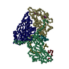
|
|---|---|
| 1 | x 60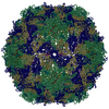
|
| 2 |
|
| 3 | x 5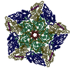
|
| 4 | x 6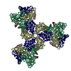
|
| 5 | 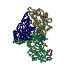
|
| 対称性 | 点対称性: (ヘルマン・モーガン記号: 532 / シェーンフリース記号: I (正20面体型対称)) |
-要素
-Genome polyprotein, Coat protein ... , 7種, 7分子 1235678
| #1: タンパク質 | 分子量: 26135.506 Da / 分子数: 1 / 断片: residues 649-880 / 由来タイプ: 天然 / 詳細: Virus was propogated in HELA cell suspension / 由来: (天然) Human poliovirus 1 (ポリオウイルス) / 属: Enterovirusエンテロウイルス / 生物種: Poliovirusポリオウイルス / 株: Type 1 Mahoney / 参照: UniProt: P03300 |
|---|---|
| #2: タンパク質 | 分子量: 26244.518 Da / 分子数: 1 / 断片: residues 96-332 / 由来タイプ: 天然 / 詳細: Virus was propogated in HELA cell suspension / 由来: (天然) Human poliovirus 1 (ポリオウイルス) / 属: Enterovirusエンテロウイルス / 生物種: Poliovirusポリオウイルス / 株: Type 1 Mahoney / 参照: UniProt: P03300 |
| #3: タンパク質 | 分子量: 20488.623 Da / 分子数: 1 / 断片: residues 390-571 / 由来タイプ: 天然 / 詳細: Virus was propogated in HELA cell suspension / 由来: (天然) Human poliovirus 1 (ポリオウイルス) / 属: Enterovirusエンテロウイルス / 生物種: Poliovirusポリオウイルス / 株: Type 1 Mahoney / 参照: UniProt: P03300 |
| #4: タンパク質・ペプチド | 分子量: 1214.349 Da / 分子数: 1 / 断片: residues 341-352 / 由来タイプ: 天然 / 詳細: Virus was propogated in HELA cell suspension / 由来: (天然) Human poliovirus 1 (ポリオウイルス) / 属: Enterovirusエンテロウイルス / 生物種: Poliovirusポリオウイルス / 株: Type 1 Mahoney / 参照: UniProt: P03300 |
| #5: タンパク質・ペプチド | 分子量: 3947.482 Da / 分子数: 1 / 断片: residues 354-389 / 由来タイプ: 天然 / 詳細: Virus was propogated in HELA cell suspension / 由来: (天然) Human poliovirus 1 (ポリオウイルス) / 属: Enterovirusエンテロウイルス / 生物種: Poliovirusポリオウイルス / 株: Type 1 Mahoney / 参照: UniProt: P03300 |
| #6: タンパク質・ペプチド | 分子量: 1488.682 Da / 分子数: 1 / 断片: residues 81-94 / 由来タイプ: 天然 / 詳細: Virus was propogated in HELA cell suspension / 由来: (天然) Human poliovirus 1 (ポリオウイルス) / 属: Enterovirusエンテロウイルス / 生物種: Poliovirusポリオウイルス / 株: Type 1 Mahoney / 参照: UniProt: P03300 |
| #7: タンパク質・ペプチド | 分子量: 1030.129 Da / 分子数: 1 / 断片: residues 620-630 / 由来タイプ: 天然 / 詳細: Virus was propogated in HELA cell suspension / 由来: (天然) Human poliovirus 1 (ポリオウイルス) / 属: Enterovirusエンテロウイルス / 生物種: Poliovirusポリオウイルス / 株: Type 1 Mahoney / 参照: UniProt: P03300 |
-実験情報
-実験
| 実験 | 手法: 電子顕微鏡法 |
|---|---|
| EM実験 | 試料の集合状態: PARTICLE / 3次元再構成法: 単粒子再構成法 |
- 試料調製
試料調製
| 構成要素 | 名称: POLIOVIRUS 135S CELL ENTRY INTERMEDIATE / タイプ: VIRUS 詳細: 135S was made by heating native virus in 20mM HEPES pH 7.4, 2mM CaCl2 at 50 degrees for 3 minutes |
|---|---|
| ウイルスについての詳細 | ホストのカテゴリ: VERTEBRATES / タイプ: VIRION |
| 天然宿主 | 生物種: Homo sapiens / 株: HELA |
| 緩衝液 | 名称: 20mM HEPES, 2mM CaCl2 / pH: 7.4 / 詳細: 20mM HEPES, 2mM CaCl2 |
| 試料 | 濃度: 0.7 mg/ml / 包埋: NO / シャドウイング: NO / 染色: NO / 凍結: YES |
| 試料支持 | 詳細: Quantifoil Holey Carbon Grids (R2/2) |
| 急速凍結 | 凍結剤: ETHANE / 詳細: Plunge freezing into liquid ethane |
- 電子顕微鏡撮影
| 実験機器 |  モデル: Tecnai F20 / 画像提供: FEI Company |
|---|---|
| 顕微鏡 | モデル: FEI TECNAI F20 / 日付: 2002年9月17日 / 詳細: Focal pairs were taken |
| 電子銃 | 電子線源: FIELD EMISSION GUN / 加速電圧: 200 kV / 照射モード: FLOOD BEAM |
| 電子レンズ | モード: BRIGHT FIELDBright-field microscopy / 倍率(公称値): 50000 X / 倍率(補正後): 50000 X / 最大 デフォーカス(公称値): 3000 nm / 最小 デフォーカス(公称値): 900 nm / Cs: 2 mm |
| 試料ホルダ | 温度: 77 K / 傾斜角・最大: 0 ° / 傾斜角・最小: 0 ° |
| 撮影 | 電子線照射量: 1 e/Å2 / フィルム・検出器のモデル: KODAK SO-163 FILM |
- 解析
解析
| EMソフトウェア |
| ||||||||||||
|---|---|---|---|---|---|---|---|---|---|---|---|---|---|
| CTF補正 | 詳細: CTF correction of each particle. Phase flipped correction for particles included in the orientation search, and deconvoluted and gaussian decay corrected for the particles used in the reconstruction | ||||||||||||
| 対称性 | 点対称性: I (正20面体型対称) | ||||||||||||
| 3次元再構成 | 手法: icosahedrally symmetric model-based orientation search (PFT and EM3DR) 解像度: 11 Å / 粒子像の数: 3641 / ピクセルサイズ(公称値): 2.69 Å / ピクセルサイズ(実測値): 2.69 Å 倍率補正: radial scaling to previous low-resolution reconstruction which had been calibrated to crystal structure of native virus 対称性のタイプ: POINT | ||||||||||||
| 原子モデル構築 | プロトコル: RIGID BODY FIT / 空間: RECIPROCAL Target criteria: amplitude-weighted phase error, and the summation of the squared model-based amplitudes. 詳細: METHOD--gradient search REFINEMENT PROTOCOL--Rigid body (chains 1 and 6 as one rigid body, chains 3 and 7 as one rigid body, chains 2 and 8 as individual rigid bodies, chain 5 placed along 5fold axis visually) | ||||||||||||
| 原子モデル構築 | PDB-ID: 1HXS Accession code: 1HXS / Source name: PDB / タイプ: experimental model | ||||||||||||
| 精密化ステップ | サイクル: LAST
|