 ムービー
ムービー コントローラー


+ データを開く

- 基本情報
基本情報
| 登録情報 | データベース: EMDB / ID: EMD-8727 | ||||||||||||
|---|---|---|---|---|---|---|---|---|---|---|---|---|---|
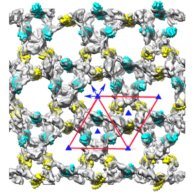
| タイトル | The cryo-ET structure of frozen-hydrated honey bee Z-disk | ||||||||||||
 マップデータ マップデータ | frozen-hydrated honey bee Z-disk- filtered to 60 Angstrom | ||||||||||||
 試料 試料 |
| ||||||||||||
| 生物種 |   Apis mellifera (セイヨウミツバチ) Apis mellifera (セイヨウミツバチ) | ||||||||||||
| 手法 | サブトモグラム平均法 / クライオ電子顕微鏡法 / 解像度: 60.0 Å | ||||||||||||
 データ登録者 データ登録者 | Taylor KA / Hu Z | ||||||||||||
| 資金援助 | European Union,  米国, 3件 米国, 3件
| ||||||||||||
 引用 引用 | ジャーナル: J Muscle Res Cell Motil / 年: 2017 タイトル: Structure of isolated Z-disks from honeybee flight muscle. 著者: Mara Rusu / Zhongjun Hu / Kenneth A Taylor / John Trinick /  要旨: The Z-disk is a complex structure comprising some 40 proteins that are involved in the transmission of force developed during muscle contraction and in important signalling pathways that govern ...The Z-disk is a complex structure comprising some 40 proteins that are involved in the transmission of force developed during muscle contraction and in important signalling pathways that govern muscle homeostasis. In the Z-disk the ends of antiparallel thin filaments from adjacent sarcomeres are crosslinked by α-actinin. The structure of the Z-disk lattice varies greatly throughout the animal kingdom. In vertebrates the thin filaments form a tetragonal lattice, whereas invertebrate flight muscle has a hexagonal lattice. The width of the Z-disk varies considerably and correlates with the number of α-actinin bridges. A detailed description at a high resolution of the Z-disk lattice is needed in order to better understand muscle function and disease. The molecular architecture of the Z-disk lattice in honeybee (Apis mellifera) is known from plastic embedded thin sections to a resolution of 7 nm, which is not sufficient to dock component protein crystal structures. It has been shown that sectioning is a damaging process that leads to the loss of finer details present in biological specimens. However, the Apis Z-disk is a thin structure (120 nm) suitable for cryo EM. We have isolated intact honeybee Z-disks from indirect flight muscle, thus obviating the need of plastic sectioning. We have employed cryo electron tomography and image processing to investigate the arrangement of proteins within the hexagonal lattice of the Apis Z-disk. The resolution obtained, ~6 nm, was probably limited by damage caused by the harshness of the conditions used to extract the myofibrils and isolate the Z-disks. | ||||||||||||
| 履歴 |
|
- 構造の表示
構造の表示
| ムービー |
 ムービービューア ムービービューア |
|---|---|
| 構造ビューア | EMマップ: SurfViewMolmilJmol/JSmol |
| 添付画像 |
- ダウンロードとリンク
-EMDBアーカイブ
| マップデータ | emd_8727.map.gz | 24.9 MB | EMDBマップデータ形式 | |
|---|---|---|---|---|
| ヘッダ (付随情報) | emd-8727-v30.xmlemd-8727.xml | 19.7 KB 19.7 KB | 表示 表示 | EMDBヘッダ |
| FSC (解像度算出) | emd_8727_fsc.xml | 6.3 KB | 表示 | FSCデータファイル |
| 画像 |  emd_8727.png emd_8727.png | 255.3 KB | ||
| その他 | emd_8727_additional.map.gzemd_8727_additional_1.map.gz | 2.1 GB 2.1 GB | ||
| アーカイブディレクトリ |  http://ftp.pdbj.org/pub/emdb/structures/EMD-8727ftp://ftp.pdbj.org/pub/emdb/structures/EMD-8727 http://ftp.pdbj.org/pub/emdb/structures/EMD-8727ftp://ftp.pdbj.org/pub/emdb/structures/EMD-8727 | HTTPS FTP |
-関連構造データ
| 関連構造データ | |
|---|---|
| 類似構造データ | |
| 電子顕微鏡画像生データ | EMPIAR-10095 (タイトル: Structure of the Z-disk isolated from the indirect flight muscle of the honey bee Data size: 3.0 / Data #1: tomo9.mrc [class averages]) |
-リンク
| EMDBのページ | EMDB (EBI/PDBe) / EMDataResource |
|---|
-マップ
| ファイル | ダウンロード / ファイル: emd_8727.map.gz / 形式: CCP4 / 大きさ: 27 MB / タイプ: IMAGE STORED AS FLOATING POINT NUMBER (4 BYTES) | ||||||||||||||||||||||||||||||||||||||||||||||||||||||||||||||||||||
|---|---|---|---|---|---|---|---|---|---|---|---|---|---|---|---|---|---|---|---|---|---|---|---|---|---|---|---|---|---|---|---|---|---|---|---|---|---|---|---|---|---|---|---|---|---|---|---|---|---|---|---|---|---|---|---|---|---|---|---|---|---|---|---|---|---|---|---|---|---|
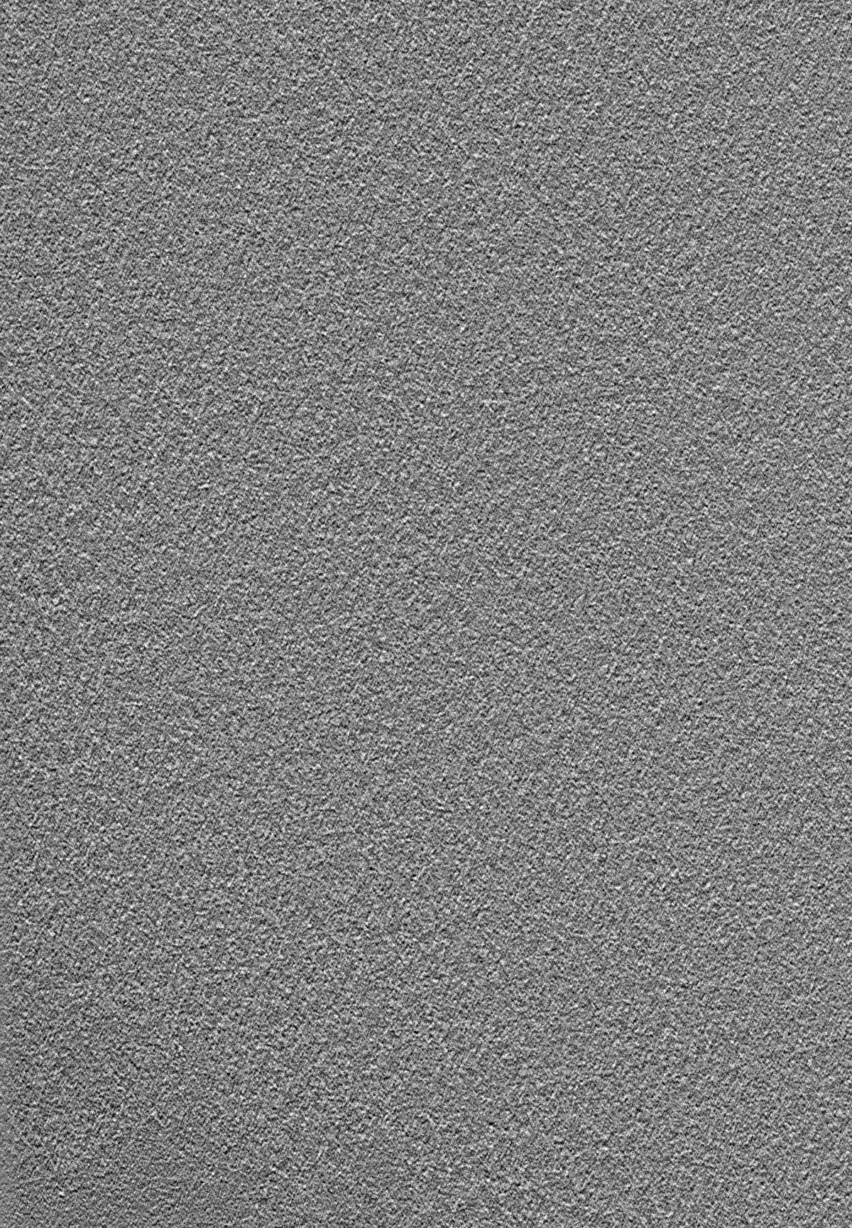
| 注釈 | frozen-hydrated honey bee Z-disk- filtered to 60 Angstrom | ||||||||||||||||||||||||||||||||||||||||||||||||||||||||||||||||||||
| ボクセルのサイズ | X=Y=Z: 7.6 Å | ||||||||||||||||||||||||||||||||||||||||||||||||||||||||||||||||||||
| 密度 |
| ||||||||||||||||||||||||||||||||||||||||||||||||||||||||||||||||||||
| 対称性 | 空間群: 1 | ||||||||||||||||||||||||||||||||||||||||||||||||||||||||||||||||||||
| 詳細 | EMDB XML:
CCP4マップ ヘッダ情報:
| ||||||||||||||||||||||||||||||||||||||||||||||||||||||||||||||||||||
-添付データ
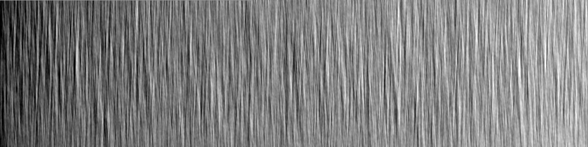
-追加マップ: frozen-hydrated honey bee Z-disk - raw tomogram. The...
| ファイル | emd_8727_additional.map | ||||||||||||
|---|---|---|---|---|---|---|---|---|---|---|---|---|---|
| 注釈 | frozen-hydrated honey bee Z-disk - raw tomogram. The contour level here is non-sense. The map is raw tomogram, which is used to make the subvolume. Layer line data is not actually layer line, instead, it is a coordiates file for extracting subvolume | ||||||||||||
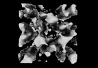
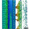
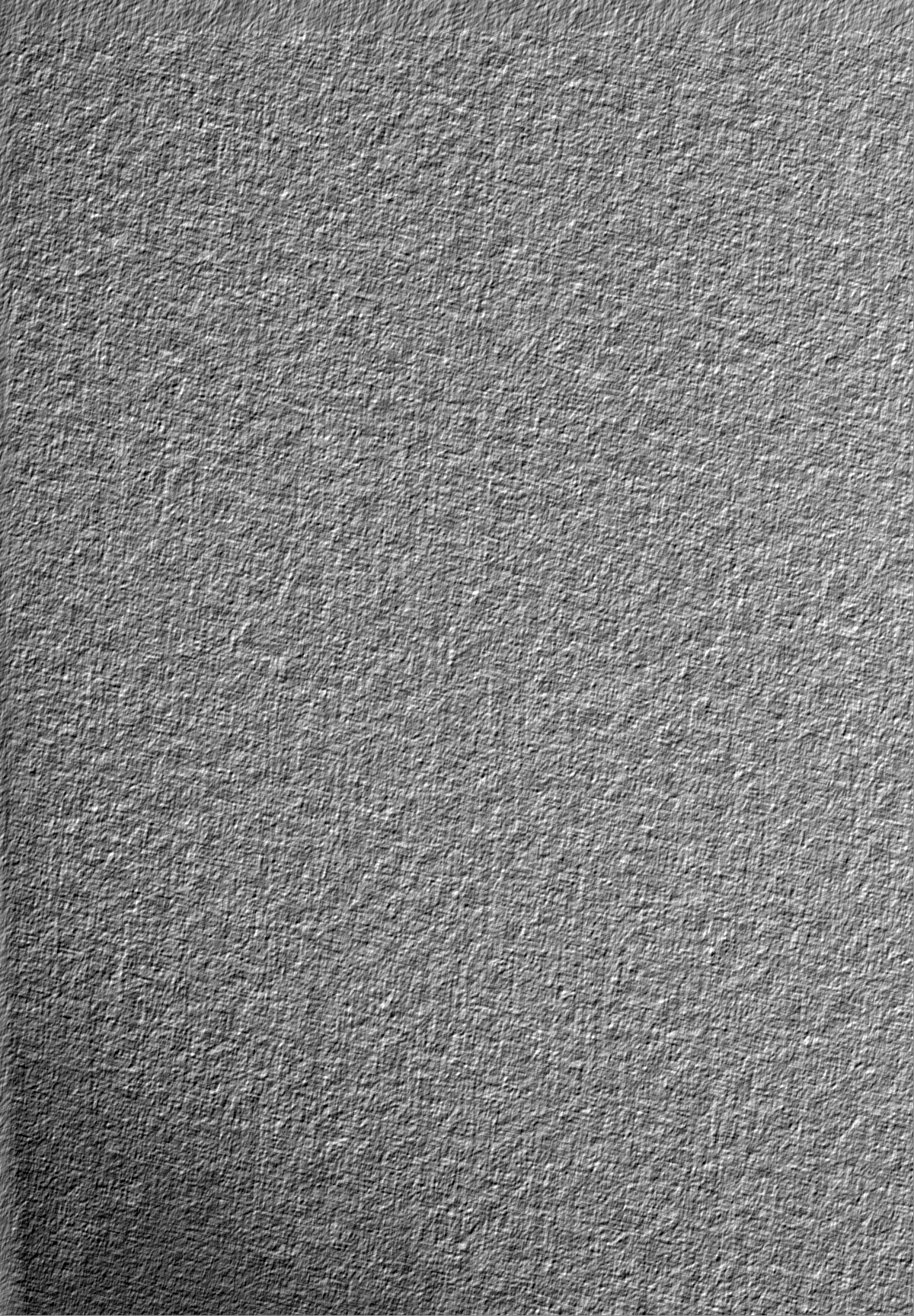
| 投影像・断面図 |
| ||||||||||||
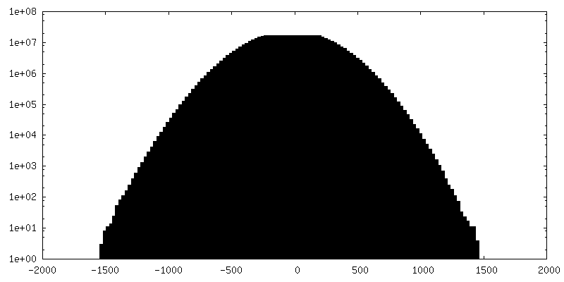
| 密度ヒストグラム |
 Z
Z Y
Y X
X


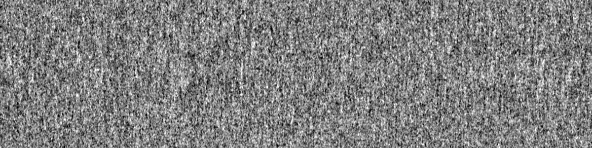
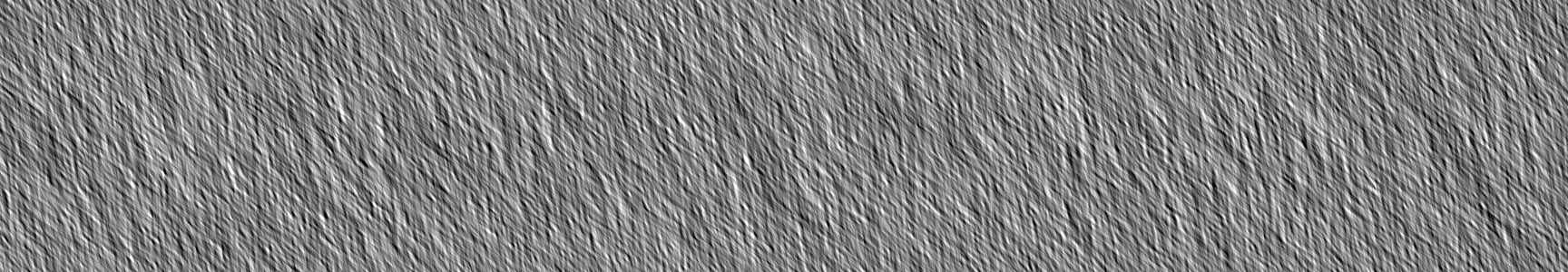
-追加マップ: frozen-hydrated honey bee Z-disk - raw tomogram. The...
| ファイル | emd_8727_additional_1.map | ||||||||||||
|---|---|---|---|---|---|---|---|---|---|---|---|---|---|
| 注釈 | frozen-hydrated honey bee Z-disk - raw tomogram. The contour level here is non-sense. The map is raw tomogram, which is used to make the subvolume. Layer line data is not actually layer line, instead, it is a coordiates file for extracting subvolume | ||||||||||||
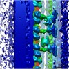
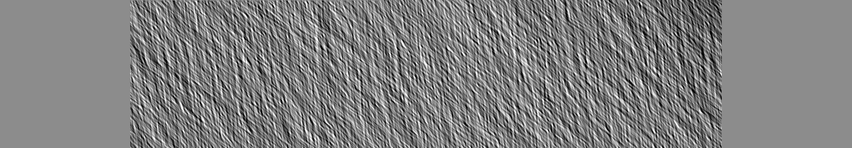
| 投影像・断面図 |
| ||||||||||||
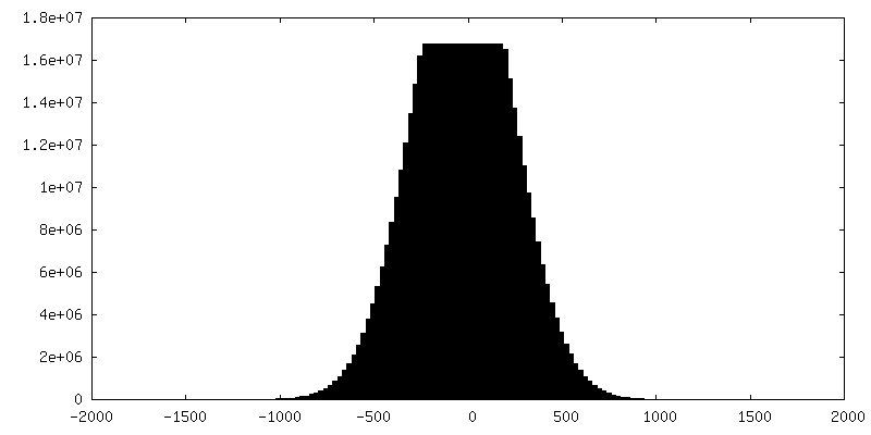
| 密度ヒストグラム |


- 試料の構成要素
-全体 : Z-disc
| 全体 | 名称: Z-disc |
|---|---|
| 要素 |
|
-超分子 #1: Z-disc
| 超分子 | 名称: Z-disc / タイプ: organelle_or_cellular_component / ID: 1 / 親要素: 0 / 含まれる分子: all 詳細: Z-disc directly isolated from Apis mellifera (honey bee) indirect flight muscle |
|---|---|
| 由来(天然) | 生物種: Apis mellifera (セイヨウミツバチ) / 器官: flight muscle / 組織: muscle / Organelle: Z-disc / 細胞中の位置: sarcomere |
-分子 #1: Actin
| 分子 | 名称: Actin / タイプ: protein_or_peptide / ID: 1 / 光学異性体: LEVO |
|---|---|
| 由来(天然) | 生物種: Apis mellifera (セイヨウミツバチ) / 器官: flight muscle / 組織: muscle |
| 配列 | 文字列: MVGMGQKDSY VGDEAQSKRG ILTLKYPIEH GIITNWDDME KIWHHTFYNE LRVAPEEHPV LLTEAPLNPK ANREKMTQIM FETFNSPAMY VAIQAVLSLY ASGRTTGIVL DSGDGVSHTV PIYEGYALPH AILRLDLAGR DLTDYLMKIL TERGYSFTTT AEREIVRDIK ...文字列: MVGMGQKDSY VGDEAQSKRG ILTLKYPIEH GIITNWDDME KIWHHTFYNE LRVAPEEHPV LLTEAPLNPK ANREKMTQIM FETFNSPAMY VAIQAVLSLY ASGRTTGIVL DSGDGVSHTV PIYEGYALPH AILRLDLAGR DLTDYLMKIL TERGYSFTTT AEREIVRDIK EKLCYVALDF EQEMATAAAS TSLEKSYELP DGQVITIGNE RFRCPEALFQ PSFLGMESCG IHETVYNSIM KCDVDIRKDL YANNVLSGGT TMYPGIADRM QKEITALAPS TIKIKIIAPP ERKYSVWIGG SILASLSTFQ QMWISKQEYD ESGPGIVHRK CF |
-分子 #2: alpha-actinin
| 分子 | 名称: alpha-actinin / タイプ: protein_or_peptide / ID: 2 / 光学異性体: LEVO |
|---|---|
| 由来(天然) | 生物種: Apis mellifera (セイヨウミツバチ) / 器官: Flight muscle / 組織: muscle |
| 配列 | 文字列: MEEYERLASD LLEWIRRTMP WLASRQTDNS LAGCQKKLEE YRTYRRKHKP PRVEQKAKLE TNFNTLQTKL RLSNRPAYMP TEGKMVSDIN KAWKGLELAE KSFEEWLLSE MMRLERLEHL AQKFKHKADA HEEWTAGKEE MLTSQHFRQC KLNELKALKK KHEAFESDLA ...文字列: MEEYERLASD LLEWIRRTMP WLASRQTDNS LAGCQKKLEE YRTYRRKHKP PRVEQKAKLE TNFNTLQTKL RLSNRPAYMP TEGKMVSDIN KAWKGLELAE KSFEEWLLSE MMRLERLEHL AQKFKHKADA HEEWTAGKEE MLTSQHFRQC KLNELKALKK KHEAFESDLA AHQDRVEQIA AIAQELNTLE YHDSASVNAR CQRICDQWDR LGTLTQRRRQ ALDEAERILE KIDVLHLEFA KRAAPFNNWL DGTREDLVDM FIVHTMEEIQ GLMDAHAAFK ATLGEADKEY NAIVGLVREV ESIVKQFQIP GGLENPYTTL TALDLTKKWS DVRQLVPQRD GTLQAELRKQ QNNELLRRQF AEKANAVGPW IERQLDAVTA IGLGLQGTLE DQLHRLKEYE QAVYQYKVHL EELEKIHQAV QEGMIFENRY TQYTMETLRV GWEQLLTSIN RNINEVENQI LTRDSKGITQ EQLNEFRSSF NHFDKNRTGR LAPDEFKSCL VSLGYSIGKD RQGDIDFQRI LAIVDPNNSG YVHFDAFLDF MTRESTDTDT AEQVIDSFRI LAGDKPYILA DELRRELPPD QAEYCIQRMP PYKGPNAIPG ALDYRSFSTA LYGESDL |
-実験情報
-構造解析
| 手法 | クライオ電子顕微鏡法 |
|---|---|
 解析 解析 | サブトモグラム平均法 |
| 試料の集合状態 | tissue |
-試料調製
| 緩衝液 | pH: 7.2 構成要素:
| |||||||||
|---|---|---|---|---|---|---|---|---|---|---|
| グリッド | モデル: Quantifoil R3.5/1 / 材質: COPPER / メッシュ: 200 / 支持フィルム - 材質: CARBON / 支持フィルム - トポロジー: HOLEY ARRAY / 前処理 - タイプ: GLOW DISCHARGE / 前処理 - 時間: 20 sec. / 前処理 - 雰囲気: OTHER 詳細: Carbon coated grids were made hydrophilic by glow discharging for 40 seconds at a high tension of 10 kV in a Cressington 208 Carbon Coater. | |||||||||
| 凍結 | 凍結剤: ETHANE / チャンバー内湿度: 100 % / チャンバー内温度: 277 K / 装置: FEI VITROBOT MARK IV 詳細: After the Z-disk suspension was added to the grid it was left to settle for 10-15 seconds followed by washing with low salt buffer (25 mM HEPES, 100 mM NaCl). The washing step is crucial for ...詳細: After the Z-disk suspension was added to the grid it was left to settle for 10-15 seconds followed by washing with low salt buffer (25 mM HEPES, 100 mM NaCl). The washing step is crucial for the removal of salts present in the extraction buffer, ensuring that plunge frozen grids were free of contamination. Plunge freezing was carried out using Quantifoil grids (Agar Scientific) in a Vitrobot Mark IV (FEI) at 4-5 degrees C, 90-100% humidity using a blotting force of 3-5 for 4-6 seconds.. | |||||||||
| 詳細 | Indirect flight muscle was harvested from the thorax of Apis mellifera obtained from a local bee keeper. Myofibrils in suspension were prepared according to previously published methods (Bullard et al., 1973; Saide and Ullrick, 1974) with the following modifications. The IFM were collected in ice cold sucrose containing buffer (0.3 M sucrose, 0.1 M KCl, 0.01 M potassium phosphate pH 7, 1 mM MgCl2, 2 mM EGTA, 0.02 M NaN3 and EDTA-free Protease Inhibitor Cocktail Tablets followed by homogenization using a Wheaton tissue grinder. Soluble proteins and sucrose were washed away by centrifugation in 0.1 M KCl, 0.01 M potassium phosphate pH 7 buffer. Myofibrils in suspension were stored in 75% glycerol at -80 degrees C. Intact Z-disks were isolated by incubating myofibrils on ice in a high ionic strength extraction solution containing 0.7 M KCl, 0.6 M KI, 0.08 M NaHCO3 pH 8 for 60 minutes. Following extraction Z-disks were either negatively stained or plunge frozen for further electron microscopic investigation. |
- 電子顕微鏡法
| 顕微鏡 | FEI TITAN KRIOS |
|---|---|
| 電子線 | 加速電圧: 300 kV / 電子線源: FIELD EMISSION GUN |
| 電子光学系 | 倍率(補正後): 22500 / 照射モード: FLOOD BEAM / 撮影モード: BRIGHT FIELDBright-field microscopy / 倍率(公称値): 22500 |
| 試料ステージ | 試料ホルダーモデル: FEI TITAN KRIOS AUTOGRID HOLDER ホルダー冷却材: NITROGEN |
| 詳細 | Tilt series were recorded using the Saxton acquisition scheme (Saxton et al., 1984). Tilt angles ranged from -69.87 to +67.66 degrees starting at 0 degrees, 96 total images, with an initial step of 2 degrees. The average electron dose/micrograph was ~0.7 electrons/Angstrom2. A total of 12 tilt series, designated tomo1 - tomo12, were collected but only one proved to be useful, tomo9. The others had poor resolution due to either low intrinsic order, excessive extraction of actin or other structural Z-disk proteins, or due to excessive radiation damage. |
| 撮影 | フィルム・検出器のモデル: FEI FALCON II (4k x 4k) 検出モード: INTEGRATING / 平均電子線量: 1.0 e/Å2 / 詳細: Electron dose may not be accurate. |
| 実験機器 |  モデル: Titan Krios / 画像提供: FEI Company |
-画像解析
| 結晶パラメータ | 単位格子 - A: 520 Å / 単位格子 - B: 520 Å / 単位格子 - C: 425 Å / 単位格子 - C sampling length: 950 Å / 単位格子 - γ: 60 ° / 面群: P 3 1 2 |
|---|---|
| 抽出 | トモグラム数: 1 / 使用した粒子像数: 399 / 参照モデル: cross correlation / 手法: lattice fitting / ソフトウェア - 名称: I3 (ver. 0.9.4) 詳細: Lattice positions were determined from a cross correlation map computed between the tomogram and a reference masked from within it. The Fourier transform was first filtered using a reciprocal ...詳細: Lattice positions were determined from a cross correlation map computed between the tomogram and a reference masked from within it. The Fourier transform was first filtered using a reciprocal lattice that contained 3 orders in a* and 5 orders in b*. Lattice positions were determined by peak fitting along a grid of predicted positions. Points on the predicted grid that fell outside of the actual Z-disk were removed as were any points that occurred too close to the edge of the tomogram. In total we extracted 399 subvolumes. |
| 最終 3次元分類 | クラス数: 1 / 平均メンバー数/クラス: 399 / 詳細: Here only shows result of global average. |
| 最終 角度割当 | タイプ: NOT APPLICABLE |
| 最終 再構成 | 使用したクラス数: 1 / アルゴリズム: EXACT BACK PROJECTION / 解像度のタイプ: BY AUTHOR / 解像度: 60.0 Å / 解像度の算出法: FSC 0.143 CUT-OFF / ソフトウェア - 名称: I3 (ver. 0.94) 詳細: Base on structure feature, and the fitting model, the resolution was estimated manually. Also, the FSC yielded a value of 60 A. 使用したサブトモグラム数: 399 |
| FSC曲線 (解像度の算出) | 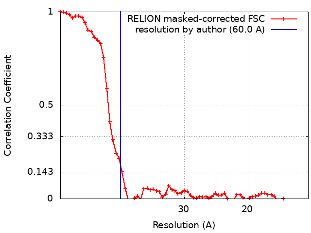 |
-原子モデル構築 1
| 詳細 | For actin, filaments of 28 subunits having the helical symmetry of 28/13 were constructed and maps computed using the software pdb2mrc. These maps were then placed manually within the Z-disk thin filaments and fit quantitatively using chimera. |
|---|---|
| 精密化 | 空間: REAL / プロトコル: RIGID BODY FIT |
