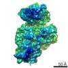
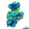
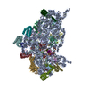
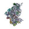
- EMDB-8621: The cryo-EM structure of YjeQ bound to the 30S subunit suggests a... -
+
データを開く
IDまたはキーワード:
読み込み中...
-
基本情報
登録情報
データベース: EMDB / ID: EMD-8621
タイトル
The cryo-EM structure of YjeQ bound to the 30S subunit suggests a fidelity checkpoint function for this protein in ribosome assembly
マップデータ
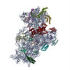
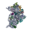
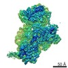
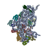
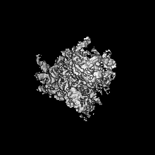
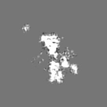
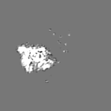
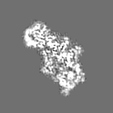
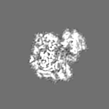
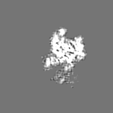
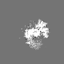
Structure of the mature 30S subunit in complex with YjeQ (RsgA) GTPase
試料
複合体: Structure of the 30S subunit in complex with YjeQ GTPase
RNA: x 1種
タンパク質・ペプチド: x 20種
リガンド: x 2種
機能・相同性
機能・相同性情報
guanosine tetraphosphate binding / mRNA base-pairing translational repressor activity / ornithine decarboxylase inhibitor activity / 加水分解酵素; 酸無水物に作用; リン含有酸無水物に作用 / transcription antitermination factor activity, RNA binding / four-way junction DNA binding / negative regulation of translational initiation / mRNA regulatory element binding translation repressor activity / transcription elongation factor complex / DNA endonuclease activity ...guanosine tetraphosphate binding / mRNA base-pairing translational repressor activity / ornithine decarboxylase inhibitor activity / 加水分解酵素; 酸無水物に作用; リン含有酸無水物に作用 / transcription antitermination factor activity, RNA binding / four-way junction DNA binding / negative regulation of translational initiation / mRNA regulatory element binding translation repressor activity / transcription elongation factor complex / DNA endonuclease activity / regulation of DNA-templated transcription elongation / transcription antitermination / maintenance of translational fidelity / DNA-templated transcription termination / mRNA 5'-UTR binding / ribosomal small subunit biogenesis / small ribosomal subunit rRNA binding / ribosomal small subunit assembly / GDP binding / cytosolic small ribosomal subunit / リボソーム生合成 / small ribosomal subunit / cytoplasmic translation / tRNA binding / negative regulation of translation / rRNA binding / リボソーム / structural constituent of ribosome / 翻訳 (生物学) / ribonucleoprotein complex / response to antibiotic / mRNA binding / GTPase activity / GTP binding / zinc ion binding / 生体膜 / metal ion binding / 細胞質基質 / 細胞質 類似検索 - 分子機能
Ribosome biogenesis GTPase RsgA / RsgA GTPase domain / RsgA GTPase / EngC GTPase domain profile. / Circularly permuted (CP)-type guanine nucleotide-binding (G) domain / Circularly permuted (CP)-type guanine nucleotide-binding (G) domain profile. / Ribosomal protein S14, bacterial/plastid / Ribosomal protein S16, conserved site / Ribosomal protein S16 signature. / Ribosomal protein S3, bacterial-type ...Ribosome biogenesis GTPase RsgA / RsgA GTPase domain / RsgA GTPase / EngC GTPase domain profile. / Circularly permuted (CP)-type guanine nucleotide-binding (G) domain / Circularly permuted (CP)-type guanine nucleotide-binding (G) domain profile. / Ribosomal protein S14, bacterial/plastid / Ribosomal protein S16, conserved site / Ribosomal protein S16 signature. / Ribosomal protein S3, bacterial-type / Ribosomal protein S6, conserved site / Ribosomal protein S6 signature. / Ribosomal protein S19, bacterial-type / Ribosomal protein S7, bacterial/organellar-type / Ribosomal protein S11, bacterial-type / Ribosomal protein S13, bacterial-type / Ribosomal protein S20 / Ribosomal protein S20 superfamily / Ribosomal protein S20 / Ribosomal protein S9, bacterial/plastid / Ribosomal protein S4, bacterial-type / 30S ribosomal protein S17 / Ribosomal protein S5, bacterial-type / Ribosomal protein S6, plastid/chloroplast / Ribosomal protein S2, bacteria/mitochondria/plastid / Ribosomal protein S18, conserved site / Ribosomal protein S18 signature. / Ribosomal protein S16 / Ribosomal protein S16 / Ribosomal protein S16 domain superfamily / Ribosomal protein S15, bacterial-type / Ribosomal protein S2 signature 2. / Ribosomal protein S6 / Ribosomal protein S6 / Ribosomal protein S6 superfamily / Ribosomal protein S12, bacterial-type / Translation elongation factor EF1B/ribosomal protein S6 / Ribosomal protein S18 / Ribosomal protein S18 / Ribosomal protein S18 superfamily / KHドメイン / K homology RNA-binding domain / Ribosomal protein S3, conserved site / Ribosomal protein S14, conserved site / Ribosomal protein S10, conserved site / : / K Homology domain, type 2 / Ribosomal protein S3, C-terminal / Ribosomal protein S3, C-terminal domain superfamily / Ribosomal protein S15/S19, conserved site / KHドメイン / Ribosomal protein S19/S15 / Ribosomal protein S19/S15, superfamily / Ribosomal protein S10 / Ribosomal protein S3, C-terminal domain / Ribosomal protein S3 signature. / Ribosomal protein S10 signature. / Ribosomal protein S14 signature. / Ribosomal protein S7, conserved site / K homology domain superfamily, prokaryotic type / Ribosomal protein S17, conserved site / Ribosomal protein S19 / Ribosomal protein S2 signature 1. / Ribosomal protein S2, conserved site / Ribosomal protein S13, conserved site / Ribosomal protein S2 / Ribosomal protein S2, flavodoxin-like domain superfamily / Ribosomal protein S13 / 30s ribosomal protein S13, C-terminal / Ribosomal protein S2 / Ribosomal protein S4/S9 N-terminal domain / Ribosomal protein S14 / Ribosomal protein S4/S9, N-terminal / Ribosomal protein S4, conserved site / Type-2 KH domain profile. / Ribosomal protein S4/S9 N-terminal domain / Ribosomal protein S13/S18 / Ribosomal protein S4/S9 / Ribosomal protein S19 signature. / K homology domain-like, alpha/beta / Ribosomal protein S14p/S29e / Ribosomal protein S8 / Ribosomal protein S8 superfamily / Ribosomal protein S5, N-terminal, conserved site / Ribosomal protein S5 signature. / Ribosomal protein S5 / Ribosomal protein S5, N-terminal / Ribosomal protein S7 signature. / Ribosomal protein S10p/S20e / S5 double stranded RNA-binding domain profile. / Ribosomal protein S5, C-terminal / Ribosomal protein S9, conserved site / Ribosomal protein S5, N-terminal domain / Ribosomal protein S8 / Ribosomal protein S10 domain / Ribosomal protein S10 domain superfamily / Ribosomal protein S13-like, H2TH / Ribosomal protein S17 signature. / Ribosomal protein S5, C-terminal domain / S4 RNA-binding domain 類似検索 - ドメイン・相同性
Small ribosomal subunit protein bS20 / Small ribosomal subunit protein uS2 / Small ribosomal subunit protein uS9 / Small ribosomal subunit protein uS4 / Small ribosomal subunit protein uS11 / Small ribosomal subunit protein uS8 / Small ribosomal subunit protein uS14 / Small ribosomal subunit protein uS17 / Small ribosomal subunit protein uS3 / Small ribosomal subunit protein uS19 ...Small ribosomal subunit protein bS20 / Small ribosomal subunit protein uS2 / Small ribosomal subunit protein uS9 / Small ribosomal subunit protein uS4 / Small ribosomal subunit protein uS11 / Small ribosomal subunit protein uS8 / Small ribosomal subunit protein uS14 / Small ribosomal subunit protein uS17 / Small ribosomal subunit protein uS3 / Small ribosomal subunit protein uS19 / Small ribosomal subunit protein uS10 / Small ribosomal subunit protein uS12 / Small ribosomal subunit protein bS16 / Small ribosomal subunit protein bS18 / Small ribosomal subunit protein bS6 / Small ribosomal subunit protein uS7 / Small ribosomal subunit protein uS10 / Small ribosomal subunit protein uS13 / Small ribosomal subunit protein uS13 / Small ribosomal subunit protein bS16 / Small ribosomal subunit protein uS19 / Small ribosomal subunit protein bS20 / Small ribosomal subunit protein uS2 / Small ribosomal subunit protein uS3 / Small ribosomal subunit protein uS4 / Small ribosomal subunit protein uS5 / Small ribosomal subunit protein uS5 / Small ribosomal subunit protein uS15 / Small ribosomal subunit protein uS14 / Small ribosomal subunit biogenesis GTPase RsgA / Small ribosomal subunit protein uS15 類似検索 - 構成要素
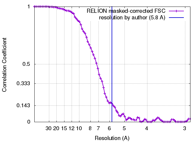
ジャーナル: Proc Natl Acad Sci U S A / 年: 2017 タイトル: The cryo-EM structure of YjeQ bound to the 30S subunit suggests a fidelity checkpoint function for this protein in ribosome assembly. 著者: Aida Razi / Alba Guarné / Joaquin Ortega / 要旨: Recent work suggests that bacterial YjeQ (RsgA) participates in the late stages of assembly of the 30S subunit and aids the assembly of the decoding center but also binds the mature 30S subunit with ...Recent work suggests that bacterial YjeQ (RsgA) participates in the late stages of assembly of the 30S subunit and aids the assembly of the decoding center but also binds the mature 30S subunit with high affinity. To determine the function and mechanisms of YjeQ in the context of the mature subunit, we determined the cryo-EM structure of the fully assembled 30S subunit in complex with YjeQ at 5.8-Å resolution. We found that binding of YjeQ stabilizes helix 44 into a conformation similar to that adopted by the subunit during proofreading. This finding indicates that, along with acting as an assembly factor, YjeQ has a role as a checkpoint protein, consisting of testing the proofreading ability of the 30S subunit. The structure also informs the mechanism by which YjeQ implements the release from the 30S subunit of a second assembly factor, called RbfA. Finally, it reveals how the 30S subunit stimulates YjeQ GTPase activity and leads to release of the protein. Checkpoint functions have been described for eukaryotic ribosome assembly factors; however, this work describes an example of a bacterial assembly factor that tests a specific translation mechanism of the 30S subunit.
 ムービー
ムービー コントローラー


 データを開く
 基本情報
基本情報 マップデータ
 試料
試料 機能・相同性情報
 guanosine tetraphosphate binding / mRNA base-pairing translational repressor activity / ornithine decarboxylase inhibitor activity /
 データ登録者
 カナダ, 1件
  引用
引用 構造の表示
構造の表示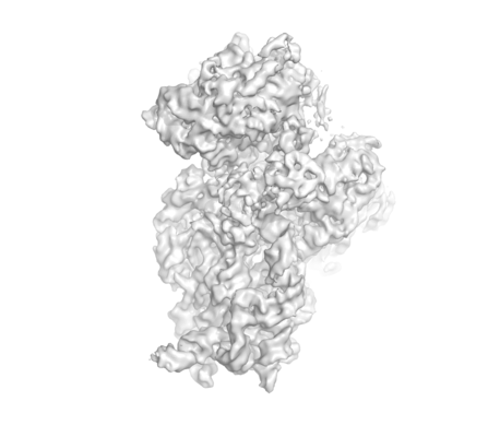
 ダウンロードとリンク
 emd_8621.png
 http://ftp.pdbj.org/pub/emdb/structures/EMD-8621
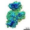


 Z (Sec.)
 Y (Row.)
 X (Col.)


 試料の構成要素
 解析
解析 電子顕微鏡法