glutamate-gated calcium ion channel activity / NMDA glutamate receptor activity / NMDA selective glutamate receptor complex / calcium ion transmembrane import into cytosol / protein heterotetramerization / response to zinc ion / response to magnesium ion / late endosome / postsynaptic membrane / リソソーム ...glutamate-gated calcium ion channel activity / NMDA glutamate receptor activity / NMDA selective glutamate receptor complex / calcium ion transmembrane import into cytosol / protein heterotetramerization / response to zinc ion / response to magnesium ion / late endosome / postsynaptic membrane / リソソーム / metal ion binding / 細胞膜 類似検索 - 分子機能
Glutamate [NMDA] receptor, epsilon subunit, C-terminal / N-methyl D-aspartate receptor 2B3 C-terminus / Bacterial extracellular solute-binding proteins, family 3 / Solute-binding protein family 3/N-terminal domain of MltF / Ionotropic glutamate receptor, metazoa / Ligated ion channel L-glutamate- and glycine-binding site / : / リガンド依存性イオンチャネル / Ionotropic glutamate receptor, L-glutamate and glycine-binding domain / Ligated ion channel L-glutamate- and glycine-binding site ...Glutamate [NMDA] receptor, epsilon subunit, C-terminal / N-methyl D-aspartate receptor 2B3 C-terminus / Bacterial extracellular solute-binding proteins, family 3 / Solute-binding protein family 3/N-terminal domain of MltF / Ionotropic glutamate receptor, metazoa / Ligated ion channel L-glutamate- and glycine-binding site / : / リガンド依存性イオンチャネル / Ionotropic glutamate receptor, L-glutamate and glycine-binding domain / Ligated ion channel L-glutamate- and glycine-binding site / Ionotropic glutamate receptor / Eukaryotic homologues of bacterial periplasmic substrate binding proteins. / Receptor, ligand binding region / Receptor family ligand binding region / Periplasmic binding protein-like I 類似検索 - ドメイン・相同性
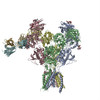
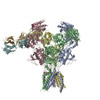
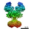
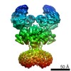
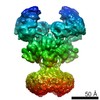
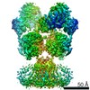
ジャーナル: Science / 年: 2017 タイトル: Cryo-EM structures of the triheteromeric NMDA receptor and its allosteric modulation. 著者: Wei Lü / Juan Du / April Goehring / Eric Gouaux / 要旨: -methyl-d-aspartate receptors (NMDARs) are heterotetrameric ion channels assembled as diheteromeric or triheteromeric complexes. Here, we report structures of the triheteromeric GluN1/GluN2A/GluN2B ...-methyl-d-aspartate receptors (NMDARs) are heterotetrameric ion channels assembled as diheteromeric or triheteromeric complexes. Here, we report structures of the triheteromeric GluN1/GluN2A/GluN2B receptor in the absence or presence of the GluN2B-specific allosteric modulator Ro 25-6981 (Ro), determined by cryogenic electron microscopy (cryo-EM). In the absence of Ro, the GluN2A and GluN2B amino-terminal domains (ATDs) adopt "closed" and "open" clefts, respectively. Upon binding Ro, the GluN2B ATD clamshell transitions from an open to a closed conformation. Consistent with a predominance of the GluN2A subunit in ion channel gating, the GluN2A subunit interacts more extensively with GluN1 subunits throughout the receptor, in comparison with the GluN2B subunit. Differences in the conformation of the pseudo-2-fold-related GluN1 subunits further reflect receptor asymmetry. The triheteromeric NMDAR structures provide the first view of the most common NMDA receptor assembly and show how incorporation of two different GluN2 subunits modifies receptor symmetry and subunit interactions, allowing each subunit to uniquely influence receptor structure and function, thus increasing receptor complexity.
 ムービー
ムービー コントローラー


 データを開く
 基本情報
基本情報 膜タンパク質
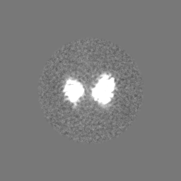
 マップデータ
 試料
試料 機能・相同性情報
 データ登録者
 引用
引用
 構造の表示
構造の表示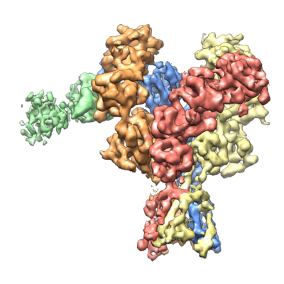
 ダウンロードとリンク
 emd_8580.png
 http://ftp.pdbj.org/pub/emdb/structures/EMD-8580


 Z (Sec.)
 Y (Row.)
 X (Col.)


 試料の構成要素
 解析
解析 電子顕微鏡法
