 ムービー
ムービー コントローラー


+ データを開く

- 基本情報
基本情報
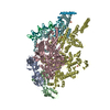
| 登録情報 | データベース: EMDB / ID: EMD-8483 | |||||||||
|---|---|---|---|---|---|---|---|---|---|---|
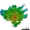
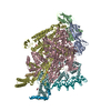
| タイトル | Structure of higher-order HIV-1 strand transfer complex intasome | |||||||||
 マップデータ マップデータ | structure of higher-order HIV-1 Strand Transfer Complex Intasome | |||||||||
 試料 試料 |
| |||||||||
| 機能・相同性 |  機能・相同性情報 機能・相同性情報RNA endonuclease activity /  nucleotidyltransferase activity / HIV-1 retropepsin / : / retroviral ribonuclease H / exoribonuclease H / : / exoribonuclease H activity / host multivesicular body / DNA integration ...RNA endonuclease activity / nucleotidyltransferase activity / HIV-1 retropepsin / : / retroviral ribonuclease H / exoribonuclease H / : / exoribonuclease H activity / host multivesicular body / DNA integration / 逆転写酵素 / viral genome integration into host DNA / viral penetration into host nucleus / establishment of integrated proviral latency / RNA-directed DNA polymerase activity / 転移酵素; リンを含む基を移すもの; 核酸を移すもの / RNA-DNA hybrid ribonuclease activity / viral nucleocapsid / endonuclease activity / DNA recombination / 加水分解酵素; エステル加水分解酵素 / nucleic acid binding / DNAポリメラーゼ / aspartic-type endopeptidase activity / DNA-directed DNA polymerase activity / symbiont entry into host cell / symbiont-mediated suppression of host gene expression / lipid binding / host cell nucleus / structural molecule activity / host cell plasma membrane / virion membrane / タンパク質分解 / DNA binding / RNA binding / zinc ion binding / 生体膜 nucleotidyltransferase activity / HIV-1 retropepsin / : / retroviral ribonuclease H / exoribonuclease H / : / exoribonuclease H activity / host multivesicular body / DNA integration ...RNA endonuclease activity / nucleotidyltransferase activity / HIV-1 retropepsin / : / retroviral ribonuclease H / exoribonuclease H / : / exoribonuclease H activity / host multivesicular body / DNA integration / 逆転写酵素 / viral genome integration into host DNA / viral penetration into host nucleus / establishment of integrated proviral latency / RNA-directed DNA polymerase activity / 転移酵素; リンを含む基を移すもの; 核酸を移すもの / RNA-DNA hybrid ribonuclease activity / viral nucleocapsid / endonuclease activity / DNA recombination / 加水分解酵素; エステル加水分解酵素 / nucleic acid binding / DNAポリメラーゼ / aspartic-type endopeptidase activity / DNA-directed DNA polymerase activity / symbiont entry into host cell / symbiont-mediated suppression of host gene expression / lipid binding / host cell nucleus / structural molecule activity / host cell plasma membrane / virion membrane / タンパク質分解 / DNA binding / RNA binding / zinc ion binding / 生体膜類似検索 - 分子機能 | |||||||||
| 生物種 |   Human immunodeficiency virus 1 (ヒト免疫不全ウイルス) / Human immunodeficiency virus 1 (ヒト免疫不全ウイルス) /  Homo sapiens (ヒト) Homo sapiens (ヒト) | |||||||||
| 手法 | 単粒子再構成法 / クライオ電子顕微鏡法 / 解像度: 4.6 Å | |||||||||
 データ登録者 データ登録者 | Lyumkis D / Passos D | |||||||||
 引用 引用 | ジャーナル: Science / 年: 2017 タイトル: Cryo-EM structures and atomic model of the HIV-1 strand transfer complex intasome. 著者: Dario Oliveira Passos / Min Li / Renbin Yang / Stephanie V Rebensburg / Rodolfo Ghirlando / Youngmin Jeon / Nikoloz Shkriabai / Mamuka Kvaratskhelia / Robert Craigie / Dmitry Lyumkis /  要旨: Like all retroviruses, HIV-1 irreversibly inserts a viral DNA (vDNA) copy of its RNA genome into host target DNA (tDNA). The intasome, a higher-order nucleoprotein complex composed of viral integrase ...Like all retroviruses, HIV-1 irreversibly inserts a viral DNA (vDNA) copy of its RNA genome into host target DNA (tDNA). The intasome, a higher-order nucleoprotein complex composed of viral integrase (IN) and the ends of linear vDNA, mediates integration. Productive integration into host chromatin results in the formation of the strand transfer complex (STC) containing catalytically joined vDNA and tDNA. HIV-1 intasomes have been refractory to high-resolution structural studies. We used a soluble IN fusion protein to facilitate structural studies, through which we present a high-resolution cryo-electron microscopy (cryo-EM) structure of the core tetrameric HIV-1 STC and a higher-order form that adopts carboxyl-terminal domain rearrangements. The distinct STC structures highlight how HIV-1 can use the common retroviral intasome core architecture to accommodate different IN domain modules for assembly. | |||||||||
| 履歴 |
|
- 構造の表示
構造の表示
| ムービー |
ムービービューア |
|---|---|
| 構造ビューア | EMマップ: SurfViewMolmilJmol/JSmol |
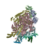
| 添付画像 |

- ダウンロードとリンク
-EMDBアーカイブ
| マップデータ | emd_8483.map.gz | 7.2 MB | EMDBマップデータ形式 | |
|---|---|---|---|---|
| ヘッダ (付随情報) | emd-8483-v30.xmlemd-8483.xml | 18.1 KB 18.1 KB | 表示 表示 | EMDBヘッダ |
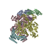
| 画像 | 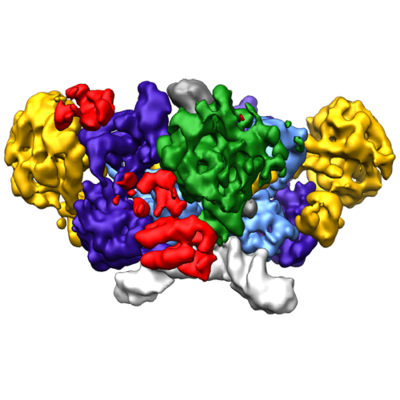 emd_8483.png emd_8483.png | 145 KB | ||
| アーカイブディレクトリ |  http://ftp.pdbj.org/pub/emdb/structures/EMD-8483ftp://ftp.pdbj.org/pub/emdb/structures/EMD-8483 http://ftp.pdbj.org/pub/emdb/structures/EMD-8483ftp://ftp.pdbj.org/pub/emdb/structures/EMD-8483 | HTTPS FTP |
-関連構造データ

-リンク
| EMDBのページ | EMDB (EBI/PDBe) / EMDataResource |
|---|---|
| 「今月の分子」の関連する項目 |

-マップ
| ファイル | ダウンロード / ファイル: emd_8483.map.gz / 形式: CCP4 / 大きさ: 27 MB / タイプ: IMAGE STORED AS FLOATING POINT NUMBER (4 BYTES) | ||||||||||||||||||||||||||||||||||||||||||||||||||||||||||||||||||||
|---|---|---|---|---|---|---|---|---|---|---|---|---|---|---|---|---|---|---|---|---|---|---|---|---|---|---|---|---|---|---|---|---|---|---|---|---|---|---|---|---|---|---|---|---|---|---|---|---|---|---|---|---|---|---|---|---|---|---|---|---|---|---|---|---|---|---|---|---|---|
| 注釈 | structure of higher-order HIV-1 Strand Transfer Complex Intasome | ||||||||||||||||||||||||||||||||||||||||||||||||||||||||||||||||||||
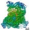
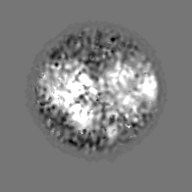
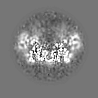
| 投影像・断面図 | 画像のコントロール
画像は Spider により作成 | ||||||||||||||||||||||||||||||||||||||||||||||||||||||||||||||||||||
| ボクセルのサイズ | X=Y=Z: 1.31 Å | ||||||||||||||||||||||||||||||||||||||||||||||||||||||||||||||||||||
| 密度 |
| ||||||||||||||||||||||||||||||||||||||||||||||||||||||||||||||||||||
| 対称性 | 空間群: 1 | ||||||||||||||||||||||||||||||||||||||||||||||||||||||||||||||||||||
| 詳細 | EMDB XML:
CCP4マップ ヘッダ情報:
| ||||||||||||||||||||||||||||||||||||||||||||||||||||||||||||||||||||
 Z (Sec.)
 Y (Row.)
 X (Col.)


-添付データ
- 試料の構成要素
-全体 : complex formed by a higher-order assembly of Sso7d-fusion HIV-1 I...
| 全体 | 名称: complex formed by a higher-order assembly of Sso7d-fusion HIV-1 Integrase with with IN-binding domain of LEDGF/p75, and the product of DNA strand transfer |
|---|---|
| 要素 |
|
-超分子 #1: complex formed by a higher-order assembly of Sso7d-fusion HIV-1 I...
| 超分子 | 名称: complex formed by a higher-order assembly of Sso7d-fusion HIV-1 Integrase with with IN-binding domain of LEDGF/p75, and the product of DNA strand transfer タイプ: complex / ID: 1 / 親要素: 0 / 含まれる分子: #1 |
|---|---|
| 由来(天然) | 生物種: Human immunodeficiency virus 1 (ヒト免疫不全ウイルス) |
| 組換発現 | 生物種:  Escherichia coli (大腸菌) / 組換プラスミド: pSca355 Escherichia coli (大腸菌) / 組換プラスミド: pSca355 |
| 分子量 | 実験値: 500 KDa |
-分子 #1: HIV-1 Integrase, Sso7d chimera
| 分子 | 名称: HIV-1 Integrase, Sso7d chimera / タイプ: protein_or_peptide / ID: 1 詳細: Sso7d protein fused to the integrase N-terminus via an 11-glycine linker 光学異性体: LEVO |
|---|---|
| 由来(天然) | 生物種: Human immunodeficiency virus 1 (ヒト免疫不全ウイルス) |
| 組換発現 | 生物種: Escherichia coli (大腸菌) |
| 配列 | 文字列: MGSSHHHHHH SSGLVPRGSH MATVKFKYKG EEKEVDISKI KKVWRVGKMI SFTYDEGGGK TGRGAVSEKD APKELLQMLE KQKKGGGGGG GGGGGFLDGI DKAQEEHEKY HSNWRAMASD FNLPPVVAKE IVASCDKCQL KGEAMHGQVD CSPGIWQLDC THLEGKVILV ...文字列: MGSSHHHHHH SSGLVPRGSH MATVKFKYKG EEKEVDISKI KKVWRVGKMI SFTYDEGGGK TGRGAVSEKD APKELLQMLE KQKKGGGGGG GGGGGFLDGI DKAQEEHEKY HSNWRAMASD FNLPPVVAKE IVASCDKCQL KGEAMHGQVD CSPGIWQLDC THLEGKVILV AVHVASGYIE AEVIPAETGQ ETAYFLLKLA GRWPVKTVHT DNGSNFTSTT VKAACWWAGI KQEFGIPYNP QSQGVIQSMN KELKKIIGQV RDQAEHLKTA VQMAVFIHNF KRKGGIGGYS AGERIVDIIA TDIQTKELQK QITKIQNFRV YYRDSRDPVW KGPAKLLWKG EGAVVIQDNS DIKVVPRRKA KIIRDYGKQM AGDDCVASRQ DED |
-分子 #2: Integrase binding domain of LEDGF/p75
| 分子 | 名称: Integrase binding domain of LEDGF/p75 / タイプ: protein_or_peptide / ID: 2 / 光学異性体: LEVO |
|---|---|
| 由来(天然) | 生物種: Homo sapiens (ヒト) |
| 配列 | 文字列: SMDSRLQRIH AEIKNSLKID NLDVNRCIEA LDELASLQVT MQQAQKHTEM ITTLKKIRRF KVSQVIMEKS TMLYNKFKNM FLV |
-分子 #3: DNA (11-MER)
| 分子 | 名称: DNA (11-MER) / タイプ: dna / ID: 3 / 分類: DNA |
|---|---|
| 由来(天然) | 生物種: Homo sapiens (ヒト) |
| 配列 | 文字列: GTACGCTGAC T |
-分子 #4: DNA (23-MER)
| 分子 | 名称: DNA (23-MER) / タイプ: dna / ID: 4 / 分類: DNA |
|---|---|
| 由来(天然) | 生物種: Human immunodeficiency virus 1 (ヒト免疫不全ウイルス) |
| 配列 | 文字列: ACTGCTAGAG ATTTTCCACA CTG |
-分子 #5: DNA (37-MER)
| 分子 | 名称: DNA (37-MER) / タイプ: dna / ID: 5 / 分類: DNA |
|---|---|
| 由来(天然) | 生物種: Homo sapiens (ヒト) |
| 配列 | 文字列: CAGTGTGGAA AATCTCTAGC AGTTACAGTC AGCGTAC |
-実験情報
-構造解析
| 手法 | クライオ電子顕微鏡法 |
|---|---|
 解析 解析 | 単粒子再構成法 |
| 試料の集合状態 | particle |
-試料調製
| 濃度 | 0.5 mg/mL | |||||||||||||||
|---|---|---|---|---|---|---|---|---|---|---|---|---|---|---|---|---|
| 緩衝液 | pH: 8 構成要素:
| |||||||||||||||
| グリッド | モデル: Quantifoil / 材質: GOLD / メッシュ: 400 / 前処理 - タイプ: PLASMA CLEANING / 前処理 - 雰囲気: OTHER | |||||||||||||||
| 凍結 | 凍結剤: ETHANE / チャンバー内湿度: 50 % / チャンバー内温度: 277 K / 装置: HOMEMADE PLUNGER 詳細: Sample containing HIV STC intasomes in SEC buffer was applied onto freshly plasma-treated (6 seconds, Gatan Solarus plasma cleaner) holey gold UltrAuFoil grids (Quantifoil), adsorbed for 30 ...詳細: Sample containing HIV STC intasomes in SEC buffer was applied onto freshly plasma-treated (6 seconds, Gatan Solarus plasma cleaner) holey gold UltrAuFoil grids (Quantifoil), adsorbed for 30 seconds, then plunged into liquid ethane using a manual cryo-plunger in an ambient environment of 4 degrees C.. |
- 電子顕微鏡法
| 顕微鏡 | FEI TITAN KRIOS |
|---|---|
| 電子線 | 加速電圧: 300 kV / 電子線源: FIELD EMISSION GUN |
| 電子光学系 | C2レンズ絞り径: 100.0 µm / 最大 デフォーカス(補正後): 3.5 µm / 最小 デフォーカス(補正後): 1.5 µm / 倍率(補正後): 38167 / 照射モード: FLOOD BEAM / 撮影モード: BRIGHT FIELDBright-field microscopy / Cs: 2.7 mm / 倍率(公称値): 22500 |
| 試料ステージ | 試料ホルダーモデル: FEI TITAN KRIOS AUTOGRID HOLDER ホルダー冷却材: NITROGEN |
| 温度 | 最低: 90.0 K / 最高: 90.0 K |
| 撮影 | フィルム・検出器のモデル: GATAN K2 SUMMIT (4k x 4k) 検出モード: COUNTING / デジタル化 - サイズ - 横: 3838 pixel / デジタル化 - サイズ - 縦: 3710 pixel / デジタル化 - サンプリング間隔: 5.0 µm / デジタル化 - 画像ごとのフレーム数: 1-100 / 撮影したグリッド数: 1 / 実像数: 1598 / 平均露光時間: 20.0 sec. / 平均電子線量: 95.0 e/Å2 詳細: Individual frames were gain-corrected, aligned, and summed with the application of an exposure filter using MotionCor2, according to the nominal dose rate. |
| 実験機器 |  モデル: Titan Krios / 画像提供: FEI Company |
-画像解析
| 粒子像選択 | 選択した数: 154445 |
|---|---|
| CTF補正 | ソフトウェア - 名称: CTFFIND (ver. 3) / 詳細: performed internally in Relion and Frealign |
| 初期モデル | モデルのタイプ: INSILICO MODEL / In silico モデル: common lines model using OptiMod 詳細: An initial model was generated directly from the class averages using OptiMod. |
| 初期 角度割当 | タイプ: PROJECTION MATCHING Projection matching processing - Angular sampling: 7.5 degrees ソフトウェア - 名称: RELION (ver. 1.3) / 詳細: Relion 3D classification, auto mode |
| 最終 3次元分類 | ソフトウェア - 名称: FREALIGN (ver. 3.11) |
| 最終 角度割当 | タイプ: PROJECTION MATCHING / ソフトウェア - 名称: FREALIGN (ver. 9.11) / 詳細: Frealign 3D classification and refinement |
| 最終 再構成 | 想定した対称性 - 点群: C2 (2回回転対称) / アルゴリズム: FOURIER SPACE / 解像度のタイプ: BY AUTHOR / 解像度: 4.6 Å / 解像度の算出法: FSC 0.143 CUT-OFF / ソフトウェア - 名称: FREALIGN (ver. 9.11) / 詳細: Resolution-limited refinement used throughout / 使用した粒子像数: 11099 |
-原子モデル構築 1
| 精密化 | プロトコル: RIGID BODY FIT / 温度因子: 200 |
|---|
