 ムービー
ムービー コントローラー


+ データを開く

- 基本情報
基本情報
| 登録情報 | データベース: EMDB / ID: EMD-8251 | |||||||||
|---|---|---|---|---|---|---|---|---|---|---|
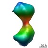
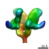
| タイトル | Subtomogram-averaged map of in vitro assembled rubella virus capsid protein tetramer complex | |||||||||
 マップデータ マップデータ | Subtomogram-averaged map of in vitro assembled rubella virus capsid protein tetramer complex, unfiltered and unmasked | |||||||||
 試料 試料 |
| |||||||||
| 機能・相同性 |  機能・相同性情報 機能・相同性情報T=4 icosahedral viral capsid / host cell Golgi membrane / host cell mitochondrion / clathrin-dependent endocytosis of virus by host cell / viral nucleocapsid / fusion of virus membrane with host endosome membrane /  エンベロープ (ウイルス) / virion attachment to host cell / virion membrane / RNA binding ...T=4 icosahedral viral capsid / host cell Golgi membrane / host cell mitochondrion / clathrin-dependent endocytosis of virus by host cell / viral nucleocapsid / fusion of virus membrane with host endosome membrane / エンベロープ (ウイルス) / virion attachment to host cell / virion membrane / RNA binding / 生体膜 / metal ion binding エンベロープ (ウイルス) / virion attachment to host cell / virion membrane / RNA binding ...T=4 icosahedral viral capsid / host cell Golgi membrane / host cell mitochondrion / clathrin-dependent endocytosis of virus by host cell / viral nucleocapsid / fusion of virus membrane with host endosome membrane / エンベロープ (ウイルス) / virion attachment to host cell / virion membrane / RNA binding / 生体膜 / metal ion binding類似検索 - 分子機能 | |||||||||
| 生物種 |  Rubella virus strain M33 (風疹ウイルス) Rubella virus strain M33 (風疹ウイルス) | |||||||||
| 手法 | サブトモグラム平均法 / クライオ電子顕微鏡法 / 解像度: 35.0 Å | |||||||||
 データ登録者 データ登録者 | Mangala Prasad V / Klose T / Rossmann MG | |||||||||
 引用 引用 | ジャーナル: PLoS Pathog / 年: 2017 タイトル: Assembly, maturation and three-dimensional helical structure of the teratogenic rubella virus. 著者: Vidya Mangala Prasad / Thomas Klose / Michael G Rossmann /  要旨: Viral infections during pregnancy are a significant cause of infant morbidity and mortality. Of these, rubella virus infection is a well-substantiated example that leads to miscarriages or severe ...Viral infections during pregnancy are a significant cause of infant morbidity and mortality. Of these, rubella virus infection is a well-substantiated example that leads to miscarriages or severe fetal defects. However, structural information about the rubella virus has been lacking due to the pleomorphic nature of the virions. Here we report a helical structure of rubella virions using cryo-electron tomography. Sub-tomogram averaging of the surface spikes established the relative positions of the viral glycoproteins, which differed from the earlier icosahedral models of the virus. Tomographic analyses of in vitro assembled nucleocapsids and virions provide a template for viral assembly. Comparisons of immature and mature virions show large rearrangements in the glycoproteins that may be essential for forming the infectious virions. These results present the first known example of a helical membrane-enveloped virus, while also providing a structural basis for its assembly and maturation pathway. | |||||||||
| 履歴 |
|
- 構造の表示
構造の表示
| ムービー |
ムービービューア |
|---|---|
| 構造ビューア | EMマップ: SurfViewMolmilJmol/JSmol |
| 添付画像 |

- ダウンロードとリンク
-EMDBアーカイブ
| マップデータ | emd_8251.map.gz | 454 KB | EMDBマップデータ形式 | |
|---|---|---|---|---|
| ヘッダ (付随情報) | emd-8251-v30.xmlemd-8251.xml | 11.4 KB 11.4 KB | 表示 表示 | EMDBヘッダ |
| 画像 | 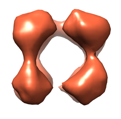 emd_8251.png emd_8251.png | 88.3 KB | ||
| アーカイブディレクトリ |  http://ftp.pdbj.org/pub/emdb/structures/EMD-8251ftp://ftp.pdbj.org/pub/emdb/structures/EMD-8251 http://ftp.pdbj.org/pub/emdb/structures/EMD-8251ftp://ftp.pdbj.org/pub/emdb/structures/EMD-8251 | HTTPS FTP |
-関連構造データ


-リンク
| EMDBのページ | EMDB (EBI/PDBe) / EMDataResource |
|---|---|
| 「今月の分子」の関連する項目 |

-マップ
| ファイル | ダウンロード / ファイル: emd_8251.map.gz / 形式: CCP4 / 大きさ: 489.3 KB / タイプ: IMAGE STORED AS FLOATING POINT NUMBER (4 BYTES) | ||||||||||||||||||||||||||||||||||||||||||||||||||||||||||||
|---|---|---|---|---|---|---|---|---|---|---|---|---|---|---|---|---|---|---|---|---|---|---|---|---|---|---|---|---|---|---|---|---|---|---|---|---|---|---|---|---|---|---|---|---|---|---|---|---|---|---|---|---|---|---|---|---|---|---|---|---|---|
| 注釈 | Subtomogram-averaged map of in vitro assembled rubella virus capsid protein tetramer complex, unfiltered and unmasked | ||||||||||||||||||||||||||||||||||||||||||||||||||||||||||||
| ボクセルのサイズ | X=Y=Z: 5.28 Å | ||||||||||||||||||||||||||||||||||||||||||||||||||||||||||||
| 密度 |
| ||||||||||||||||||||||||||||||||||||||||||||||||||||||||||||
| 対称性 | 空間群: 1 | ||||||||||||||||||||||||||||||||||||||||||||||||||||||||||||
| 詳細 | EMDB XML:
CCP4マップ ヘッダ情報:
| ||||||||||||||||||||||||||||||||||||||||||||||||||||||||||||
-添付データ
- 試料の構成要素
-全体 : Rubella virus capsid protein
| 全体 | 名称: Rubella virus capsid protein |
|---|---|
| 要素 |
|
-超分子 #1: Rubella virus capsid protein
| 超分子 | 名称: Rubella virus capsid protein / タイプ: complex / ID: 1 / 親要素: 0 / 含まれる分子: all |
|---|---|
| 由来(天然) | 生物種: Rubella virus strain M33 (風疹ウイルス) |
| 組換発現 | 生物種:  Escherichia coli BL21(DE3) (大腸菌) / 組換株: Rosetta-2 (DE3) / 組換プラスミド: pTXB1 Escherichia coli BL21(DE3) (大腸菌) / 組換株: Rosetta-2 (DE3) / 組換プラスミド: pTXB1 |
| 分子量 | 実験値: 256 KDa |
-分子 #1: Rubella virus capsid protein
| 分子 | 名称: Rubella virus capsid protein / タイプ: protein_or_peptide / ID: 1 / 光学異性体: LEVO |
|---|---|
| 由来(天然) | 生物種: Rubella virus strain M33 (風疹ウイルス) |
| 組換発現 | 生物種: Escherichia coli BL21(DE3) (大腸菌) |
| 配列 | 文字列: MEDLQKALEA QSRALRAELA AGASQSRRPR PPRQRDSSTS GDDSGRDSGG PRRRRGNRGR GQRRDWSRAP PPPEERQETR SQTPAPKPSR APPQQPQPPR MQTGRGGSAP RPELGPPTNP FQAAVARGLR PPLHDPDTEA PTEACVTSWL WSEGEGAVFY RVDLHFTNLG ...文字列: MEDLQKALEA QSRALRAELA AGASQSRRPR PPRQRDSSTS GDDSGRDSGG PRRRRGNRGR GQRRDWSRAP PPPEERQETR SQTPAPKPSR APPQQPQPPR MQTGRGGSAP RPELGPPTNP FQAAVARGLR PPLHDPDTEA PTEACVTSWL WSEGEGAVFY RVDLHFTNLG TPPLDEDGRW DPALMYNPCG PEPPAHVVRA YNQPAGDVRG VWGKGERTYA EQDFRVGGTR WHRLLRMPVR GLDGDSAPLP PHTTERIETR SARHPWRIR |
-実験情報
-構造解析
| 手法 | クライオ電子顕微鏡法 |
|---|---|
 解析 解析 | サブトモグラム平均法 |
| 試料の集合状態 | particle |
-試料調製
| 濃度 | 1 mg/mL | |||||||||
|---|---|---|---|---|---|---|---|---|---|---|
| 緩衝液 | pH: 7.2 構成要素:
| |||||||||
| グリッド | モデル: Quantifoil R1.2/1.3 / 材質: COPPER / メッシュ: 200 / 支持フィルム - 材質: CARBON | |||||||||
| 凍結 | 凍結剤: ETHANE |
- 電子顕微鏡法
| 顕微鏡 | FEI TITAN KRIOS |
|---|---|
| 電子線 | 加速電圧: 300 kV / 電子線源: FIELD EMISSION GUN |
| 電子光学系 | 照射モード: SPOT SCAN / 撮影モード: BRIGHT FIELDBright-field microscopy / 最大 デフォーカス(公称値): 0.005 µm / 倍率(公称値): 11000 |
| 撮影 | フィルム・検出器のモデル: GATAN K2 SUMMIT (4k x 4k) 検出モード: SUPER-RESOLUTION / 平均電子線量: 90.0 e/Å2 |
| 実験機器 |  モデル: Titan Krios / 画像提供: FEI Company |
-画像解析
| 抽出 | トモグラム数: 15 / 使用した粒子像数: 20 / 手法: manual picking / ソフトウェア - 名称: IMOD/PEEt (ver. 4.8.40/1.11.0) |
|---|---|
| CTF補正 | ソフトウェア - 名称: IMOD (ver. 4.8.40) |
| 最終 角度割当 | タイプ: OTHER |
| 最終 再構成 | 想定した対称性 - 点群: C1 (非対称) / 解像度のタイプ: BY AUTHOR / 解像度: 35.0 Å / 解像度の算出法: OTHER / ソフトウェア - 名称: PEET (ver. 1.11.0) / 使用したサブトモグラム数: 18 |
