 ムービー
ムービー コントローラー


+ データを開く

- 基本情報
基本情報
| 登録情報 | データベース: EMDB / ID: EMD-6914 | |||||||||
|---|---|---|---|---|---|---|---|---|---|---|
| タイトル | Serial cryotomogram of a metaphase budding yeast cell. | |||||||||
 マップデータ マップデータ | Serial cryotomogram of a metaphase budding yeast cell | |||||||||
 試料 試料 |
| |||||||||
| 生物種 |   Saccharomyces cerevisiae (パン酵母) Saccharomyces cerevisiae (パン酵母) | |||||||||
| 手法 | 電子線トモグラフィー法 / クライオ電子顕微鏡法 | |||||||||
 データ登録者 データ登録者 | Ng C / Deng L / Chen C / Lim H / Shi J / Surana U / Gan L | |||||||||
 引用 引用 | ジャーナル: J Cell Biol / 年: 2019 タイトル: Electron cryotomography analysis of Dam1C/DASH at the kinetochore-spindle interface in situ. 著者: Cai Tong Ng / Li Deng / Chen Chen / Hong Hwa Lim / Jian Shi / Uttam Surana / Lu Gan /  要旨: In dividing cells, depolymerizing spindle microtubules move chromosomes by pulling at their kinetochores. While kinetochore subcomplexes have been studied extensively in vitro, little is known about ...In dividing cells, depolymerizing spindle microtubules move chromosomes by pulling at their kinetochores. While kinetochore subcomplexes have been studied extensively in vitro, little is known about their in vivo structure and interactions with microtubules or their response to spindle damage. Here we combine electron cryotomography of serial cryosections with genetic and pharmacological perturbation to study the yeast chromosome segregation machinery in vivo. Each kinetochore microtubule has one (rarely, two) Dam1C/DASH outer kinetochore assemblies. Dam1C/DASH contacts the microtubule walls and does so with its flexible "bridges"; there are no contacts with the protofilaments' curved tips. In metaphase, ∼40% of the Dam1C/DASH assemblies are complete rings; the rest are partial rings. Ring completeness and binding position along the microtubule are sensitive to kinetochore attachment and tension, respectively. Our study and those of others support a model in which each kinetochore must undergo cycles of conformational change to couple microtubule depolymerization to chromosome movement. | |||||||||
| 履歴 |
|
- 構造の表示
構造の表示
| ムービー |
 ムービービューア ムービービューア |
|---|---|
| 添付画像 |

- ダウンロードとリンク
-EMDBアーカイブ
| マップデータ | emd_6914.map.gz | 5.1 GB | EMDBマップデータ形式 | |
|---|---|---|---|---|
| ヘッダ (付随情報) | emd-6914-v30.xmlemd-6914.xml | 10.8 KB 10.8 KB | 表示 表示 | EMDBヘッダ |
| 画像 | 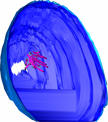 emd_6914.png emd_6914.png | 133.2 KB | ||
| アーカイブディレクトリ |  http://ftp.pdbj.org/pub/emdb/structures/EMD-6914ftp://ftp.pdbj.org/pub/emdb/structures/EMD-6914 http://ftp.pdbj.org/pub/emdb/structures/EMD-6914ftp://ftp.pdbj.org/pub/emdb/structures/EMD-6914 | HTTPS FTP |
-関連構造データ
| 関連構造データ |  6912C C: 同じ文献を引用 ( |
|---|---|
| 電子顕微鏡画像生データ | EMPIAR-10159 (タイトル: A multi-scale model of the yeast chromosome-segregation system Data size: 95.4 Data #1: Tilt series, tomograms, and models of cryosections of mitotic budding yeast and reconstituted DASH rings around microtubules [tilt series]) |
-リンク
| EMDBのページ | EMDB (EBI/PDBe) / EMDataResource |
|---|
-マップ
| ファイル | ダウンロード / ファイル: emd_6914.map.gz / 形式: CCP4 / 大きさ: 11.4 GB / タイプ: IMAGE STORED AS SIGNED BYTE | ||||||||||||||||||||||||||||||||||||||||||||||||||||||||||||||||||||
|---|---|---|---|---|---|---|---|---|---|---|---|---|---|---|---|---|---|---|---|---|---|---|---|---|---|---|---|---|---|---|---|---|---|---|---|---|---|---|---|---|---|---|---|---|---|---|---|---|---|---|---|---|---|---|---|---|---|---|---|---|---|---|---|---|---|---|---|---|---|
| 注釈 | Serial cryotomogram of a metaphase budding yeast cell | ||||||||||||||||||||||||||||||||||||||||||||||||||||||||||||||||||||
| ボクセルのサイズ | X=Y=Z: 8.93 Å | ||||||||||||||||||||||||||||||||||||||||||||||||||||||||||||||||||||
| 密度 |
| ||||||||||||||||||||||||||||||||||||||||||||||||||||||||||||||||||||
| 対称性 | 空間群: 1 | ||||||||||||||||||||||||||||||||||||||||||||||||||||||||||||||||||||
| 詳細 | EMDB XML:
CCP4マップ ヘッダ情報:
| ||||||||||||||||||||||||||||||||||||||||||||||||||||||||||||||||||||
-添付データ
- 試料の構成要素
-全体 : S. cerevisiae cell, arrested in metaphase
| 全体 | 名称: S. cerevisiae cell, arrested in metaphase |
|---|---|
| 要素 |
|
-超分子 #1: S. cerevisiae cell, arrested in metaphase
| 超分子 | 名称: S. cerevisiae cell, arrested in metaphase / タイプ: cell / ID: 1 / 親要素: 0 |
|---|---|
| 由来(天然) | 生物種: Saccharomyces cerevisiae (パン酵母) / 株: W303 |
-実験情報
-構造解析
| 手法 | クライオ電子顕微鏡法 |
|---|---|
 解析 解析 | 電子線トモグラフィー法 |
| 試料の集合状態 | cell |
-試料調製
| 緩衝液 | pH: 7 / 詳細: YEPD |
|---|---|
| グリッド | モデル: C-flat-2/0.5 4C / 材質: COPPER / メッシュ: 150 / 支持フィルム - 材質: CARBON / 支持フィルム - トポロジー: CONTINUOUS / 前処理 - タイプ: PLASMA CLEANING / 前処理 - 雰囲気: AIR 詳細: plasma cleaned at 15 mA for 45 seconds, carbon side pre-coated with BSA-Gold mixture |
| 凍結 | 凍結剤: ETHANE |
| 加圧凍結法 | 装置: OTHER 詳細: Self-pressurized freezing. The value given for _emd_high_pressure_freezing.instrument is Vitrobot cup. This is not in a list of allowed values set(['LEICA EM PACT2', 'LEICA EM PACT', 'EMS-002 ...詳細: Self-pressurized freezing. The value given for _emd_high_pressure_freezing.instrument is Vitrobot cup. This is not in a list of allowed values set(['LEICA EM PACT2', 'LEICA EM PACT', 'EMS-002 RAPID IMMERSION FREEZER', 'OTHER', 'LEICA EM HPM100', 'BAL-TEC HPM 010']) so OTHER is written into the XML file. |
| Cryo protectant | 25% dextran |
| 切片作成 | ウルトラミクロトーム - 装置: Leica UC7/FC7 / ウルトラミクロトーム - 温度: 123 K / ウルトラミクロトーム - 最終 厚さ: 100 nm |
| 位置合わせマーカー | Manufacturer: BBI / 直径: 10 nm |
- 電子顕微鏡法
| 顕微鏡 | FEI TITAN KRIOS |
|---|---|
| 電子線 | 加速電圧: 300 kV / 電子線源: FIELD EMISSION GUN |
| 電子光学系 | 倍率(補正後): 15678 / 照射モード: FLOOD BEAM / 撮影モード: BRIGHT FIELDBright-field microscopy / Cs: 2.7 mm / 最小 デフォーカス(公称値): 10.0 µm / 倍率(公称値): 8700 |
| 試料ステージ | 試料ホルダーモデル: FEI TITAN KRIOS AUTOGRID HOLDER ホルダー冷却材: NITROGEN |
| 撮影 | フィルム・検出器のモデル: FEI FALCON II (4k x 4k) 検出モード: INTEGRATING / デジタル化 - サイズ - 横: 4096 pixel / デジタル化 - サイズ - 縦: 4096 pixel / デジタル化 - サンプリング間隔: 14.0 µm / 撮影したグリッド数: 1 / 実像数: 61 / 平均露光時間: 1.0 sec. / 平均電子線量: 1.6 e/Å2 |
| 実験機器 |  モデル: Titan Krios / 画像提供: FEI Company |
-画像解析
| CTF補正 | ソフトウェア - 名称: eTomo (ver. 4.10) / 詳細: Phase flipping done in Etomo 4.10 |
|---|---|
| 最終 再構成 | アルゴリズム: BACK PROJECTION / ソフトウェア - 名称: IMOD (ver. 4.10) / 使用した粒子像数: 61 |
