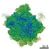
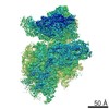
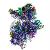
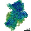
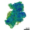
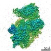
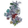
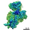
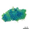
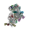
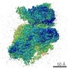
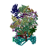
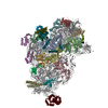
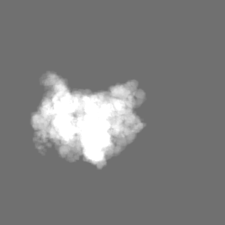
- EMDB-6780: Small subunit of Toxoplasma gondii ribosome -
+
データを開く
IDまたはキーワード:
読み込み中...
-
基本情報
登録情報
データベース: EMDB / ID: EMD-6780
タイトル
Small subunit of Toxoplasma gondii ribosome
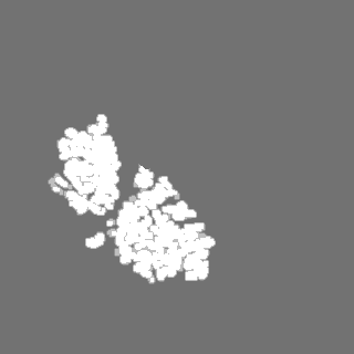
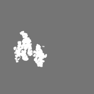
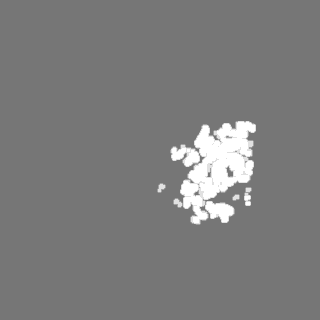
マップデータ
試料
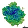
複合体: Small subunit of Toxoplasma gondii ribosome
RNA: x 1種
タンパク質・ペプチド: x 33種
機能・相同性
機能・相同性情報
葉緑体 / ribosomal small subunit assembly / cytosolic small ribosomal subunit / small ribosomal subunit / membrane => GO:0016020 / rRNA binding / リボソーム / structural constituent of ribosome / 翻訳 (生物学) / RNA binding ...葉緑体 / ribosomal small subunit assembly / cytosolic small ribosomal subunit / small ribosomal subunit / membrane => GO:0016020 / rRNA binding / リボソーム / structural constituent of ribosome / 翻訳 (生物学) / RNA binding / zinc ion binding / metal ion binding 類似検索 - 分子機能
: / Ribosomal protein S12e / S27a-like superfamily / Ribosomal protein S10, eukaryotic/archaeal / Ribosomal protein S25 / Ribosomal protein S26e signature. / Ribosomal protein S17e, conserved site / Ribosomal protein S30 / 40S ribosomal protein S29/30S ribosomal protein S14 type Z / Ribosomal protein S27a ...: / Ribosomal protein S12e / S27a-like superfamily / Ribosomal protein S10, eukaryotic/archaeal / Ribosomal protein S25 / Ribosomal protein S26e signature. / Ribosomal protein S17e, conserved site / Ribosomal protein S30 / 40S ribosomal protein S29/30S ribosomal protein S14 type Z / Ribosomal protein S27a / Ribosomal protein S27a / Ribosomal protein S3, eukaryotic/archaeal / S25 ribosomal protein / Ribosomal protein L41 / Ribosomal protein L41 / Ribosomal protein S26e / Ribosomal protein S26e superfamily / Ribosomal protein S26e / Ribosomal protein S19A/S15e / Ribosomal protein S21e, conserved site / Ribosomal protein S21e signature. / Ribosomal protein S30 / Ribosomal protein S12e signature. / Ribosomal protein S17e / Ribosomal protein S17e-like superfamily / Ribosomal protein S27a / Ribosomal protein S19e / Ribosomal_S19e / Ribosomal protein S5, eukaryotic/archaeal / 40S ribosomal protein S11, N-terminal / 40S ribosomal protein S1/3, eukaryotes / Ribosomal protein S6, eukaryotic / Ribosomal protein S2, eukaryotic / Ribosomal protein S7e / 40S ribosomal protein S4, C-terminal domain / Ribosomal protein S4e, N-terminal, conserved site / Ribosomal S17 / Ribosomal protein S21e / Ribosomal protein S21e superfamily / Ribosomal protein S21e / Ribosomal protein S19e / 40S Ribosomal protein S10 / Ribosomal protein S17, archaeal/eukaryotic / Ribosomal protein S27 / Ribosomal protein S28e conserved site / Ribosomal protein S6/S6e/A/B/2, conserved site / Ribosomal protein S28e / 40S ribosomal protein S4 C-terminus / Ribosomal protein S4e, N-terminal / Ribosomal protein S23, eukaryotic/archaeal / Ribosomal_S17 N-terminal / Plectin/S10, N-terminal / Ribosomal protein S3Ae / Ribosomal S3Ae family / Ribosomal protein S7e / Plectin/S10 domain / Ribosomal protein S2, eukaryotic/archaeal / Ribosomal protein S8e / Ribosomal protein S4, KOW domain / Ribosomal protein S5/S7, eukaryotic/archaeal / Ribosomal protein S4e / Ribosomal protein S4e, central region / Ribosomal protein S4e, central domain superfamily / Ribosomal protein S6e / Ribosomal protein S13/S15, N-terminal / Ribosomal protein S15P / Ribosomal S13/S15 N-terminal domain / Ribosomal protein S6e / Ribosomal protein S4/S9, eukaryotic/archaeal / RS4NT (NUC023) domain / Ribosomal protein S27 / Ribosomal S3Ae family / Ribosomal protein S17e signature. / Ribosomal protein S28e / Ribosomal family S4e / Ribosomal S13/S15 N-terminal domain / Ribosomal protein S6e / Ribosomal protein S4e signature. / Ribosomal S24e conserved site / Ribosomal protein S24e signature. / Ribosomal protein S24e / Ribosomal protein S24e / Ribosomal protein S6e signature. / Ribosomal protein S28e signature. / Ribosomal protein S8e/ribosomal biogenesis NSA2 / Ribosomal protein S8e / Ribosomal protein L7Ae/L30e/S12e/Gadd45 / Ribosomal protein S14/S29 / Ribosomal protein L7Ae/L30e/S12e/Gadd45 family / 50S ribosomal protein L30e-like / Ubiquitin domain / Ribosomal protein S2 signature 2. / Ribosomal protein S14, conserved site / K Homology domain, type 2 / Ribosomal protein S3, C-terminal / Ribosomal protein S3, C-terminal domain superfamily / Ribosomal protein S15/S19, conserved site / KHドメイン / Ribosomal protein S19/S15 / Ribosomal protein S19/S15, superfamily 類似検索 - ドメイン・相同性
40S ribosomal protein S3, putative / 40S ribosomal protein S29, putative / Ribosomal protein RPS28 / Ribosomal protein RPS15A / 40S ribosomal protein S24 / Ribosomal protein RPS19 / 40S ribosomal protein S21 / 40S ribosomal protein S13, putative / Ribosomal protein RPS27 / 40s ribosomal protein S14, putative ...40S ribosomal protein S3, putative / 40S ribosomal protein S29, putative / Ribosomal protein RPS28 / Ribosomal protein RPS15A / 40S ribosomal protein S24 / Ribosomal protein RPS19 / 40S ribosomal protein S21 / 40S ribosomal protein S13, putative / Ribosomal protein RPS27 / 40s ribosomal protein S14, putative / Ribosomal protein RPS16 / 40S ribosomal protein S8 / 40s ribosomal protein S20, putative / 40S ribosomal protein S3a / Ribosomal protein RPS23 / 40S ribosomal protein S18, putative / 40S ribosomal protein S11, putative / 40S ribosomal protein S7 / 40S ribosomal protein S26 / 40S ribosomal protein S5, putative / Ribosomal-ubiquitin protein RPS27A / 40S ribosomal protein S9, putative / 40S ribosomal protein S15, putative / 40S ribosomal protein S30 / 40S ribosomal protein S6 / Ribosomal protein RPS10 / 40s ribosomal protein s17, putative / 40S ribosomal protein S2, putative / 40S ribosomal protein S12 / Uncharacterized protein / 40S ribosomal protein S4 / 40S ribosomal protein SA / 40S ribosomal protein S25 類似検索 - 構成要素
ジャーナル: Cell Res / 年: 2017 タイトル: Cryo-EM structures of the 80S ribosomes from human parasites Trichomonas vaginalis and Toxoplasma gondii. 著者: Zhifei Li / Qiang Guo / Lvqin Zheng / Yongsheng Ji / Yi-Ting Xie / De-Hua Lai / Zhao-Rong Lun / Xun Suo / Ning Gao / 要旨: As an indispensable molecular machine universal in all living organisms, the ribosome has been selected by evolution to be the natural target of many antibiotics and small-molecule inhibitors. High- ...As an indispensable molecular machine universal in all living organisms, the ribosome has been selected by evolution to be the natural target of many antibiotics and small-molecule inhibitors. High-resolution structures of pathogen ribosomes are crucial for understanding the general and unique aspects of translation control in disease-causing microbes. With cryo-electron microscopy technique, we have determined structures of the cytosolic ribosomes from two human parasites, Trichomonas vaginalis and Toxoplasma gondii, at resolution of 3.2-3.4 Å. Although the ribosomal proteins from both pathogens are typical members of eukaryotic families, with a co-evolution pattern between certain species-specific insertions/extensions and neighboring ribosomal RNA (rRNA) expansion segments, the sizes of their rRNAs are sharply different. Very interestingly, rRNAs of T. vaginalis are in size comparable to prokaryotic counterparts, with nearly all the eukaryote-specific rRNA expansion segments missing. These structures facilitate the dissection of evolution path for ribosomal proteins and RNAs, and may aid in design of novel translation inhibitors.
 ムービー
ムービー コントローラー


 データを開く
 基本情報
基本情報 マップデータ
 試料
試料 機能・相同性情報
 葉緑体 /
葉緑体 / 
 データ登録者
 引用
引用
 構造の表示
構造の表示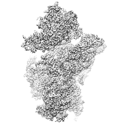
 ダウンロードとリンク
 emd_6780.png
 http://ftp.pdbj.org/pub/emdb/structures/EMD-6780
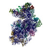


 Z
Z Y
Y X
X


 試料の構成要素
 解析
解析 電子顕微鏡法