 ムービー
ムービー コントローラー


+ データを開く

- 基本情報
基本情報
| 登録情報 | データベース: EMDB / ID: EMD-6715 | |||||||||
|---|---|---|---|---|---|---|---|---|---|---|
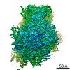
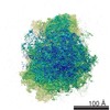
| タイトル | FliF ring of Salmonella flagellar motor | |||||||||
 マップデータ マップデータ | FliF ring assembly | |||||||||
 試料 試料 |
| |||||||||
| 機能・相同性 |  機能・相同性情報 機能・相同性情報bacterial-type flagellum basal body, MS ring / cytoskeletal motor activity / bacterial-type flagellum-dependent cell motility /  細胞膜 細胞膜類似検索 - 分子機能 | |||||||||
| 生物種 |  Salmonella enterica subsp. enterica serovar Typhimurium (サルモネラ菌) Salmonella enterica subsp. enterica serovar Typhimurium (サルモネラ菌) | |||||||||
| 手法 | 単粒子再構成法 / クライオ電子顕微鏡法 / ネガティブ染色法 / 解像度: 22.0 Å | |||||||||
 データ登録者 データ登録者 | Suzuki H / Yonekura K | |||||||||
 引用 引用 | ジャーナル: J Mol Biol / 年: 2004 タイトル: Structure of the rotor of the bacterial flagellar motor revealed by electron cryomicroscopy and single-particle image analysis. 著者: Hirofumi Suzuki / Koji Yonekura / Keiichi Namba /  要旨: The FliF ring is the base for self-assembly of the bacterial flagellum and the FliF/FliG ring complex is the core of the rotor of the flagellar motor. We report the structures of these two ring ...The FliF ring is the base for self-assembly of the bacterial flagellum and the FliF/FliG ring complex is the core of the rotor of the flagellar motor. We report the structures of these two ring complexes obtained by electron cryomicroscopy and single-particle image analysis at 22A and 25A resolution, respectively. Direct comparison of these structures with the flagellar basal body made by superimposing the density maps on the central section reveals many interesting features, such as how the mechanically stable connection between the ring and the rod is formed, how directly FliF domains are involved in the near axial density of the basal body forming the proximal end of the central channel for a potential gating mechanism, some indication of flexibility in the connection of FliF and FliG, and structural and functional similarities to the head-to-tail connectors of bacteriophages. | |||||||||
| 履歴 |
|
- 構造の表示
構造の表示
| ムービー |
ムービービューア |
|---|---|
| 構造ビューア | EMマップ: SurfViewMolmilJmol/JSmol |
| 添付画像 |

- ダウンロードとリンク
-EMDBアーカイブ
| マップデータ | emd_6715.map.gz | 469.7 KB | EMDBマップデータ形式 | |
|---|---|---|---|---|
| ヘッダ (付随情報) | emd-6715-v30.xmlemd-6715.xml | 10.3 KB 10.3 KB | 表示 表示 | EMDBヘッダ |
| 画像 | 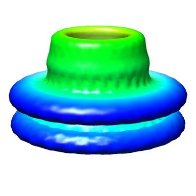 emd_6715.png emd_6715.png | 76.4 KB | ||
| アーカイブディレクトリ |  http://ftp.pdbj.org/pub/emdb/structures/EMD-6715ftp://ftp.pdbj.org/pub/emdb/structures/EMD-6715 http://ftp.pdbj.org/pub/emdb/structures/EMD-6715ftp://ftp.pdbj.org/pub/emdb/structures/EMD-6715 | HTTPS FTP |
-関連構造データ

-リンク
| EMDBのページ | EMDB (EBI/PDBe) / EMDataResource |
|---|---|
| 「今月の分子」の関連する項目 |

-マップ
| ファイル | ダウンロード / ファイル: emd_6715.map.gz / 形式: CCP4 / 大きさ: 844.7 KB / タイプ: IMAGE STORED AS FLOATING POINT NUMBER (4 BYTES) | ||||||||||||||||||||||||||||||||||||||||||||||||||||||||||||
|---|---|---|---|---|---|---|---|---|---|---|---|---|---|---|---|---|---|---|---|---|---|---|---|---|---|---|---|---|---|---|---|---|---|---|---|---|---|---|---|---|---|---|---|---|---|---|---|---|---|---|---|---|---|---|---|---|---|---|---|---|---|
| 注釈 | FliF ring assembly | ||||||||||||||||||||||||||||||||||||||||||||||||||||||||||||
| ボクセルのサイズ | X=Y=Z: 4.8 Å | ||||||||||||||||||||||||||||||||||||||||||||||||||||||||||||
| 密度 |
| ||||||||||||||||||||||||||||||||||||||||||||||||||||||||||||
| 対称性 | 空間群: 1 | ||||||||||||||||||||||||||||||||||||||||||||||||||||||||||||
| 詳細 | EMDB XML:
CCP4マップ ヘッダ情報:
| ||||||||||||||||||||||||||||||||||||||||||||||||||||||||||||
-添付データ
- 試料の構成要素
-全体 : FliF ring
| 全体 | 名称: FliF ring |
|---|---|
| 要素 |
|
-超分子 #1: FliF ring
| 超分子 | 名称: FliF ring / タイプ: complex / ID: 1 / 親要素: 0 / 詳細: FliF ring oligomer from Salmonella |
|---|---|
| 由来(天然) | 生物種: Salmonella enterica subsp. enterica serovar Typhimurium (サルモネラ菌) |
| 組換発現 | 生物種: Escherichia coli (大腸菌) / 組換株: BL21 |
| 分子量 | 理論値: 1.6 MDa |
-実験情報
-構造解析
| 手法 | ネガティブ染色法, クライオ電子顕微鏡法 |
|---|---|
 解析 解析 | 単粒子再構成法 |
| 試料の集合状態 | particle |
-試料調製
| 濃度 | 5 mg/mL |
|---|---|
| 緩衝液 | pH: 8 |
| 染色 | タイプ: POSITIVE / 材質: ammonium molybdate / 詳細: 8% (w/v) ammonium molybdate, 2% (w/v) trehalose |
| グリッド | モデル: Quantifoil R2/2 / 支持フィルム - 材質: CARBON / 支持フィルム - トポロジー: HOLEY / 前処理 - タイプ: GLOW DISCHARGE |
| 凍結 | 凍結剤: ETHANE |
- 電子顕微鏡法
| 顕微鏡 | HITACHI EF2000 |
|---|---|
| 電子線 | 加速電圧: 200 kV / 電子線源: FIELD EMISSION GUN |
| 電子光学系 | 倍率(補正後): 62500 / 照射モード: FLOOD BEAM / 撮影モード: BRIGHT FIELDBright-field microscopy / 最大 デフォーカス(公称値): 0.003 µm / 最小 デフォーカス(公称値): 0.0008 µm |
| 特殊光学系 | エネルギーフィルター - 名称: Hitachi gamma-type energy filter |
| 試料ステージ | 試料ホルダーモデル: OTHER / ホルダー冷却材: NITROGEN |
| 温度 | 最低: 93.0 K / 最高: 93.0 K |
| 撮影 | フィルム・検出器のモデル: GATAN MULTISCAN / 平均電子線量: 2.0 e/Å2 |
-画像解析
| 初期モデル | モデルのタイプ: OTHER / 詳細: model by EMAN startCsym |
|---|---|
| 初期 角度割当 | タイプ: PROJECTION MATCHING / ソフトウェア - 名称: EMAN (ver. 1) |
| 最終 角度割当 | タイプ: PROJECTION MATCHING / ソフトウェア - 名称: EMAN (ver. 1) |
| 最終 再構成 | 使用したクラス数: 33 / 想定した対称性 - 点群: C26 (26回回転対称) / 解像度のタイプ: BY AUTHOR / 解像度: 22.0 Å / 解像度の算出法: FSC 0.5 CUT-OFF / ソフトウェア - 名称: EMAN (ver. 1) / 使用した粒子像数: 4800 |
