 ムービー
ムービー コントローラー


+ データを開く

- 基本情報
基本情報
| 登録情報 | データベース: EMDB / ID: EMD-6037 | |||||||||
|---|---|---|---|---|---|---|---|---|---|---|
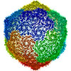
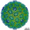
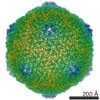
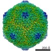
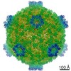
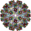
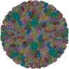
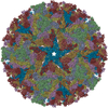
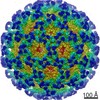
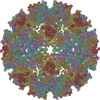
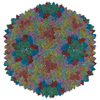
| タイトル | Capsid Expansion Mechanism Of Bacteriophage T7 Revealed By Multi-State Atomic Models Derived From Cryo-EM Reconstructions | |||||||||
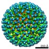
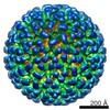
 マップデータ マップデータ | Reconstruction of bacteriophage T7 mature capsid with icosahedral symmetry averaging | |||||||||
 試料 試料 |
| |||||||||
 キーワード キーワード |  Bacteriophage T7 (T7ファージ) / Maturation / DNA packaging (染色体) / Procapsid (カプシド) / Non-covalent topological linking / Single particle cryo-EM Bacteriophage T7 (T7ファージ) / Maturation / DNA packaging (染色体) / Procapsid (カプシド) / Non-covalent topological linking / Single particle cryo-EM | |||||||||
| 機能・相同性 | Capsid Gp10A/Gp10B / : / Major capsid protein / カプシド / identical protein binding / Major capsid protein 機能・相同性情報 機能・相同性情報 | |||||||||
| 生物種 |   Enterobacteria phage T7 (ファージ) Enterobacteria phage T7 (ファージ) | |||||||||
| 手法 | 単粒子再構成法 / クライオ電子顕微鏡法 / 解像度: 3.6 Å | |||||||||
 データ登録者 データ登録者 | Guo F / Liu Z / Fang PA / Zhang Q / Wright ET / Wu W / Zhang C / Vago F / Ren Y / Jakata J ...Guo F / Liu Z / Fang PA / Zhang Q / Wright ET / Wu W / Zhang C / Vago F / Ren Y / Jakata J / Chiu W / Serwer P / Jiang W | |||||||||
 引用 引用 | ジャーナル: Proc Natl Acad Sci U S A / 年: 2014 タイトル: Capsid expansion mechanism of bacteriophage T7 revealed by multistate atomic models derived from cryo-EM reconstructions. 著者: Fei Guo / Zheng Liu / Ping-An Fang / Qinfen Zhang / Elena T Wright / Weimin Wu / Ci Zhang / Frank Vago / Yue Ren / Joanita Jakana / Wah Chiu / Philip Serwer / Wen Jiang /  要旨: Many dsDNA viruses first assemble a DNA-free procapsid, using a scaffolding protein-dependent process. The procapsid, then, undergoes dramatic conformational maturation while packaging DNA. For ...Many dsDNA viruses first assemble a DNA-free procapsid, using a scaffolding protein-dependent process. The procapsid, then, undergoes dramatic conformational maturation while packaging DNA. For bacteriophage T7 we report the following four single-particle cryo-EM 3D reconstructions and the derived atomic models: procapsid (4.6-Å resolution), an early-stage DNA packaging intermediate (3.5 Å), a later-stage packaging intermediate (6.6 Å), and the final infectious phage (3.6 Å). In the procapsid, the N terminus of the major capsid protein, gp10, has a six-turn helix at the inner surface of the shell, where each skewed hexamer of gp10 interacts with two scaffolding proteins. With the exit of scaffolding proteins during maturation the gp10 N-terminal helix unfolds and swings through the capsid shell to the outer surface. The refolded N-terminal region has a hairpin that forms a novel noncovalent, joint-like, intercapsomeric interaction with a pocket formed during shell expansion. These large conformational changes also result in a new noncovalent, intracapsomeric topological linking. Both interactions further stabilize the capsids by interlocking all pentameric and hexameric capsomeres in both DNA packaging intermediate and phage. Although the final phage shell has nearly identical structure to the shell of the DNA-free intermediate, surprisingly we found that the icosahedral faces of the phage are slightly (∼4 Å) contracted relative to the faces of the intermediate, despite the internal pressure from the densely packaged DNA genome. These structures provide a basis for understanding the capsid maturation process during DNA packaging that is essential for large numbers of dsDNA viruses. | |||||||||
| 履歴 |
|
- 構造の表示
構造の表示
| ムービー |
ムービービューア |
|---|---|
| 構造ビューア | EMマップ: SurfViewMolmilJmol/JSmol |
| 添付画像 |

- ダウンロードとリンク
-EMDBアーカイブ
| マップデータ | emd_6037.map.gz | 542 MB | EMDBマップデータ形式 | |
|---|---|---|---|---|
| ヘッダ (付随情報) | emd-6037-v30.xmlemd-6037.xml | 13 KB 13 KB | 表示 表示 | EMDBヘッダ |
| 画像 | 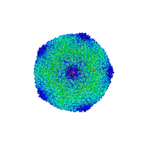 emd_6037.png emd_6037.png | 131.4 KB | ||
| Filedesc structureFactors | emd_6037_sf.cif.gz | 3.4 MB | ||
| アーカイブディレクトリ |  http://ftp.pdbj.org/pub/emdb/structures/EMD-6037ftp://ftp.pdbj.org/pub/emdb/structures/EMD-6037 http://ftp.pdbj.org/pub/emdb/structures/EMD-6037ftp://ftp.pdbj.org/pub/emdb/structures/EMD-6037 | HTTPS FTP |
-関連構造データ

-リンク
| EMDBのページ | EMDB (EBI/PDBe) / EMDataResource |
|---|
-マップ
| ファイル | ダウンロード / ファイル: emd_6037.map.gz / 形式: CCP4 / 大きさ: 1.9 GB / タイプ: IMAGE STORED AS FLOATING POINT NUMBER (4 BYTES) | ||||||||||||||||||||||||||||||||||||||||||||||||||||||||||||||||||||
|---|---|---|---|---|---|---|---|---|---|---|---|---|---|---|---|---|---|---|---|---|---|---|---|---|---|---|---|---|---|---|---|---|---|---|---|---|---|---|---|---|---|---|---|---|---|---|---|---|---|---|---|---|---|---|---|---|---|---|---|---|---|---|---|---|---|---|---|---|---|
| 注釈 | Reconstruction of bacteriophage T7 mature capsid with icosahedral symmetry averaging | ||||||||||||||||||||||||||||||||||||||||||||||||||||||||||||||||||||
| ボクセルのサイズ | X=Y=Z: 1.1 Å | ||||||||||||||||||||||||||||||||||||||||||||||||||||||||||||||||||||
| 密度 |
| ||||||||||||||||||||||||||||||||||||||||||||||||||||||||||||||||||||
| 対称性 | 空間群: 1 | ||||||||||||||||||||||||||||||||||||||||||||||||||||||||||||||||||||
| 詳細 | EMDB XML:
CCP4マップ ヘッダ情報:
| ||||||||||||||||||||||||||||||||||||||||||||||||||||||||||||||||||||
-添付データ
- 試料の構成要素
-全体 : Bacteriophage T7 mature phage capsid
| 全体 | 名称: Bacteriophage T7 mature phage capsid |
|---|---|
| 要素 |
|
-超分子 #1000: Bacteriophage T7 mature phage capsid
| 超分子 | 名称: Bacteriophage T7 mature phage capsid / タイプ: sample / ID: 1000 集合状態: 415 copies of gp10A form T=7 icosahedral shell Number unique components: 1 |
|---|---|
| 分子量 | 理論値: 15.1 MDa |
-超分子 #1: Enterobacteria phage T7
| 超分子 | 名称: Enterobacteria phage T7 / タイプ: virus / ID: 1 / NCBI-ID: 10760 / 生物種: Enterobacteria phage T7 / データベース: NCBI / ウイルスタイプ: VIRION / ウイルス・単離状態: SPECIES / ウイルス・エンベロープ: No / ウイルス・中空状態: No |
|---|---|
| 宿主 | 生物種:  Escherichia coli (大腸菌) / 別称: BACTERIA(EUBACTERIA) Escherichia coli (大腸菌) / 別称: BACTERIA(EUBACTERIA) |
| 分子量 | 理論値: 15.1 MDa |
| ウイルス殻 | Shell ID: 1 / 名称: mature phage capsid / 直径: 564 Å / T番号(三角分割数): 7 |
-実験情報
-構造解析
| 手法 | クライオ電子顕微鏡法 |
|---|---|
 解析 解析 | 単粒子再構成法 |
| 試料の集合状態 | particle |
-試料調製
| 緩衝液 | pH: 7.4 / 詳細: 200 mM NaCl, 10 mM Tris-HCl, 1 mM MgCl2 |
|---|---|
| グリッド | 詳細: 400 mesh copper grid with one lacy carbon layer |
| 凍結 | 凍結剤: ETHANE / チャンバー内湿度: 90 % / チャンバー内温度: 120 K / 装置: FEI VITROBOT MARK I 手法: Blot for 2 seconds twice with 2 mm offset before plunging. |
- 電子顕微鏡法
| 顕微鏡 | FEI TITAN KRIOS |
|---|---|
| 電子線 | 加速電圧: 300 kV / 電子線源: FIELD EMISSION GUN |
| 電子光学系 | 倍率(補正後): 57727 / 照射モード: FLOOD BEAM / 撮影モード: BRIGHT FIELDBright-field microscopy / Cs: 2.7 mm / 最大 デフォーカス(公称値): 2.4 µm / 最小 デフォーカス(公称値): 0.6 µm / 倍率(公称値): 59000 |
| 試料ステージ | 試料ホルダー: Liquid nitrogen-cooled 試料ホルダーモデル: FEI TITAN KRIOS AUTOGRID HOLDER |
| 温度 | 最低: 80 K / 最高: 100 K / 平均: 95 K |
| 日付 | 2010年8月9日 |
| 撮影 | カテゴリ: FILM / フィルム・検出器のモデル: KODAK SO-163 FILM デジタル化 - スキャナー: NIKON SUPER COOLSCAN 9000 デジタル化 - サンプリング間隔: 6.35 µm / 実像数: 364 / 平均電子線量: 25 e/Å2 / Od range: 1 / ビット/ピクセル: 16 |
| 実験機器 |  モデル: Titan Krios / 画像提供: FEI Company |
-画像解析
| CTF補正 | 詳細: Each particle |
|---|---|
| 最終 再構成 | アルゴリズム: OTHER / 解像度のタイプ: BY AUTHOR / 解像度: 3.6 Å / 解像度の算出法: OTHER / ソフトウェア - 名称: jspr, EMAN, EMAN2 詳細: For 3D reconstruction, whole datasets were divided into even and odd halves and the initial de novo models and subsequent iterative refinements were all independently performed for each half dataset. 使用した粒子像数: 33952 |
| 詳細 | Particles were selected from scanned micrograph images, first automatically by the ethan method and then by manual screening with the boxer program in EMAN. The TEM instrument contrast transfer function parameters were determined automatically using fitctf2.py and were then visually validated using the EMAN ctfit program. The datasets were then divided into two subsets (even and odd) and processed completely independently, including both initial models and refinements. For 3D reconstructions, the whole datasets were divided into even-odd halves and the initial de novo models and subsequent iterative refinements were all independently performed for each half dataset. The images were first binned 4x to obtain initial models and particle parameters assuming icosahedral symmetry. De novo initial models were built using the random model approach. Random subsets of particles were assigned random initial orientations and iteratively refined until convergence. Consistent icosahedral capsid structures (other than occasional differences in handedness) were obtained by repeating the random model process. Particles with inconsistent/unstable view parameters in the initial refinements were excluded in further image processing. The orientation and center parameters were then transferred to the un-binned images for high-resolution refinements which included Simplex method-based orientation/center optimization and grid search-based refinement of defocus, astigmatism, and magnification of the images. All image refinement and reconstructions were performed with in-house developed programs jspr.py (for overall work-flow), jalign (for 2D alignment) and j3dr (for 3D reconstruction), which use EMAN and EMAN2 library functions. |
