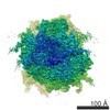
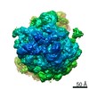
ジャーナル: Nat Methods / 年: 2019 タイトル: A cryo-FIB lift-out technique enables molecular-resolution cryo-ET within native Caenorhabditis elegans tissue. 著者: Miroslava Schaffer / Stefan Pfeffer / Julia Mahamid / Stephan Kleindiek / Tim Laugks / Sahradha Albert / Benjamin D Engel / Andreas Rummel / Andrew J Smith / Wolfgang Baumeister / Juergen M Plitzko / 要旨: Cryo-focused ion beam milling of frozen-hydrated cells has recently provided unprecedented insights into the inner space of cells. In combination with cryo-electron tomography, this method allows ...Cryo-focused ion beam milling of frozen-hydrated cells has recently provided unprecedented insights into the inner space of cells. In combination with cryo-electron tomography, this method allows access to native structures deep inside cells, enabling structural studies of macromolecules in situ. However, this approach has been mainly limited to individual cells that can be completely vitrified by plunge-freezing. Here, we describe a preparation method that is based on the targeted extraction of material from high-pressure-frozen bulk specimens with a cryo-gripper tool. This lift-out technique enables cryo-electron tomography to be performed on multicellular organisms and tissue, extending the range of applications for in situ structural biology. We demonstrate the potential of the lift-out technique with a structural study of cytosolic 80S ribosomes in a Caenorhabditis elegans worm. The preparation quality allowed for subtomogram analysis with sufficient resolution to distinguish individual ribosomal translocation states and revealed significant cell-to-cell variation in ribosome structure.
 ムービー
ムービー コントローラー


 データを開く
 基本情報
基本情報 マップデータ
 試料
試料
 Caenorhabditis elegans (センチュウ)
 データ登録者
 ドイツ, 2件
  引用
引用 構造の表示
構造の表示 ムービービューア
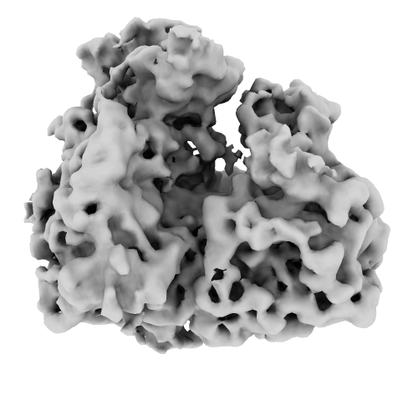
 ダウンロードとリンク
 emd_4756.png
 http://ftp.pdbj.org/pub/emdb/structures/EMD-4756


 試料の構成要素
 解析
解析 電子顕微鏡法
