 ムービー
ムービー コントローラー


+ データを開く

- 基本情報
基本情報
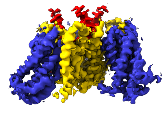
| 登録情報 | データベース: EMDB / ID: EMD-4386 | |||||||||
|---|---|---|---|---|---|---|---|---|---|---|
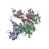
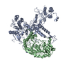
| タイトル | cryo-EM structure of the human neutral amino acid transporter ASCT2 | |||||||||
 マップデータ マップデータ | ||||||||||
 試料 試料 |
| |||||||||
| 機能・相同性 |  機能・相同性情報 機能・相同性情報glutamine secretion / L-glutamine import across plasma membrane / L-serine transmembrane transporter activity / glutamine transport / L-glutamine transmembrane transporter activity /  ligand-gated channel activity / neutral amino acid transport / amino acid transmembrane transporter activity / Amino acid transport across the plasma membrane / neutral L-amino acid transmembrane transporter activity ...glutamine secretion / L-glutamine import across plasma membrane / L-serine transmembrane transporter activity / glutamine transport / L-glutamine transmembrane transporter activity / ligand-gated channel activity / neutral amino acid transport / amino acid transmembrane transporter activity / Amino acid transport across the plasma membrane / neutral L-amino acid transmembrane transporter activity / L-aspartate transmembrane transporter activity / L-aspartate import across plasma membrane / symporter activity / amino acid transport / antiporter activity / RHOJ GTPase cycle / RHOQ GTPase cycle / protein homotrimerization / RHOH GTPase cycle / transport across blood-brain barrier / RAC3 GTPase cycle / RAC1 GTPase cycle / basal plasma membrane / 赤血球形成 / メラノソーム / virus receptor activity / signaling receptor activity / extracellular exosome / 生体膜 / metal ion binding / 細胞膜 ligand-gated channel activity / neutral amino acid transport / amino acid transmembrane transporter activity / Amino acid transport across the plasma membrane / neutral L-amino acid transmembrane transporter activity ...glutamine secretion / L-glutamine import across plasma membrane / L-serine transmembrane transporter activity / glutamine transport / L-glutamine transmembrane transporter activity / ligand-gated channel activity / neutral amino acid transport / amino acid transmembrane transporter activity / Amino acid transport across the plasma membrane / neutral L-amino acid transmembrane transporter activity / L-aspartate transmembrane transporter activity / L-aspartate import across plasma membrane / symporter activity / amino acid transport / antiporter activity / RHOJ GTPase cycle / RHOQ GTPase cycle / protein homotrimerization / RHOH GTPase cycle / transport across blood-brain barrier / RAC3 GTPase cycle / RAC1 GTPase cycle / basal plasma membrane / 赤血球形成 / メラノソーム / virus receptor activity / signaling receptor activity / extracellular exosome / 生体膜 / metal ion binding / 細胞膜類似検索 - 分子機能 | |||||||||
| 生物種 |  Homo sapiens (ヒト) Homo sapiens (ヒト) | |||||||||
| 手法 | 単粒子再構成法 / クライオ電子顕微鏡法 / 解像度: 3.85 Å | |||||||||
 データ登録者 データ登録者 | Garaeva AA / Oostergetel GT / Gati C / Guskov A / Paulino C / Slotboom DJ | |||||||||
| 資金援助 |  オランダ, 2件 オランダ, 2件
| |||||||||
 引用 引用 | ジャーナル: Nat Struct Mol Biol / 年: 2018 タイトル: Cryo-EM structure of the human neutral amino acid transporter ASCT2. 著者: Alisa A Garaeva / Gert T Oostergetel / Cornelius Gati / Albert Guskov / Cristina Paulino / Dirk J Slotboom /  要旨: Human ASCT2 belongs to the SLC1 family of secondary transporters and is specific for the transport of small neutral amino acids. ASCT2 is upregulated in cancer cells and serves as the receptor for ...Human ASCT2 belongs to the SLC1 family of secondary transporters and is specific for the transport of small neutral amino acids. ASCT2 is upregulated in cancer cells and serves as the receptor for many retroviruses; hence, it has importance as a potential drug target. Here we used single-particle cryo-EM to determine a structure of the functional and unmodified human ASCT2 at 3.85-Å resolution. ASCT2 forms a homotrimeric complex in which each subunit contains a transport and a scaffold domain. Prominent extracellular extensions on the scaffold domain form the predicted docking site for retroviruses. Relative to structures of other SLC1 members, ASCT2 is in the most extreme inward-oriented state, with the transport domain largely detached from the central scaffold domain on the cytoplasmic side. This domain detachment may be required for substrate binding and release on the intracellular side of the membrane. | |||||||||
| 履歴 |
|
- 構造の表示
構造の表示
| ムービー |
ムービービューア |
|---|---|
| 構造ビューア | EMマップ: SurfViewMolmilJmol/JSmol |
| 添付画像 |
- ダウンロードとリンク
-EMDBアーカイブ
| マップデータ | emd_4386.map.gz | 48.8 MB | EMDBマップデータ形式 | |
|---|---|---|---|---|
| ヘッダ (付随情報) | emd-4386-v30.xmlemd-4386.xml | 25 KB 25 KB | 表示 表示 | EMDBヘッダ |
| FSC (解像度算出) | emd_4386_fsc.xml | 8.6 KB | 表示 | FSCデータファイル |
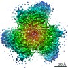
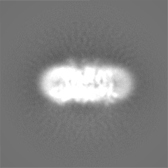
| 画像 |  emd_4386.png emd_4386.png | 223.3 KB | ||
| マスクデータ | emd_4386_msk_1.map | 52.7 MB | マスクマップ | |
| その他 | emd_4386_additional.map.gzemd_4386_half_map_1.map.gzemd_4386_half_map_2.map.gz | 49.1 MB 40.6 MB 40.6 MB | ||
| アーカイブディレクトリ |  http://ftp.pdbj.org/pub/emdb/structures/EMD-4386ftp://ftp.pdbj.org/pub/emdb/structures/EMD-4386 http://ftp.pdbj.org/pub/emdb/structures/EMD-4386ftp://ftp.pdbj.org/pub/emdb/structures/EMD-4386 | HTTPS FTP |
-関連構造データ

-リンク
| EMDBのページ | EMDB (EBI/PDBe) / EMDataResource |
|---|---|
| 「今月の分子」の関連する項目 |


-マップ
| ファイル | ダウンロード / ファイル: emd_4386.map.gz / 形式: CCP4 / 大きさ: 52.7 MB / タイプ: IMAGE STORED AS FLOATING POINT NUMBER (4 BYTES) | ||||||||||||||||||||||||||||||||||||||||||||||||||||||||||||
|---|---|---|---|---|---|---|---|---|---|---|---|---|---|---|---|---|---|---|---|---|---|---|---|---|---|---|---|---|---|---|---|---|---|---|---|---|---|---|---|---|---|---|---|---|---|---|---|---|---|---|---|---|---|---|---|---|---|---|---|---|---|
| ボクセルのサイズ | X=Y=Z: 1.012 Å | ||||||||||||||||||||||||||||||||||||||||||||||||||||||||||||
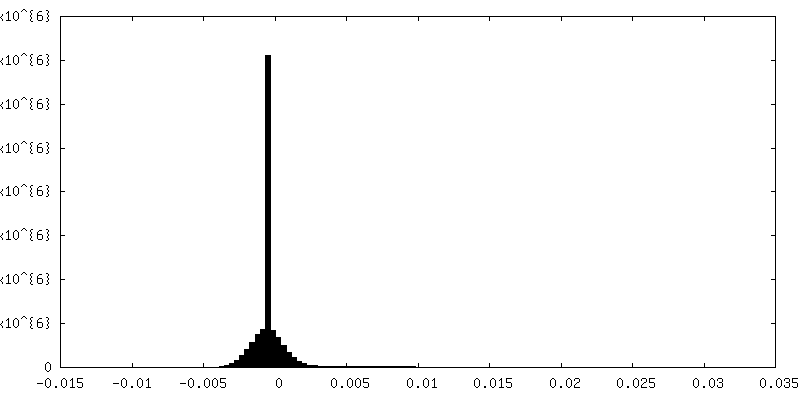
| 密度 |
| ||||||||||||||||||||||||||||||||||||||||||||||||||||||||||||
| 対称性 | 空間群: 1 | ||||||||||||||||||||||||||||||||||||||||||||||||||||||||||||
| 詳細 | EMDB XML:
CCP4マップ ヘッダ情報:
| ||||||||||||||||||||||||||||||||||||||||||||||||||||||||||||
-添付データ
-マスク #1
| ファイル | emd_4386_msk_1.map | ||||||||||||
|---|---|---|---|---|---|---|---|---|---|---|---|---|---|
| 投影像・断面図 |
| ||||||||||||
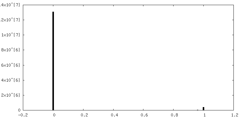
| 密度ヒストグラム |
 Z
Z Y
Y X
X


-追加マップ: #1
| ファイル | emd_4386_additional.map | ||||||||||||
|---|---|---|---|---|---|---|---|---|---|---|---|---|---|
| 投影像・断面図 |
| ||||||||||||
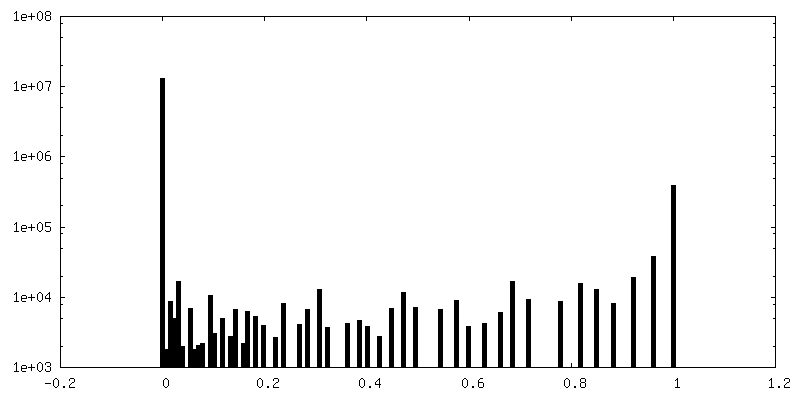
| 密度ヒストグラム |


-ハーフマップ: half-map 1 used for post processing step and...
| ファイル | emd_4386_half_map_1.map | ||||||||||||
|---|---|---|---|---|---|---|---|---|---|---|---|---|---|
| 注釈 | half-map 1 used for post processing step and FSC resolution calculation. | ||||||||||||
| 投影像・断面図 |
| ||||||||||||
| 密度ヒストグラム |


-ハーフマップ: half-map 2 used for post processing step and...
| ファイル | emd_4386_half_map_2.map | ||||||||||||
|---|---|---|---|---|---|---|---|---|---|---|---|---|---|
| 注釈 | half-map 2 used for post processing step and FSC resolution calculation. | ||||||||||||
| 投影像・断面図 |
| ||||||||||||
| 密度ヒストグラム |


- 試料の構成要素
-全体 : human ASCT2 SLC1A5
| 全体 | 名称: human ASCT2 SLC1A5 |
|---|---|
| 要素 |
|
-超分子 #1: human ASCT2 SLC1A5
| 超分子 | 名称: human ASCT2 SLC1A5 / タイプ: complex / ID: 1 / 親要素: 0 / 含まれる分子: #1 / 詳細: human ASCT2 SLC1A5 |
|---|---|
| 由来(天然) | 生物種: Homo sapiens (ヒト) |
| 組換発現 | 生物種:  Komagataella pastoris (菌類) Komagataella pastoris (菌類) |
| 分子量 | 実験値: 172 KDa |
-分子 #1: Neutral amino acid transporter B(0)
| 分子 | 名称: Neutral amino acid transporter B(0) / タイプ: protein_or_peptide / ID: 1 / コピー数: 3 / 光学異性体: LEVO |
|---|---|
| 由来(天然) | 生物種: Homo sapiens (ヒト) |
| 分子量 | 理論値: 56.638902 KDa |
| 組換発現 | 生物種: Komagataella pastoris (菌類) |
| 配列 | 文字列: MVADPPRDSK GLAAAEPTAN GGLALASIED QGAAAGGYCG SRDQVRRCLR ANLLVLLTVV AVVAGVALGL GVSGAGGALA LGPERLSAF VFPGELLLRL LRMIILPLVV CSLIGGAASL DPGALGRLGA WALLFFLVTT LLASALGVGL ALALQPGAAS A AINASVGA ...文字列: MVADPPRDSK GLAAAEPTAN GGLALASIED QGAAAGGYCG SRDQVRRCLR ANLLVLLTVV AVVAGVALGL GVSGAGGALA LGPERLSAF VFPGELLLRL LRMIILPLVV CSLIGGAASL DPGALGRLGA WALLFFLVTT LLASALGVGL ALALQPGAAS A AINASVGA AGSAENAPSK EVLDSFLDLA RNIFPSNLVS AAFRSYSTTY EERNITGTRV KVPVGQEVEG MNILGLVVFA IV FGVALRK LGPEGELLIR FFNSFNEATM VLVSWIMWYA PVGIMFLVAG KIVEMEDVGL LFARLGKYIL CCLLGHAIHG LLV LPLIYF LFTRKNPYRF LWGIVTPLAT AFGTSSSSAT LPLMMKCVEE NNGVAKHISR FILPIGATVN MDGAALFQCV AAVF IAQLS QQSLDFVKII TILVTATASS VGAAGIPAGG VLTLAIILEA VNLPVDHISL ILAVDWLVDR SCTVLNVEGD ALGAG LLQN YVDRTESRST EPELIQVKSE LPLDPLPVPT EEGNPLLKHY RGPAGDATVA SEKESVM |
-分子 #2: GLUTAMINE
| 分子 | 名称: GLUTAMINE / タイプ: ligand / ID: 2 / コピー数: 3 / 式: GLN |
|---|---|
| 分子量 | 理論値: 146.144 Da |
| Chemical component information |  ChemComp-GLN: |
-実験情報
-構造解析
| 手法 | クライオ電子顕微鏡法 |
|---|---|
 解析 解析 | 単粒子再構成法 |
| 試料の集合状態 | particle |
-試料調製
| 濃度 | 2.5 mg/mL |
|---|---|
| 緩衝液 | pH: 7 詳細: 20mM Tris-HCl pH 7.4 300mM NaCl 1mM L-glutamine 0.05% DDM 0.005% CHS |
| グリッド | モデル: Quantifoil, UltrAuFoil, R1.2/1.3 / 材質: GOLD / メッシュ: 200 / 支持フィルム - 材質: CARBON / 支持フィルム - トポロジー: HOLEY / 前処理 - タイプ: GLOW DISCHARGE |
| 凍結 | 凍結剤: ETHANE / チャンバー内湿度: 100 % / チャンバー内温度: 278 K / 装置: FEI VITROBOT MARK II |
- 電子顕微鏡法
| 顕微鏡 | FEI TALOS ARCTICA |
|---|---|
| 電子線 | 加速電圧: 200 kV / 電子線源: FIELD EMISSION GUN |
| 電子光学系 | C2レンズ絞り径: 100.0 µm / 最大 デフォーカス(補正後): 0.0025 µm / 最小 デフォーカス(補正後): 0.0004 µm / 倍率(補正後): 49407 / 照射モード: FLOOD BEAM / 撮影モード: BRIGHT FIELDBright-field microscopy / Cs: 2.7 mm / 最大 デフォーカス(公称値): 0.0025 µm / 最小 デフォーカス(公称値): 0.0004 µm / 倍率(公称値): 49407 |
| 特殊光学系 | エネルギーフィルター - 名称: GIF Quantum LS エネルギーフィルター - エネルギー下限: 0 eV エネルギーフィルター - エネルギー上限: 20 eV |
| 試料ステージ | 試料ホルダーモデル: FEI TITAN KRIOS AUTOGRID HOLDER ホルダー冷却材: NITROGEN |
| 温度 | 最低: 70.0 K / 最高: 90.0 K |
| 撮影 | フィルム・検出器のモデル: GATAN K2 SUMMIT (4k x 4k) 検出モード: COUNTING / デジタル化 - 画像ごとのフレーム数: 1-60 / 平均露光時間: 9.0 sec. / 平均電子線量: 0.87 e/Å2 詳細: Freshly purified protein was concentrated using Vivaspin concentrating devices with a molecular weight cutoff of 100kDa to 2-2.5 mg ml-1. 2.8 ul were applied on holey-carbon cryo-EM grids ...詳細: Freshly purified protein was concentrated using Vivaspin concentrating devices with a molecular weight cutoff of 100kDa to 2-2.5 mg ml-1. 2.8 ul were applied on holey-carbon cryo-EM grids (Quantifoil Au R1.2-1.3, 200 and 300 mesh), which were prior glow-discharged at 5 mA for 20 s. Grids were blotted for 3-5 s in a Vitrobot (Mark 3, Thermo Fisher) at 20C temperature and 100% humidity, subsequently plunge-frozen in liquid ethane and stored in liquid nitrogen. Cryo-EM data were collected on a 200 keV Talos Arctica microscope (Thermo Fisher) using a post-column energy filter (Gatan) in zero-loss mode, using a 20 eV slit, a 100 um objective aperture, in an automated fashion using EPU software (Thermo Fisher) on a K2 summit detector (Gatan) in counting mode. Cryo-EM images were acquired at a pixel size of 1.012A (calibrated magnification of 49,407x), a defocus range from -0.4 to 2.5 um, an exposure time of 9 sec and a sub-frame exposure time of 150 ms (60 frames), and a total electron dose on the specimen level of about 52 electrons per A2. Best regions on the grid were screened with a self-written script to calculate the ice thickness and data quality was monitored on the fly using the software FOCUS |
| 実験機器 |  モデル: Talos Arctica / 画像提供: FEI Company |
-画像解析
| CTF補正 | ソフトウェア - 名称: CTFFIND (ver. 4.1) |
|---|---|
| 初期モデル | モデルのタイプ: PDB ENTRY PDBモデル - PDB ID: |
| 初期 角度割当 | タイプ: ANGULAR RECONSTITUTION / ソフトウェア - 名称: RELION (ver. 2.1) |
| 最終 角度割当 | タイプ: ANGULAR RECONSTITUTION / ソフトウェア - 名称: RELION (ver. 2.1) |
| 最終 再構成 | 想定した対称性 - 点群: C3 (3回回転対称) / 解像度のタイプ: BY AUTHOR / 解像度: 3.85 Å / 解像度の算出法: FSC 0.143 CUT-OFF / ソフトウェア - 名称: RELION (ver. 2.1) 詳細: A total of 6345 dose-fractionated cryo-EM images were recorded and subjected to motion-correction and dose-weighting of frames by MotionCor2. The CTF parameters were estimated on the movie ...詳細: A total of 6345 dose-fractionated cryo-EM images were recorded and subjected to motion-correction and dose-weighting of frames by MotionCor2. The CTF parameters were estimated on the movie frames by ctffind4.1. Bad images showing contamination, a defocus below or above 0.4 and -3um or a bad CTF estimation were discarded, resulting in 4863 images used for further analysis with the software package RELION2.1. About 3000 particles were picked manually to generate 2D references which where improved in several rounds of autopick. A low threshold was used during the final autopick step to ensure that no particles are missed yielding more than a million particles. Particles were extracted with a box size of 240 pixels, and initial classification steps were performed with three-fold binned data. False positives or bad particles were removed in first rounds of 2D classification, resulting in 628,015 particles that were further sorted in several rounds of 3D classification. A map generated from the GltPh structure (PDB ID 3KBC) was used as reference for the first round, and the best output class was used in subsequent jobs in an iterative way. The best 3D class, comprising 184,080 particles from 4859 images, was subjected to auto-refinement, yielding a map with a resolution of 4.26 A before masking and 3.91 A after masking. Particles were further polished in RELION version 2.1 and subjected to another round of 2D and 3D classification resulting in a final dataset of 133,437 particles. The final polished map had a resolution of 4.26 A before masking and 3.85 A after masking. The map was sharpened using an isotropic B-factor of -171 A2, for manual inspection a B-factor of -225 A2 was used. The approach of focused refinement, where the less-resolved detergent micelle was subtracted from the particle images, did not improve resolution. During 3D classification and auto-refinement jobs a C3-symmetry was imposed. To check for conformational heterogeneity of the data, where single protomers within the trimer might adopt a different conformation, 3D classifications with no symmetry imposed were performed at different stages of image processing. We further performed 3D classification on the individual protomers of a single transporter using symmetry expansion and signal subtraction. Both approaches showed no indication of the existence of a different conformation. Local resolution estimates were estimated by RELION. All resolutions were estimated using the 0.143 cut-off criterion with gold-standard Fourier shell correlation (FSC) between two independently refined half maps. During post-processing, the approach of high-resolution noise substitution was used to correct for convolution effects of real-space masking on the FSC curve. 使用した粒子像数: 184080 |
| FSC曲線 (解像度の算出) |  |

-原子モデル構築 1
| 精密化 | 空間: REAL / プロトコル: AB INITIO MODEL |
|---|---|
| 得られたモデル | 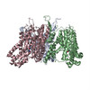 PDB-6gct: |
