 ムービー
ムービー コントローラー


+ データを開く

- 基本情報
基本情報
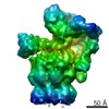
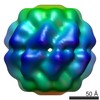
| 登録情報 | データベース: EMDB / ID: EMD-4191 | |||||||||
|---|---|---|---|---|---|---|---|---|---|---|
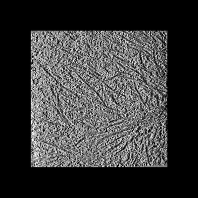
| タイトル | In situ cryo-electron tomogram from Rat neuron with C9ORF72 Poly-GA aggregates | |||||||||
 マップデータ マップデータ | Tomogram of Rat neuron cell | |||||||||
 試料 試料 |
| |||||||||
| 生物種 |   Rattus norvegicus (ドブネズミ) Rattus norvegicus (ドブネズミ) | |||||||||
| 手法 | 電子線トモグラフィー法 / クライオ電子顕微鏡法 | |||||||||
 データ登録者 データ登録者 | Guo Q / Lehmer C / Martinez-Sanchez A / Rudack T / Beck F / Hartmann H / Hipp MS / Hartl FU / Edbauer D / Baumeister W / Fernandez-Busnadiego F | |||||||||
 引用 引用 | ジャーナル: Cell / 年: 2018 タイトル: In Situ Structure of Neuronal C9orf72 Poly-GA Aggregates Reveals Proteasome Recruitment. 著者: Qiang Guo / Carina Lehmer / Antonio Martínez-Sánchez / Till Rudack / Florian Beck / Hannelore Hartmann / Manuela Pérez-Berlanga / Frédéric Frottin / Mark S Hipp / F Ulrich Hartl / Dieter ...著者: Qiang Guo / Carina Lehmer / Antonio Martínez-Sánchez / Till Rudack / Florian Beck / Hannelore Hartmann / Manuela Pérez-Berlanga / Frédéric Frottin / Mark S Hipp / F Ulrich Hartl / Dieter Edbauer / Wolfgang Baumeister / Rubén Fernández-Busnadiego /   要旨: Protein aggregation and dysfunction of the ubiquitin-proteasome system are hallmarks of many neurodegenerative diseases. Here, we address the elusive link between these phenomena by employing cryo- ...Protein aggregation and dysfunction of the ubiquitin-proteasome system are hallmarks of many neurodegenerative diseases. Here, we address the elusive link between these phenomena by employing cryo-electron tomography to dissect the molecular architecture of protein aggregates within intact neurons at high resolution. We focus on the poly-Gly-Ala (poly-GA) aggregates resulting from aberrant translation of an expanded GGGGCC repeat in C9orf72, the most common genetic cause of amyotrophic lateral sclerosis and frontotemporal dementia. We find that poly-GA aggregates consist of densely packed twisted ribbons that recruit numerous 26S proteasome complexes, while other macromolecules are largely excluded. Proximity to poly-GA ribbons stabilizes a transient substrate-processing conformation of the 26S proteasome, suggesting stalled degradation. Thus, poly-GA aggregates may compromise neuronal proteostasis by driving the accumulation and functional impairment of a large fraction of cellular proteasomes. | |||||||||
| 履歴 |
|
- 構造の表示
構造の表示
| ムービー |
 ムービービューア ムービービューア |
|---|---|
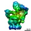
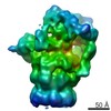
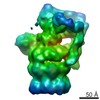
| 添付画像 |
- ダウンロードとリンク
-EMDBアーカイブ
| マップデータ | emd_4191.map.gz | 215.5 MB | EMDBマップデータ形式 | |
|---|---|---|---|---|
| ヘッダ (付随情報) | emd-4191-v30.xmlemd-4191.xml | 14.9 KB 14.9 KB | 表示 表示 | EMDBヘッダ |
| 画像 |  emd_4191.png emd_4191.png | 75.4 KB | ||
| アーカイブディレクトリ |  http://ftp.pdbj.org/pub/emdb/structures/EMD-4191ftp://ftp.pdbj.org/pub/emdb/structures/EMD-4191 http://ftp.pdbj.org/pub/emdb/structures/EMD-4191ftp://ftp.pdbj.org/pub/emdb/structures/EMD-4191 | HTTPS FTP |
-関連構造データ
-リンク
| EMDBのページ | EMDB (EBI/PDBe) / EMDataResource |
|---|
-マップ
| ファイル | ダウンロード / ファイル: emd_4191.map.gz / 形式: CCP4 / 大きさ: 232.2 MB / タイプ: IMAGE STORED AS FLOATING POINT NUMBER (4 BYTES) | ||||||||||||||||||||||||||||||||||||||||||||||||||||||||||||
|---|---|---|---|---|---|---|---|---|---|---|---|---|---|---|---|---|---|---|---|---|---|---|---|---|---|---|---|---|---|---|---|---|---|---|---|---|---|---|---|---|---|---|---|---|---|---|---|---|---|---|---|---|---|---|---|---|---|---|---|---|---|
| 注釈 | Tomogram of Rat neuron cell | ||||||||||||||||||||||||||||||||||||||||||||||||||||||||||||
| ボクセルのサイズ | X=Y=Z: 13.68 Å | ||||||||||||||||||||||||||||||||||||||||||||||||||||||||||||
| 密度 |
| ||||||||||||||||||||||||||||||||||||||||||||||||||||||||||||
| 対称性 | 空間群: 1 | ||||||||||||||||||||||||||||||||||||||||||||||||||||||||||||
| 詳細 | EMDB XML:
CCP4マップ ヘッダ情報:
| ||||||||||||||||||||||||||||||||||||||||||||||||||||||||||||
-添付データ
- 試料の構成要素
-全体 : in situ tomogram from Rat neuron
| 全体 | 名称: in situ tomogram from Rat neuron |
|---|---|
| 要素 |
|
-超分子 #1: in situ tomogram from Rat neuron
| 超分子 | 名称: in situ tomogram from Rat neuron / タイプ: cell / ID: 1 / 親要素: 0 / 含まれる分子: #1-#32 |
|---|---|
| 由来(天然) | 生物種: Rattus norvegicus (ドブネズミ) |
-実験情報
-構造解析
| 手法 | クライオ電子顕微鏡法 |
|---|---|
 解析 解析 | 電子線トモグラフィー法 |
| 試料の集合状態 | cell |
-試料調製
| 緩衝液 | pH: 7 |
|---|---|
| グリッド | モデル: Quantifoil R2/1 / 材質: GOLD / メッシュ: 200 / 支持フィルム - 材質: CARBON / 支持フィルム - トポロジー: HOLEY / 前処理 - タイプ: GLOW DISCHARGE / 前処理 - 雰囲気: AIR / 詳細: The grid was coated with C prior to use |
| 凍結 | 凍結剤: ETHANE-PROPANE / チャンバー内湿度: 100 % / チャンバー内温度: 298 K / 装置: FEI VITROBOT MARK IV |
| 詳細 | FIB milled rat primary neurons |
| 切片作成 | 集束イオンビーム - 装置: OTHER / 集束イオンビーム - イオン: OTHER / 集束イオンビーム - 電圧: 30 kV / 集束イオンビーム - 電流: 0.5 nA / 集束イオンビーム - Dose rate: 1 / 集束イオンビーム - 時間: 100 sec. / 集束イオンビーム - 温度: 90 K / 集束イオンビーム - Initial thickness: 1500 / 集束イオンビーム - 最終 厚さ: 200 集束イオンビーム - 詳細: 11nA rough milling. The value given for _emd_sectioning_focused_ion_beam.instrument is FEI Scios. This is not in a list of allowed values set(['DB235', 'OTHER']) so ...集束イオンビーム - 詳細: 11nA rough milling. The value given for _emd_sectioning_focused_ion_beam.instrument is FEI Scios. This is not in a list of allowed values set(['DB235', 'OTHER']) so OTHER is written into the XML file. |
- 電子顕微鏡法
| 顕微鏡 | FEI TITAN KRIOS |
|---|---|
| 電子線 | 加速電圧: 300 kV / 電子線源: FIELD EMISSION GUN |
| 電子光学系 | C2レンズ絞り径: 70.0 µm / 照射モード: FLOOD BEAM / 撮影モード: BRIGHT FIELDBright-field microscopy / Cs: 2.7 mm / 最大 デフォーカス(公称値): 7.0 µm / 最小 デフォーカス(公称値): 5.0 µm / 倍率(公称値): 42000 |
| 試料ステージ | 試料ホルダーモデル: FEI TITAN KRIOS AUTOGRID HOLDER ホルダー冷却材: NITROGEN |
| 撮影 | フィルム・検出器のモデル: GATAN K2 SUMMIT (4k x 4k) 検出モード: COUNTING / デジタル化 - サイズ - 横: 3838 pixel / デジタル化 - サイズ - 縦: 3710 pixel / 平均露光時間: 2.0 sec. / 平均電子線量: 1.8 e/Å2 |
| 実験機器 |  モデル: Titan Krios / 画像提供: FEI Company |
-画像解析
| 最終 再構成 | 解像度の算出法: OTHER / ソフトウェア - 名称: eTomo / 使用した粒子像数: 59 |
|---|
