 ムービー
ムービー コントローラー


+ データを開く

- 基本情報
基本情報
| 登録情報 | データベース: EMDB / ID: EMD-4120 | |||||||||
|---|---|---|---|---|---|---|---|---|---|---|
| タイトル | In situ TPPII 32mer | |||||||||
 マップデータ マップデータ | in situ TPPII 32mer | |||||||||
 試料 試料 |
| |||||||||
| 生物種 |   Rattus (クマネズミ属) Rattus (クマネズミ属) | |||||||||
| 手法 | サブトモグラム平均法 / クライオ電子顕微鏡法 / 解像度: 36.5 Å | |||||||||
 データ登録者 データ登録者 | Fukuda Y / Beck F / Baumeister W | |||||||||
 引用 引用 | ジャーナル: Proc Natl Acad Sci U S A / 年: 2017 タイトル: In situ structural studies of tripeptidyl peptidase II (TPPII) reveal spatial association with proteasomes. 著者: Yoshiyuki Fukuda / Florian Beck / Jürgen M Plitzko / Wolfgang Baumeister /  要旨: Tripeptidyl peptidase II (TPPII) is a eukaryotic protease acting downstream of the 26S proteasome; it removes tripeptides from the degradation products released by the proteasome. Structural studies ...Tripeptidyl peptidase II (TPPII) is a eukaryotic protease acting downstream of the 26S proteasome; it removes tripeptides from the degradation products released by the proteasome. Structural studies in vitro have revealed the basic architecture of TPPII, a two-stranded linear polymer that assembles to form a spindle-shaped complex of ∼6 MDa. Dependent on protein concentration, TPPII has a distinct tendency for polymorphism. Therefore, its structure in vivo has remained unclear. To resolve this issue, we have scrutinized cryo-electron tomograms of rat hippocampal neurons for the occurrence and spatial distribution of TPPII by template matching. The quality of the tomograms recorded with the Volta phase plate enabled a detailed structural analysis of TPPII despite its low abundance. Two different assembly states (36-mers and 32-mers) coexist as well as occasional extended forms with longer strands. A distance analysis of the relative locations of TPPII and 26S proteasomes confirmed the visual impression that these two complexes spatially associate in agreement with TPPII's role in postproteasomal degradation. | |||||||||
| 履歴 |
|
- 構造の表示
構造の表示
| ムービー |
 ムービービューア ムービービューア |
|---|---|
| 構造ビューア | EMマップ: SurfViewMolmilJmol/JSmol |
| 添付画像 |


- ダウンロードとリンク
-EMDBアーカイブ
| マップデータ | emd_4120.map.gz | 27 MB | EMDBマップデータ形式 | |
|---|---|---|---|---|
| ヘッダ (付随情報) | emd-4120-v30.xmlemd-4120.xml | 10.8 KB 10.8 KB | 表示 表示 | EMDBヘッダ |
| 画像 | 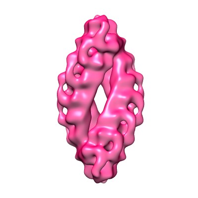 emd_4120.jpg emd_4120.jpg emd_4120.png emd_4120.png | 19.7 KB 121.9 KB | ||
| アーカイブディレクトリ |  http://ftp.pdbj.org/pub/emdb/structures/EMD-4120ftp://ftp.pdbj.org/pub/emdb/structures/EMD-4120 http://ftp.pdbj.org/pub/emdb/structures/EMD-4120ftp://ftp.pdbj.org/pub/emdb/structures/EMD-4120 | HTTPS FTP |
-関連構造データ
-リンク
| EMDBのページ | EMDB (EBI/PDBe) / EMDataResource |
|---|
-マップ
| ファイル | ダウンロード / ファイル: emd_4120.map.gz / 形式: CCP4 / 大きさ: 30.5 MB / タイプ: IMAGE STORED AS FLOATING POINT NUMBER (4 BYTES) | ||||||||||||||||||||||||||||||||||||||||||||||||||||||||||||||||||||
|---|---|---|---|---|---|---|---|---|---|---|---|---|---|---|---|---|---|---|---|---|---|---|---|---|---|---|---|---|---|---|---|---|---|---|---|---|---|---|---|---|---|---|---|---|---|---|---|---|---|---|---|---|---|---|---|---|---|---|---|---|---|---|---|---|---|---|---|---|---|
| 注釈 | in situ TPPII 32mer | ||||||||||||||||||||||||||||||||||||||||||||||||||||||||||||||||||||
| ボクセルのサイズ | X=Y=Z: 4.21 Å | ||||||||||||||||||||||||||||||||||||||||||||||||||||||||||||||||||||
| 密度 |
| ||||||||||||||||||||||||||||||||||||||||||||||||||||||||||||||||||||
| 対称性 | 空間群: 1 | ||||||||||||||||||||||||||||||||||||||||||||||||||||||||||||||||||||
| 詳細 | EMDB XML:
CCP4マップ ヘッダ情報:
| ||||||||||||||||||||||||||||||||||||||||||||||||||||||||||||||||||||
-添付データ
- 試料の構成要素
-全体 : Tripeptidyl peptidase II
| 全体 | 名称: Tripeptidyl peptidase II |
|---|---|
| 要素 |
|
-超分子 #1: Tripeptidyl peptidase II
| 超分子 | 名称: Tripeptidyl peptidase II / タイプ: complex / ID: 1 / 親要素: 0 / 詳細: 32mer |
|---|---|
| 由来(天然) | 生物種: Rattus (クマネズミ属) / 器官: brain / 組織: hippocampus |
| 分子量 | 理論値: 4.4 MDa |
-実験情報
-構造解析
| 手法 | クライオ電子顕微鏡法 |
|---|---|
 解析 解析 | サブトモグラム平均法 |
| 試料の集合状態 | cell |
-試料調製
| 緩衝液 | pH: 7 |
|---|---|
| グリッド | モデル: Quantifoil R1/4 / 材質: GOLD / メッシュ: 200 / 支持フィルム - 材質: CARBON / 支持フィルム - トポロジー: HOLEY ARRAY / 支持フィルム - Film thickness: 1000.0 nm |
| 凍結 | 凍結剤: ETHANE-PROPANE / チャンバー内湿度: 90 % / チャンバー内温度: 310 K / 装置: FEI VITROBOT MARK IV |
| 詳細 | in situ |
- 電子顕微鏡法
| 顕微鏡 | FEI TITAN KRIOS |
|---|---|
| 電子線 | 加速電圧: 300 kV / 電子線源: FIELD EMISSION GUN |
| 電子光学系 | C2レンズ絞り径: 70.0 µm / 最大 デフォーカス(補正後): 1.0 µm / 最小 デフォーカス(補正後): 0.5 µm / 照射モード: FLOOD BEAM / 撮影モード: BRIGHT FIELDBright-field microscopy / Cs: 2.7 mm / 最大 デフォーカス(公称値): 1.0 µm / 最小 デフォーカス(公称値): 0.5 µm / 倍率(公称値): 33000 |
| 特殊光学系 | 位相板: VOLTA PHASE PLATE / エネルギーフィルター - 名称: GIF エネルギーフィルター - エネルギー下限: 0 eV エネルギーフィルター - エネルギー上限: 20 eV |
| 試料ステージ | 試料ホルダーモデル: FEI TITAN KRIOS AUTOGRID HOLDER |
| 温度 | 最低: 70.0 K / 最高: 70.0 K |
| 撮影 | フィルム・検出器のモデル: GATAN K2 SUMMIT (4k x 4k) 検出モード: COUNTING / デジタル化 - サイズ - 横: 3838 pixel / デジタル化 - サイズ - 縦: 3710 pixel / デジタル化 - サンプリング間隔: 5.0 µm / デジタル化 - 画像ごとのフレーム数: 1-10 / 撮影したグリッド数: 2 / 平均露光時間: 1.4 sec. / 平均電子線量: 1.8 e/Å2 |
| 実験機器 |  モデル: Titan Krios / 画像提供: FEI Company |
-画像解析
| 抽出 | トモグラム数: 70 / 使用した粒子像数: 12 / 参照モデル: emdb2037 / 手法: template matching / ソフトウェア - 名称: PyTom (ver. 0.97) |
|---|---|
| 最終 角度割当 | タイプ: OTHER |
| 最終 再構成 | 想定した対称性 - 点群: D2 (2回x2回 2面回転対称) 解像度のタイプ: BY AUTHOR / 解像度: 36.5 Å / 解像度の算出法: FSC 0.143 CUT-OFF / ソフトウェア - 名称: PyTom (ver. 0.97) / 使用したサブトモグラム数: 12 |
