 ムービー
ムービー コントローラー


+ データを開く

- 基本情報
基本情報
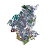
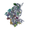
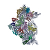
| 登録情報 | データベース: EMDB / ID: EMD-4075 | |||||||||
|---|---|---|---|---|---|---|---|---|---|---|
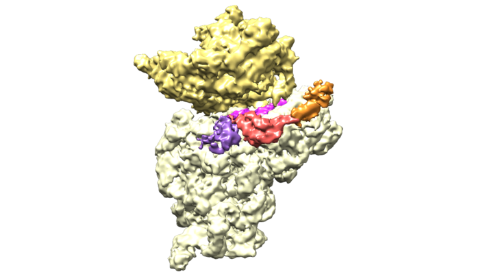
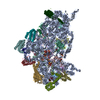
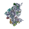
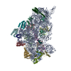
| タイトル | Structure of bacterial 30S-IF1-IF3-mRNA translation pre-initiation complex (state-1C) | |||||||||
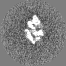
 マップデータ マップデータ | For optimal visualization of IF3 N-terminal domain, gauss-filter de map by 1.1 and display it at 0.035 contour | |||||||||
 試料 試料 |
| |||||||||
| 機能・相同性 |  機能・相同性情報 機能・相同性情報 translation initiation factor activity / ribosome binding / small ribosomal subunit / tRNA binding / rRNA binding / リボソーム / structural constituent of ribosome / 翻訳 (生物学) / ribonucleoprotein complex / mRNA binding ...translation initiation factor activity / ribosome binding / small ribosomal subunit / tRNA binding / rRNA binding / リボソーム / structural constituent of ribosome / 翻訳 (生物学) / ribonucleoprotein complex / mRNA binding / zinc ion binding / metal ion binding / 細胞質 translation initiation factor activity / ribosome binding / small ribosomal subunit / tRNA binding / rRNA binding / リボソーム / structural constituent of ribosome / 翻訳 (生物学) / ribonucleoprotein complex / mRNA binding ...translation initiation factor activity / ribosome binding / small ribosomal subunit / tRNA binding / rRNA binding / リボソーム / structural constituent of ribosome / 翻訳 (生物学) / ribonucleoprotein complex / mRNA binding / zinc ion binding / metal ion binding / 細胞質類似検索 - 分子機能 | |||||||||
| 生物種 |   Thermus thermophilus HB8 (サーマス・サーモフィルス) / Thermus thermophilus (サーマス・サーモフィルス) / Thermus thermophilus (strain HB8 / ATCC 27634 / DSM 579) (サーマス・サーモフィルス) Thermus thermophilus HB8 (サーマス・サーモフィルス) / Thermus thermophilus (サーマス・サーモフィルス) / Thermus thermophilus (strain HB8 / ATCC 27634 / DSM 579) (サーマス・サーモフィルス) | |||||||||
| 手法 | 単粒子再構成法 / クライオ電子顕微鏡法 / 解像度: 5.35 Å | |||||||||
 データ登録者 データ登録者 | Hussain T / Llacer JL / Wimberly BT / Ramakrishnan V | |||||||||
 引用 引用 | ジャーナル: Cell / 年: 2016 タイトル: Large-Scale Movements of IF3 and tRNA during Bacterial Translation Initiation. 著者: Tanweer Hussain / Jose L Llácer / Brian T Wimberly / Jeffrey S Kieft / V Ramakrishnan /   要旨: In bacterial translational initiation, three initiation factors (IFs 1-3) enable the selection of initiator tRNA and the start codon in the P site of the 30S ribosomal subunit. Here, we report 11 ...In bacterial translational initiation, three initiation factors (IFs 1-3) enable the selection of initiator tRNA and the start codon in the P site of the 30S ribosomal subunit. Here, we report 11 single-particle cryo-electron microscopy (cryoEM) reconstructions of the complex of bacterial 30S subunit with initiator tRNA, mRNA, and IFs 1-3, representing different steps along the initiation pathway. IF1 provides key anchoring points for IF2 and IF3, thereby enhancing their activities. IF2 positions a domain in an extended conformation appropriate for capturing the formylmethionyl moiety charged on tRNA. IF3 and tRNA undergo large conformational changes to facilitate the accommodation of the formylmethionyl-tRNA (fMet-tRNA(fMet)) into the P site for start codon recognition. | |||||||||
| 履歴 |
|
- 構造の表示
構造の表示
| ムービー |
ムービービューア |
|---|---|
| 構造ビューア | EMマップ: SurfViewMolmilJmol/JSmol |
| 添付画像 |
- ダウンロードとリンク
-EMDBアーカイブ
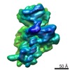
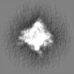
| マップデータ | emd_4075.map.gz | 59.6 MB | EMDBマップデータ形式 | |
|---|---|---|---|---|
| ヘッダ (付随情報) | emd-4075-v30.xmlemd-4075.xml | 42.2 KB 42.2 KB | 表示 表示 | EMDBヘッダ |
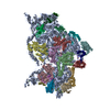
| 画像 |  emd_4075.png emd_4075.png | 167.4 KB | ||
| その他 | emd_4075_half_map_1.map.gzemd_4075_half_map_2.map.gz | 57.4 MB 58 MB | ||
| アーカイブディレクトリ |  http://ftp.pdbj.org/pub/emdb/structures/EMD-4075ftp://ftp.pdbj.org/pub/emdb/structures/EMD-4075 http://ftp.pdbj.org/pub/emdb/structures/EMD-4075ftp://ftp.pdbj.org/pub/emdb/structures/EMD-4075 | HTTPS FTP |
-関連構造データ
| 関連構造データ |  5lmpMC 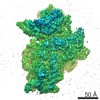 4073C  4074C 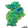 4076C 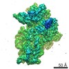 4077C 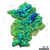 4078C 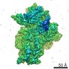 4079C 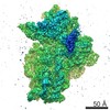 4080C 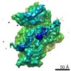 4081C 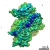 4082C  4083C 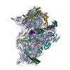 5lmnC 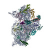 5lmoC 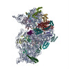 5lmqC  5lmrC 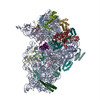 5lmsC 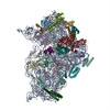 5lmtC 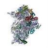 5lmuC 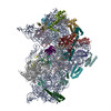 5lmvC M: このマップから作成された原子モデル C: 同じ文献を引用 ( |
|---|---|
| 類似構造データ |


-リンク
| EMDBのページ | EMDB (EBI/PDBe) / EMDataResource |
|---|---|
| 「今月の分子」の関連する項目 |


-マップ
| ファイル | ダウンロード / ファイル: emd_4075.map.gz / 形式: CCP4 / 大きさ: 67 MB / タイプ: IMAGE STORED AS FLOATING POINT NUMBER (4 BYTES) | ||||||||||||||||||||||||||||||||||||||||||||||||||||||||||||
|---|---|---|---|---|---|---|---|---|---|---|---|---|---|---|---|---|---|---|---|---|---|---|---|---|---|---|---|---|---|---|---|---|---|---|---|---|---|---|---|---|---|---|---|---|---|---|---|---|---|---|---|---|---|---|---|---|---|---|---|---|---|
| 注釈 | For optimal visualization of IF3 N-terminal domain, gauss-filter de map by 1.1 and display it at 0.035 contour | ||||||||||||||||||||||||||||||||||||||||||||||||||||||||||||
| ボクセルのサイズ | X=Y=Z: 1.34 Å | ||||||||||||||||||||||||||||||||||||||||||||||||||||||||||||
| 密度 |
| ||||||||||||||||||||||||||||||||||||||||||||||||||||||||||||
| 対称性 | 空間群: 1 | ||||||||||||||||||||||||||||||||||||||||||||||||||||||||||||
| 詳細 | EMDB XML:
CCP4マップ ヘッダ情報:
| ||||||||||||||||||||||||||||||||||||||||||||||||||||||||||||
-添付データ
-ハーフマップ: #1
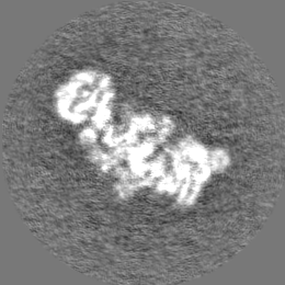
| ファイル | emd_4075_half_map_1.map | ||||||||||||
|---|---|---|---|---|---|---|---|---|---|---|---|---|---|
| 投影像・断面図 |
| ||||||||||||
| 密度ヒストグラム |
 Z
Z Y
Y X
X


-ハーフマップ: #2
| ファイル | emd_4075_half_map_2.map | ||||||||||||
|---|---|---|---|---|---|---|---|---|---|---|---|---|---|
| 投影像・断面図 |
| ||||||||||||
| 密度ヒストグラム |


- 試料の構成要素
+全体 : 30S-IF1-IF3-mRNA pre-initiation complex (state-1C)
+超分子 #1: 30S-IF1-IF3-mRNA pre-initiation complex (state-1C)
+分子 #1: 16S rRNA
+分子 #24: mRNA
+分子 #2: 30S ribosomal protein S2
+分子 #3: 30S ribosomal protein S3
+分子 #4: 30S ribosomal protein S4
+分子 #5: 30S ribosomal protein S5
+分子 #6: 30S ribosomal protein S6
+分子 #7: 30S ribosomal protein S7
+分子 #8: 30S ribosomal protein S8
+分子 #9: 30S ribosomal protein S9
+分子 #10: 30S ribosomal protein S10
+分子 #11: 30S ribosomal protein S11
+分子 #12: 30S ribosomal protein S12
+分子 #13: 30S ribosomal protein S13
+分子 #14: 30S ribosomal protein S14 type Z
+分子 #15: 30S ribosomal protein S15
+分子 #16: 30S ribosomal protein S16
+分子 #17: 30S ribosomal protein S17
+分子 #18: 30S ribosomal protein S18
+分子 #19: 30S ribosomal protein S19
+分子 #20: 30S ribosomal protein S20
+分子 #21: 30S ribosomal protein Thx
+分子 #22: Translation initiation factor IF-1
+分子 #23: Translation initiation factor IF-3
+分子 #25: MAGNESIUM ION
+分子 #26: ZINC ION
-実験情報
-構造解析
| 手法 | クライオ電子顕微鏡法 |
|---|---|
 解析 解析 | 単粒子再構成法 |
| 試料の集合状態 | particle |
-試料調製
| 濃度 | 0.08 mg/mL | ||||||||||||
|---|---|---|---|---|---|---|---|---|---|---|---|---|---|
| 緩衝液 | pH: 7.5 構成要素:
| ||||||||||||
| グリッド | モデル: Quantifoil / 材質: COPPER / メッシュ: 400 / 支持フィルム - 材質: CARBON / 支持フィルム - トポロジー: HOLEY / 前処理 - タイプ: GLOW DISCHARGE | ||||||||||||
| 凍結 | 凍結剤: ETHANE / チャンバー内湿度: 100 % / チャンバー内温度: 277 K / 装置: FEI VITROBOT MARK I |
- 電子顕微鏡法
| 顕微鏡 | FEI POLARA 300 |
|---|---|
| 電子線 | 加速電圧: 300 kV / 電子線源: FIELD EMISSION GUN |
| 電子光学系 | C2レンズ絞り径: 50.0 µm / 倍率(補正後): 104478 / 照射モード: FLOOD BEAM / 撮影モード: BRIGHT FIELDBright-field microscopy / Cs: 2.0 mm / 最大 デフォーカス(公称値): 3.5 µm / 最小 デフォーカス(公称値): 1.5 µm / 倍率(公称値): 78000 |
| 試料ステージ | ホルダー冷却材: NITROGEN |
| 温度 | 最低: 90.0 K / 最高: 100.0 K |
| 撮影 | フィルム・検出器のモデル: OTHER / 撮影したグリッド数: 5 / 実像数: 4400 / 平均露光時間: 1.1 sec. / 平均電子線量: 30.0 e/Å2 詳細: Recorded in a FEI Falcon III direct electron detector. Images were collected in movie-mode at 32 frames per second |
| 実験機器 |  モデル: Tecnai Polara / 画像提供: FEI Company |
-画像解析
| 粒子像選択 | 選択した数: 666610 |
|---|---|
| CTF補正 | ソフトウェア - 名称: CTFFIND (ver. 3) |
| 初期モデル | モデルのタイプ: PDB ENTRY PDBモデル - PDB ID: |
| 初期 角度割当 | タイプ: PROJECTION MATCHING / ソフトウェア - 名称: RELION (ver. 1.4) |
| 最終 3次元分類 | ソフトウェア - 名称: RELION (ver. 1.4) |
| 最終 角度割当 | タイプ: ANGULAR RECONSTITUTION / ソフトウェア - 名称: RELION (ver. 1.4) |
| 最終 再構成 | アルゴリズム: FOURIER SPACE / 解像度のタイプ: BY AUTHOR / 解像度: 5.35 Å / 解像度の算出法: FSC 0.143 CUT-OFF / ソフトウェア - 名称: RELION (ver. 1.4) / 使用した粒子像数: 18830 |
| 詳細 | FEI Falcon III |

-原子モデル構築 1
| 精密化 | 空間: RECIPROCAL / プロトコル: OTHER / 当てはまり具合の基準: FSC |
|---|---|
| 得られたモデル | PDB-5lmp: |
