 ムービー
ムービー コントローラー


+ データを開く

- 基本情報
基本情報
| 登録情報 | データベース: EMDB / ID: EMD-3906 | |||||||||
|---|---|---|---|---|---|---|---|---|---|---|
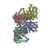
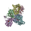
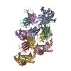
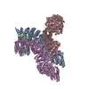
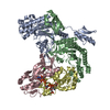
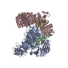
| タイトル | Cryo-EM density map of the human PLC editing module | |||||||||
 マップデータ マップデータ | CryoEM map of a protein complex | |||||||||
 試料 試料 |
| |||||||||
| 機能・相同性 |  機能・相同性情報 機能・相同性情報MHC class Ib protein complex assembly / peptide antigen stabilization / Tapasin-ERp57 complex / Calnexin/calreticulin cycle / MHC class I protein complex binding /  cytolytic granule / プロテインジスルフィドイソメラーゼ / Assembly of Viral Components at the Budding Site / cortical granule / positive regulation of dendritic cell chemotaxis ...MHC class Ib protein complex assembly / peptide antigen stabilization / Tapasin-ERp57 complex / Calnexin/calreticulin cycle / MHC class I protein complex binding / cytolytic granule / プロテインジスルフィドイソメラーゼ / Assembly of Viral Components at the Budding Site / cortical granule / positive regulation of dendritic cell chemotaxis / negative regulation of trophoblast cell migration / ATF6 (ATF6-alpha) activates chaperone genes / negative regulation of retinoic acid receptor signaling pathway / complement component C1q complex binding / cellular response to electrical stimulus / glucocorticoid receptor signaling pathway / endoplasmic reticulum quality control compartment / sequestering of calcium ion / regulation of meiotic nuclear division / peptide antigen assembly with MHC class I protein complex / response to glycoside / sarcoplasmic reticulum lumen / protein folding in endoplasmic reticulum / hormone binding / disulfide oxidoreductase activity / nuclear export signal receptor activity / negative regulation of intracellular steroid hormone receptor signaling pathway / regulation of protein complex stability / molecular sequestering activity / cardiac muscle cell differentiation / phospholipase C activity / TAP1 binding / TAP2 binding / retrograde vesicle-mediated transport, Golgi to endoplasmic reticulum / Scavenging by Class A Receptors / cellular response to interleukin-7 / positive regulation of extrinsic apoptotic signaling pathway / protein maturation by protein folding / Scavenging by Class F Receptors / cortical actin cytoskeleton organization / nuclear androgen receptor binding / response to testosterone / protein disulfide isomerase activity / cellular response to lithium ion / 小胞体 / T cell mediated cytotoxicity directed against tumor cell target / antigen processing and presentation of endogenous peptide antigen via MHC class I via ER pathway, TAP-dependent / positive regulation of memory T cell activation / TAP complex binding / protein localization to nucleus / antigen processing and presentation of exogenous peptide antigen via MHC class I / Golgi medial cisterna / negative regulation of neuron differentiation / protein-disulfide reductase activity / positive regulation of CD8-positive, alpha-beta T cell activation / CD8-positive, alpha-beta T cell activation / positive regulation of CD8-positive, alpha-beta T cell proliferation / CD8 receptor binding / MHC class I protein binding / endoplasmic reticulum exit site / beta-2-microglobulin binding / positive regulation of cell cycle / positive regulation of phagocytosis / phagocytic vesicle / : / protein folding chaperone / positive regulation of substrate adhesion-dependent cell spreading / extrinsic apoptotic signaling pathway / TAP binding / endocytic vesicle lumen / protection from natural killer cell mediated cytotoxicity / antigen processing and presentation of endogenous peptide antigen via MHC class I via ER pathway, TAP-independent / antigen processing and presentation of endogenous peptide antigen via MHC class Ib / detection of bacterium / protein export from nucleus / endoplasmic reticulum-Golgi intermediate compartment membrane / response to endoplasmic reticulum stress / T cell receptor binding / positive regulation of endothelial cell migration / acrosomal vesicle / positive regulation of ferrous iron binding / positive regulation of transferrin receptor binding / early endosome lumen / positive regulation of receptor binding / Nef mediated downregulation of MHC class I complex cell surface expression / DAP12 interactions / negative regulation of receptor binding / lumenal side of endoplasmic reticulum membrane / Endosomal/Vacuolar pathway / Antigen Presentation: Folding, assembly and peptide loading of class I MHC / peptide binding / antigen processing and presentation of exogenous protein antigen via MHC class Ib, TAP-dependent / cellular response to iron(III) ion / negative regulation of forebrain neuron differentiation / ER to Golgi transport vesicle membrane / response to molecule of bacterial origin / regulation of erythrocyte differentiation / regulation of iron ion transport / MHC class I peptide loading complex / intracellular calcium ion homeostasis cytolytic granule / プロテインジスルフィドイソメラーゼ / Assembly of Viral Components at the Budding Site / cortical granule / positive regulation of dendritic cell chemotaxis ...MHC class Ib protein complex assembly / peptide antigen stabilization / Tapasin-ERp57 complex / Calnexin/calreticulin cycle / MHC class I protein complex binding / cytolytic granule / プロテインジスルフィドイソメラーゼ / Assembly of Viral Components at the Budding Site / cortical granule / positive regulation of dendritic cell chemotaxis / negative regulation of trophoblast cell migration / ATF6 (ATF6-alpha) activates chaperone genes / negative regulation of retinoic acid receptor signaling pathway / complement component C1q complex binding / cellular response to electrical stimulus / glucocorticoid receptor signaling pathway / endoplasmic reticulum quality control compartment / sequestering of calcium ion / regulation of meiotic nuclear division / peptide antigen assembly with MHC class I protein complex / response to glycoside / sarcoplasmic reticulum lumen / protein folding in endoplasmic reticulum / hormone binding / disulfide oxidoreductase activity / nuclear export signal receptor activity / negative regulation of intracellular steroid hormone receptor signaling pathway / regulation of protein complex stability / molecular sequestering activity / cardiac muscle cell differentiation / phospholipase C activity / TAP1 binding / TAP2 binding / retrograde vesicle-mediated transport, Golgi to endoplasmic reticulum / Scavenging by Class A Receptors / cellular response to interleukin-7 / positive regulation of extrinsic apoptotic signaling pathway / protein maturation by protein folding / Scavenging by Class F Receptors / cortical actin cytoskeleton organization / nuclear androgen receptor binding / response to testosterone / protein disulfide isomerase activity / cellular response to lithium ion / 小胞体 / T cell mediated cytotoxicity directed against tumor cell target / antigen processing and presentation of endogenous peptide antigen via MHC class I via ER pathway, TAP-dependent / positive regulation of memory T cell activation / TAP complex binding / protein localization to nucleus / antigen processing and presentation of exogenous peptide antigen via MHC class I / Golgi medial cisterna / negative regulation of neuron differentiation / protein-disulfide reductase activity / positive regulation of CD8-positive, alpha-beta T cell activation / CD8-positive, alpha-beta T cell activation / positive regulation of CD8-positive, alpha-beta T cell proliferation / CD8 receptor binding / MHC class I protein binding / endoplasmic reticulum exit site / beta-2-microglobulin binding / positive regulation of cell cycle / positive regulation of phagocytosis / phagocytic vesicle / : / protein folding chaperone / positive regulation of substrate adhesion-dependent cell spreading / extrinsic apoptotic signaling pathway / TAP binding / endocytic vesicle lumen / protection from natural killer cell mediated cytotoxicity / antigen processing and presentation of endogenous peptide antigen via MHC class I via ER pathway, TAP-independent / antigen processing and presentation of endogenous peptide antigen via MHC class Ib / detection of bacterium / protein export from nucleus / endoplasmic reticulum-Golgi intermediate compartment membrane / response to endoplasmic reticulum stress / T cell receptor binding / positive regulation of endothelial cell migration / acrosomal vesicle / positive regulation of ferrous iron binding / positive regulation of transferrin receptor binding / early endosome lumen / positive regulation of receptor binding / Nef mediated downregulation of MHC class I complex cell surface expression / DAP12 interactions / negative regulation of receptor binding / lumenal side of endoplasmic reticulum membrane / Endosomal/Vacuolar pathway / Antigen Presentation: Folding, assembly and peptide loading of class I MHC / peptide binding / antigen processing and presentation of exogenous protein antigen via MHC class Ib, TAP-dependent / cellular response to iron(III) ion / negative regulation of forebrain neuron differentiation / ER to Golgi transport vesicle membrane / response to molecule of bacterial origin / regulation of erythrocyte differentiation / regulation of iron ion transport / MHC class I peptide loading complex / intracellular calcium ion homeostasis類似検索 - 分子機能 | |||||||||
| 生物種 |  Homo sapiens (ヒト) / Human (ヒト) Homo sapiens (ヒト) / Human (ヒト) | |||||||||
| 手法 | 単粒子再構成法 / クライオ電子顕微鏡法 / 解像度: 5.8 Å | |||||||||
 データ登録者 データ登録者 | Januliene D / Blees A / Trowitzsch S / Tampe R / Moeller A | |||||||||
| 資金援助 |  ドイツ, 2件 ドイツ, 2件
| |||||||||
 引用 引用 | ジャーナル: Nature / 年: 2017 タイトル: Structure of the human MHC-I peptide-loading complex. 著者: Andreas Blees / Dovile Januliene / Tommy Hofmann / Nicole Koller / Carla Schmidt / Simon Trowitzsch / Arne Moeller / Robert Tampé / 要旨: The peptide-loading complex (PLC) is a transient, multisubunit membrane complex in the endoplasmic reticulum that is essential for establishing a hierarchical immune response. The PLC coordinates ...The peptide-loading complex (PLC) is a transient, multisubunit membrane complex in the endoplasmic reticulum that is essential for establishing a hierarchical immune response. The PLC coordinates peptide translocation into the endoplasmic reticulum with loading and editing of major histocompatibility complex class I (MHC-I) molecules. After final proofreading in the PLC, stable peptide-MHC-I complexes are released to the cell surface to evoke a T-cell response against infected or malignant cells. Sampling of different MHC-I allomorphs requires the precise coordination of seven different subunits in a single macromolecular assembly, including the transporter associated with antigen processing (TAP1 and TAP2, jointly referred to as TAP), the oxidoreductase ERp57, the MHC-I heterodimer, and the chaperones tapasin and calreticulin. The molecular organization of and mechanistic events that take place in the PLC are unknown owing to the heterogeneous composition and intrinsically dynamic nature of the complex. Here, we isolate human PLC from Burkitt's lymphoma cells using an engineered viral inhibitor as bait and determine the structure of native PLC by electron cryo-microscopy. Two endoplasmic reticulum-resident editing modules composed of tapasin, calreticulin, ERp57, and MHC-I are centred around TAP in a pseudo-symmetric orientation. A multivalent chaperone network within and across the editing modules establishes the proofreading function at two lateral binding platforms for MHC-I molecules. The lectin-like domain of calreticulin senses the MHC-I glycan, whereas the P domain reaches over the MHC-I peptide-binding pocket towards ERp57. This arrangement allows tapasin to facilitate peptide editing by clamping MHC-I. The translocation pathway of TAP opens out into a large endoplasmic reticulum lumenal cavity, confined by the membrane entry points of tapasin and MHC-I. Two lateral windows channel the antigenic peptides to MHC-I. Structures of PLC captured at distinct assembly states provide mechanistic insight into the recruitment and release of MHC-I. Our work defines the molecular symbiosis of an ABC transporter and an endoplasmic reticulum chaperone network in MHC-I assembly and provides insight into the onset of the adaptive immune response. | |||||||||
| 履歴 |
|
- 構造の表示
構造の表示
| ムービー |
ムービービューア |
|---|---|
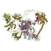
| 構造ビューア | EMマップ: SurfViewMolmilJmol/JSmol |
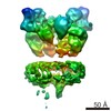
| 添付画像 |

- ダウンロードとリンク
-EMDBアーカイブ
| マップデータ | emd_3906.map.gz | 25.5 MB | EMDBマップデータ形式 | |
|---|---|---|---|---|
| ヘッダ (付随情報) | emd-3906-v30.xmlemd-3906.xml | 16 KB 16 KB | 表示 表示 | EMDBヘッダ |
| 画像 | 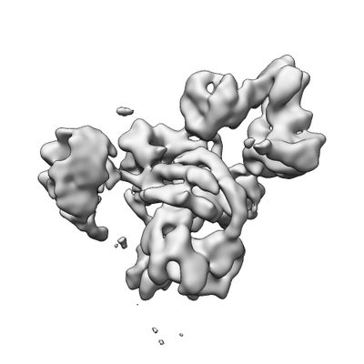 emd_3906.png emd_3906.png | 44.5 KB | ||
| アーカイブディレクトリ |  http://ftp.pdbj.org/pub/emdb/structures/EMD-3906ftp://ftp.pdbj.org/pub/emdb/structures/EMD-3906 http://ftp.pdbj.org/pub/emdb/structures/EMD-3906ftp://ftp.pdbj.org/pub/emdb/structures/EMD-3906 | HTTPS FTP |
-関連構造データ


-リンク
| EMDBのページ | EMDB (EBI/PDBe) / EMDataResource |
|---|---|
| 「今月の分子」の関連する項目 |


-マップ
| ファイル | ダウンロード / ファイル: emd_3906.map.gz / 形式: CCP4 / 大きさ: 30.5 MB / タイプ: IMAGE STORED AS FLOATING POINT NUMBER (4 BYTES) | ||||||||||||||||||||||||||||||||||||||||||||||||||||||||||||
|---|---|---|---|---|---|---|---|---|---|---|---|---|---|---|---|---|---|---|---|---|---|---|---|---|---|---|---|---|---|---|---|---|---|---|---|---|---|---|---|---|---|---|---|---|---|---|---|---|---|---|---|---|---|---|---|---|---|---|---|---|---|
| 注釈 | CryoEM map of a protein complex | ||||||||||||||||||||||||||||||||||||||||||||||||||||||||||||
| ボクセルのサイズ | X=Y=Z: 1.077 Å | ||||||||||||||||||||||||||||||||||||||||||||||||||||||||||||
| 密度 |
| ||||||||||||||||||||||||||||||||||||||||||||||||||||||||||||
| 対称性 | 空間群: 1 | ||||||||||||||||||||||||||||||||||||||||||||||||||||||||||||
| 詳細 | EMDB XML:
CCP4マップ ヘッダ情報:
| ||||||||||||||||||||||||||||||||||||||||||||||||||||||||||||
-添付データ
- 試料の構成要素
-全体 : Protein Complex
| 全体 | 名称: Protein Complexタンパク質複合体 |
|---|---|
| 要素 |
|
-超分子 #1: Protein Complex
| 超分子 | 名称: Protein Complex / タイプ: complex / ID: 1 / 親要素: 0 / 含まれる分子: all |
|---|---|
| 由来(天然) | 生物種: Homo sapiens (ヒト) |
-分子 #1: Beta-2-microglobulin
| 分子 | 名称: Beta-2-microglobulin / タイプ: protein_or_peptide / ID: 1 / コピー数: 1 / 光学異性体: LEVO |
|---|---|
| 由来(天然) | 生物種: Human (ヒト) |
| 分子量 | 理論値: 11.74816 KDa |
| 配列 | 文字列: IQRTPKIQVY SRHPAENGKS NFLNCYVSGF HPSDIEVDLL KNGERIEKVE HSDLSFSKDW SFYLLYYTEF TPTEKDEYAC RVNHVTLSQ PKIVKWDRDM |
-分子 #2: Tapasin
| 分子 | 名称: Tapasin / タイプ: protein_or_peptide / ID: 2 / コピー数: 1 / 光学異性体: LEVO |
|---|---|
| 由来(天然) | 生物種: Human (ヒト) |
| 分子量 | 理論値: 45.761184 KDa |
| 配列 | 文字列: GPAVIECWFV EDASGKGLAK RPGALLLRQG PGEPPPRPDL DPELYLSVHD PAGALQAAFR RYPRGAPAPH CEMSRFVPLP ASAKWASGL TPAQNCPRAL DGAWLMVSIS SPVLSLSSLL RPQPEPQQEP VLITMATVVL TVLTHTPAPR VRLGQDALLD L SFAYMPPT ...文字列: GPAVIECWFV EDASGKGLAK RPGALLLRQG PGEPPPRPDL DPELYLSVHD PAGALQAAFR RYPRGAPAPH CEMSRFVPLP ASAKWASGL TPAQNCPRAL DGAWLMVSIS SPVLSLSSLL RPQPEPQQEP VLITMATVVL TVLTHTPAPR VRLGQDALLD L SFAYMPPT SEAASSLAPG PPPFGLEWRR QHLGKGHLLL AATPGLNGQM PAAQEGAVAF AAWDDDEPWG PWTGNGTFWL PR VQPFQEG TYLATIHLPY LQGQVTLELA VYKPPKVSLM PATLARAAPG EAPPELLCLV SHFYPSGGLE VEWELRGGPG GRS QKAEGQ RWLSALRHHS DGSVSLSGHL QPPPVTTEQH GARYACRIHH PSLPASGRSA EVTLEVAGLS GPSLEDSVGL FLSA FLLLG LFKALGWAAV YLSTCKDSKK KAE |
-分子 #3: Protein disulfide-isomerase A3
| 分子 | 名称: Protein disulfide-isomerase A3 / タイプ: protein_or_peptide / ID: 3 / コピー数: 1 / 光学異性体: LEVO EC番号: プロテインジスルフィドイソメラーゼ |
|---|---|
| 由来(天然) | 生物種: Human (ヒト) |
| 分子量 | 理論値: 54.341102 KDa |
| 配列 | 文字列: SDVLELTDDN FESRISDTGS AGLMLVEFFA PWCGHCKRLA PEYEAAATRL KGIVPLAKVD CTANTNTCNK YGVSGYPTLK IFRDGEEAG AYDGPRTADG IVSHLKKQAG PASVPLRTEE EFKKFISDKD ASIVGFFDDS FSEAHSEFLK AASNLRDNYR F AHTNVESL ...文字列: SDVLELTDDN FESRISDTGS AGLMLVEFFA PWCGHCKRLA PEYEAAATRL KGIVPLAKVD CTANTNTCNK YGVSGYPTLK IFRDGEEAG AYDGPRTADG IVSHLKKQAG PASVPLRTEE EFKKFISDKD ASIVGFFDDS FSEAHSEFLK AASNLRDNYR F AHTNVESL VNEYDDNGEG IILFRPSHLT NKFEDKTVAY TEQKMTSGKI KKFIQENIFG ICPHMTEDNK DLIQGKDLLI AY YDVDYEK NAKGSNYWRN RVMMVAKKFL DAGHKLNFAV ASRKTFSHEL SDFGLESTAG EIPVVAIRTA KGEKFVMQEE FSR DGKALE RFLQDYFDGN LKRYLKSEPI PESNDGPVKV VVAENFDEIV NNENKDVLIE FYAPWCGHCK NLEPKYKELG EKLS KDPNI VIAKMDATAN DVPSPYEVRG FPTIYFSPAN KKLNPKKYEG GRELSDFISY LQREATNPPV IQEEKPKKKK KAQED L |
-分子 #4: HLA class I histocompatibility antigen, A-3 alpha chain
| 分子 | 名称: HLA class I histocompatibility antigen, A-3 alpha chain タイプ: protein_or_peptide / ID: 4 / コピー数: 1 / 光学異性体: LEVO |
|---|---|
| 由来(天然) | 生物種: Human (ヒト) |
| 分子量 | 理論値: 38.363535 KDa |
| 配列 | 文字列: GSHSMRYFFT SVSRPGRGEP RFIAVGYVDD TQFVRFDSDA ASQRMEPRAP WIEQEGPEYW DQETRNVKAQ SQTDRVDLGT LRGYYNQSE AGSHTIQIMY GCDVGSDGRF LRGYRQDAYD GKDYIALNED LRSWTAADMA AQITKRKWEA AHEAEQLRAY L DGTCVEWL ...文字列: GSHSMRYFFT SVSRPGRGEP RFIAVGYVDD TQFVRFDSDA ASQRMEPRAP WIEQEGPEYW DQETRNVKAQ SQTDRVDLGT LRGYYNQSE AGSHTIQIMY GCDVGSDGRF LRGYRQDAYD GKDYIALNED LRSWTAADMA AQITKRKWEA AHEAEQLRAY L DGTCVEWL RRYLENGKET LQRTDPPKTH MTHHPISDHE ATLRCWALGF YPAEITLTWQ RDGEDQTQDT ELVETRPAGD GT FQKWAAV VVPSGEEQRY TCHVQHEGLP KPLTLRWELS SQPTIPIVGI IAGLVLLGAV ITGAVVAAVM WRRKSSDRKG GSY TQAASS DSAQGSDVSL TACKV |
-分子 #5: Calreticulin
| 分子 | 名称: Calreticulin / タイプ: protein_or_peptide / ID: 5 / コピー数: 1 / 光学異性体: LEVO |
|---|---|
| 由来(天然) | 生物種: Human (ヒト) |
| 分子量 | 理論値: 46.507145 KDa |
| 配列 | 文字列: EPAVYFKEQF LDGDGWTSRW IESKHKSDFG KFVLSSGKFY GDEEKDKGLQ TSQDARFYAL SASFEPFSNK GQTLVVQFTV KHEQNIDCG GGYVKLFPNS LDQTDMHGDS EYNIMFGPDI CGPGTKKVHV IFNYKGKNVL INKDIRSKDD EFTHLYTLIV R PDNTYEVK ...文字列: EPAVYFKEQF LDGDGWTSRW IESKHKSDFG KFVLSSGKFY GDEEKDKGLQ TSQDARFYAL SASFEPFSNK GQTLVVQFTV KHEQNIDCG GGYVKLFPNS LDQTDMHGDS EYNIMFGPDI CGPGTKKVHV IFNYKGKNVL INKDIRSKDD EFTHLYTLIV R PDNTYEVK IDNSQVESGS LEDDWDFLPP KKIKDPDASK PEDWDERAKI DDPTDSKPED WDKPEHIPDP DAKKPEDWDE EM DGEWEPP VIQNPEYKGE WKPRQIDNPD YKGTWIHPEI DNPEYSPDPS IYAYDNFGVL GLDLWQVKSG TIFDNFLITN DEA YAEEFG NETWGVTKAA EKQMKDKQDE EQRLKEEEED KKRKEEEEAE DKEDDEDKDE DEEDEEDKEE DEEEDVPGQA KDEL |
-実験情報
-構造解析
| 手法 | クライオ電子顕微鏡法 |
|---|---|
 解析 解析 | 単粒子再構成法 |
| 試料の集合状態 | particle |
-試料調製
| 濃度 | 2 mg/mL |
|---|---|
| 緩衝液 | pH: 7.5 |
| グリッド | モデル: C-flat-2/2 / 前処理 - タイプ: GLOW DISCHARGE |
| 凍結 | 凍結剤: ETHANE / チャンバー内湿度: 100 % / チャンバー内温度: 277 K / 装置: FEI VITROBOT MARK IV |
- 電子顕微鏡法
| 顕微鏡 | FEI TITAN KRIOS |
|---|---|
| 電子線 | 加速電圧: 300 kV / 電子線源: FIELD EMISSION GUN |
| 電子光学系 | 照射モード: FLOOD BEAM / 撮影モード: BRIGHT FIELDBright-field microscopy |
| 撮影 | フィルム・検出器のモデル: GATAN K2 QUANTUM (4k x 4k) 検出モード: COUNTING / 平均電子線量: 55.0 e/Å2 |
| 実験機器 |  モデル: Titan Krios / 画像提供: FEI Company |
-画像解析
| CTF補正 | ソフトウェア - 名称: Gctf 詳細: CTF correction was performed internally in Relion and Frealign |
|---|---|
| 初期モデル | モデルのタイプ: OTHER 詳細: Selected particles from the best 2D class averages were subjected to 3D-classification in Relion, using a low-pass filtered global average as a starting model. The best map was then used as ...詳細: Selected particles from the best 2D class averages were subjected to 3D-classification in Relion, using a low-pass filtered global average as a starting model. The best map was then used as an initial model for subsequent 3D classification. |
| 初期 角度割当 | タイプ: ANGULAR RECONSTITUTION / ソフトウェア - 名称: RELION (ver. 2.1) |
| 最終 角度割当 | タイプ: ANGULAR RECONSTITUTION |
| 最終 再構成 | 想定した対称性 - 点群: C1 (非対称) / 解像度のタイプ: BY AUTHOR / 解像度: 5.8 Å / 解像度の算出法: FSC 0.143 CUT-OFF / ソフトウェア - 名称: FREALIGN (ver. X) / 使用した粒子像数: 141078 |
