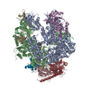
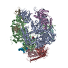
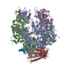
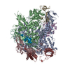
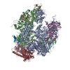
ジャーナル: Biochim Biophys Acta Biomembr / 年: 2018 タイトル: Using a SMALP platform to determine a sub-nm single particle cryo-EM membrane protein structure. 著者: Mayuriben Parmar / Shaun Rawson / Charlotte A Scarff / Adrian Goldman / Timothy R Dafforn / Stephen P Muench / Vincent L G Postis / 要旨: The field of membrane protein structural biology has been revolutionized over the last few years with a number of high profile structures being solved using cryo-EM including Piezo, Ryanodine ...The field of membrane protein structural biology has been revolutionized over the last few years with a number of high profile structures being solved using cryo-EM including Piezo, Ryanodine receptor, TRPV1 and the Glutamate receptor. Further developments in the EM field hold the promise of even greater progress in terms of greater resolution, which for membrane proteins is still typically within the 4-7Å range. One advantage of a cryo-EM approach is the ability to study membrane proteins in more "native" like environments for example proteoliposomes, amphipols and nanodiscs. Recently, styrene maleic acid co-polymers (SMA) have been used to extract membrane proteins surrounded by native lipids (SMALPs) maintaining a more natural environment. We report here the structure of the Escherichia coli multidrug efflux transporter AcrB in a SMALP scaffold to sub-nm resolution, with the resulting map being consistent with high resolution crystal structures and other EM derived maps. However, both the C-terminal helix (TM12) and TM7 are poorly defined in the map. These helices are at the exterior of the helical bundle and form the greater interaction with the native lipids and SMA polymer and may represent a more dynamic region of the protein. This work shows the promise of using an SMA approach for single particle cryo-EM studies to provide sub-nm structures.
 ムービー
ムービー コントローラー


 データを開く
 基本情報
基本情報 Acriflavine resistance protein family
 マップデータ
 試料
試料
 データ登録者
 引用
引用

 構造の表示
構造の表示 ムービービューア
 ダウンロードとリンク
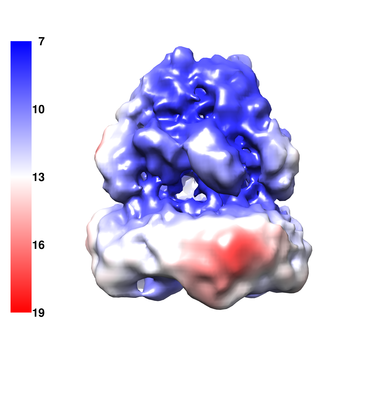 emd_3887.png
 http://ftp.pdbj.org/pub/emdb/structures/EMD-3887
 試料の構成要素
 解析
解析 電子顕微鏡法
