 ムービー
ムービー コントローラー


+ データを開く

- 基本情報
基本情報
| 登録情報 | データベース: EMDB / ID: EMD-3841 | |||||||||
|---|---|---|---|---|---|---|---|---|---|---|
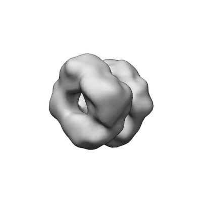
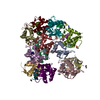
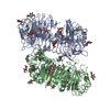
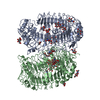
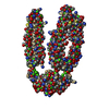
| タイトル | Negative-stain surface of low-pH sortilin dimer | |||||||||
 マップデータ マップデータ | Negative-stain surface of low-pH sortilin dimer | |||||||||
 試料 試料 |
| |||||||||
| 機能・相同性 |  機能・相同性情報 機能・相同性情報neurotensin receptor activity, non-G protein-coupled / negative regulation of lipoprotein lipase activity / myotube differentiation / cerebellar climbing fiber to Purkinje cell synapse / plasma membrane to endosome transport / retromer complex binding / maintenance of synapse structure / Golgi to endosome transport /  nerve growth factor receptor activity / Golgi to lysosome transport ...neurotensin receptor activity, non-G protein-coupled / negative regulation of lipoprotein lipase activity / myotube differentiation / cerebellar climbing fiber to Purkinje cell synapse / plasma membrane to endosome transport / retromer complex binding / maintenance of synapse structure / Golgi to endosome transport / nerve growth factor receptor activity / Golgi to lysosome transport / vesicle organization / endosome transport via multivesicular body sorting pathway / nerve growth factor binding / protein targeting to lysosome / trans-Golgi network transport vesicle / クラスリン / negative regulation of fat cell differentiation / Golgi cisterna membrane / Golgi Associated Vesicle Biogenesis / endosome to lysosome transport / glucose import / neurotrophin TRK receptor signaling pathway / extrinsic apoptotic signaling pathway via death domain receptors / neuropeptide signaling pathway / クラスリン / 骨化 / response to insulin / エンドサイトーシス / cytoplasmic vesicle / 遺伝子発現の調節 / 核膜 / リソソーム / エンドソーム / endosome membrane / G protein-coupled receptor signaling pathway / lysosomal membrane / endoplasmic reticulum membrane / perinuclear region of cytoplasm / ゴルジ体 / enzyme binding / 細胞膜 / 生体膜 / 細胞膜 / 細胞質基質 nerve growth factor receptor activity / Golgi to lysosome transport ...neurotensin receptor activity, non-G protein-coupled / negative regulation of lipoprotein lipase activity / myotube differentiation / cerebellar climbing fiber to Purkinje cell synapse / plasma membrane to endosome transport / retromer complex binding / maintenance of synapse structure / Golgi to endosome transport / nerve growth factor receptor activity / Golgi to lysosome transport / vesicle organization / endosome transport via multivesicular body sorting pathway / nerve growth factor binding / protein targeting to lysosome / trans-Golgi network transport vesicle / クラスリン / negative regulation of fat cell differentiation / Golgi cisterna membrane / Golgi Associated Vesicle Biogenesis / endosome to lysosome transport / glucose import / neurotrophin TRK receptor signaling pathway / extrinsic apoptotic signaling pathway via death domain receptors / neuropeptide signaling pathway / クラスリン / 骨化 / response to insulin / エンドサイトーシス / cytoplasmic vesicle / 遺伝子発現の調節 / 核膜 / リソソーム / エンドソーム / endosome membrane / G protein-coupled receptor signaling pathway / lysosomal membrane / endoplasmic reticulum membrane / perinuclear region of cytoplasm / ゴルジ体 / enzyme binding / 細胞膜 / 生体膜 / 細胞膜 / 細胞質基質類似検索 - 分子機能 | |||||||||
| 生物種 |  Homo sapiens (ヒト) Homo sapiens (ヒト) | |||||||||
| 手法 | 単粒子再構成法 / ネガティブ染色法 / 解像度: 13.0 Å | |||||||||
 データ登録者 データ登録者 | Januliene D / Thirup S / Moeller A | |||||||||
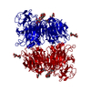
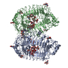
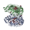
 引用 引用 | ジャーナル: Structure / 年: 2017 タイトル: Acidic Environment Induces Dimerization and Ligand Binding Site Collapse in the Vps10p Domain of Sortilin. 著者: Dovile Januliene / Jacob Lauwring Andersen / Jeppe Achton Nielsen / Esben Meldgaard Quistgaard / Maria Hansen / Dorthe Strandbygaard / Arne Moeller / Claus Munck Petersen / Peder Madsen / Søren Skou Thirup /   要旨: Sortilin is a neuronal receptor involved in transmembrane signaling, endocytosis, and intracellular sorting of proteins. It cycles through a number of cellular compartments where it encounters ...Sortilin is a neuronal receptor involved in transmembrane signaling, endocytosis, and intracellular sorting of proteins. It cycles through a number of cellular compartments where it encounters various acidic conditions. The crystal structure of the sortilin ectodomain has previously been determined at neutral pH. Here, we present the 3.5-Å resolution crystal structure of sortilin at pH 5.5, which represents an environment similar to that of late endosomes, where ligands are released. The structure reveals an overall distortion of the 10-bladed β-propeller domain. This distortion and specific conformational changes, caused by protonation of a number of histidine residues, render the currently known binding sites unavailable for ligand binding. Access to the binding sites is furthermore blocked by a reversible and pH-dependent formation of tight sortilin dimers, also confirmed by electron microscopy, size-exclusion chromatography, and mutational studies. This study reveals how sortilin binding sites are disrupted and explains pH-dependent ligand affinity. | |||||||||
| 履歴 |
|
- 構造の表示
構造の表示
| ムービー |
ムービービューア |
|---|---|
| 構造ビューア | EMマップ: SurfViewMolmilJmol/JSmol |
| 添付画像 |
- ダウンロードとリンク
-EMDBアーカイブ
| マップデータ | emd_3841.map.gz | 318.9 KB | EMDBマップデータ形式 | |
|---|---|---|---|---|
| ヘッダ (付随情報) | emd-3841-v30.xmlemd-3841.xml | 8.6 KB 8.6 KB | 表示 表示 | EMDBヘッダ |
| 画像 |  emd_3841.png emd_3841.png | 24.4 KB | ||
| アーカイブディレクトリ |  http://ftp.pdbj.org/pub/emdb/structures/EMD-3841ftp://ftp.pdbj.org/pub/emdb/structures/EMD-3841 http://ftp.pdbj.org/pub/emdb/structures/EMD-3841ftp://ftp.pdbj.org/pub/emdb/structures/EMD-3841 | HTTPS FTP |
-関連構造データ
-リンク
| EMDBのページ | EMDB (EBI/PDBe) / EMDataResource |
|---|---|
| 「今月の分子」の関連する項目 |

-マップ
| ファイル | ダウンロード / ファイル: emd_3841.map.gz / 形式: CCP4 / 大きさ: 251 KB / タイプ: IMAGE STORED AS FLOATING POINT NUMBER (4 BYTES) | ||||||||||||||||||||||||||||||||||||||||||||||||||||||||||||||||||||
|---|---|---|---|---|---|---|---|---|---|---|---|---|---|---|---|---|---|---|---|---|---|---|---|---|---|---|---|---|---|---|---|---|---|---|---|---|---|---|---|---|---|---|---|---|---|---|---|---|---|---|---|---|---|---|---|---|---|---|---|---|---|---|---|---|---|---|---|---|---|
| 注釈 | Negative-stain surface of low-pH sortilin dimer | ||||||||||||||||||||||||||||||||||||||||||||||||||||||||||||||||||||
| ボクセルのサイズ | X=Y=Z: 3.15 Å | ||||||||||||||||||||||||||||||||||||||||||||||||||||||||||||||||||||
| 密度 |
| ||||||||||||||||||||||||||||||||||||||||||||||||||||||||||||||||||||
| 対称性 | 空間群: 1 | ||||||||||||||||||||||||||||||||||||||||||||||||||||||||||||||||||||
| 詳細 | EMDB XML:
CCP4マップ ヘッダ情報:
| ||||||||||||||||||||||||||||||||||||||||||||||||||||||||||||||||||||
-添付データ
- 試料の構成要素
-全体 : Low-pH-induced dimer of human sSortilin extracellular domain
| 全体 | 名称: Low-pH-induced dimer of human sSortilin extracellular domain |
|---|---|
| 要素 |
|
-超分子 #1: Low-pH-induced dimer of human sSortilin extracellular domain
| 超分子 | 名称: Low-pH-induced dimer of human sSortilin extracellular domain タイプ: complex / ID: 1 / 親要素: 0 |
|---|---|
| 由来(天然) | 生物種: Homo sapiens (ヒト) |
| 組換発現 | 生物種: Homo sapiens (ヒト) / 組換細胞: HEK |
| 分子量 | 実験値: 200 KDa |
-実験情報
-構造解析
| 手法 | ネガティブ染色法 |
|---|---|
 解析 解析 | 単粒子再構成法 |
| 試料の集合状態 | particle |
-試料調製
| 濃度 | 0.01 mg/mL |
|---|---|
| 緩衝液 | pH: 5.5 |
| 染色 | タイプ: NEGATIVE / 材質: Uranyl Formate |
- 電子顕微鏡法
| 顕微鏡 | FEI TECNAI SPIRIT |
|---|---|
| 電子線 | 加速電圧: 120 kV / 電子線源: LAB6 |
| 電子光学系 | 照射モード: FLOOD BEAM / 撮影モード: BRIGHT FIELDBright-field microscopy |
| 撮影 | フィルム・検出器のモデル: TVIPS TEMCAM-F416 (4k x 4k) 平均電子線量: 8.0 e/Å2 |
| 実験機器 |  モデル: Tecnai Spirit / 画像提供: FEI Company |
-画像解析
| CTF補正 | ソフトウェア - 名称: CTFFIND (ver. 3) |
|---|---|
| 初期 角度割当 | タイプ: ANGULAR RECONSTITUTION |
| 最終 角度割当 | タイプ: ANGULAR RECONSTITUTION |
| 最終 再構成 | 想定した対称性 - 点群: C1 (非対称) / 解像度のタイプ: BY AUTHOR / 解像度: 13.0 Å / 解像度の算出法: FSC 0.143 CUT-OFF / ソフトウェア - 名称: RELION (ver. 2.0) / 使用した粒子像数: 55967 |
