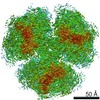
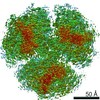
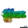
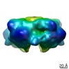
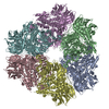
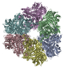
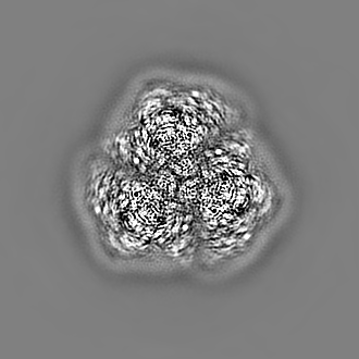
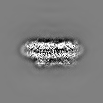
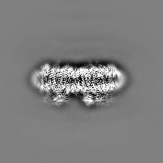
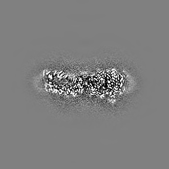
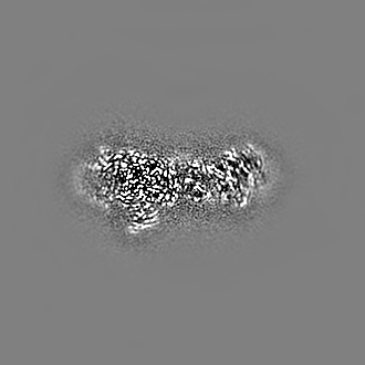
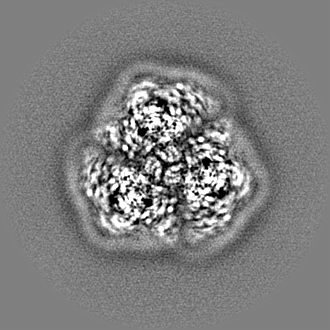
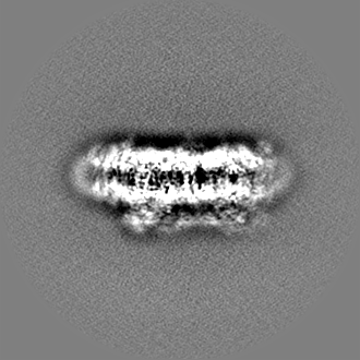
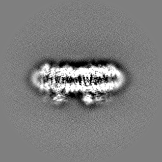
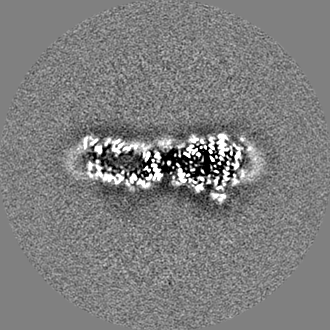
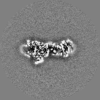
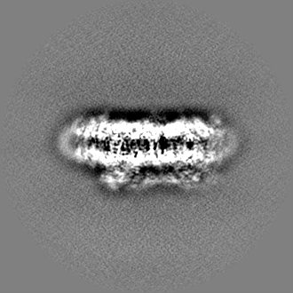
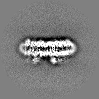
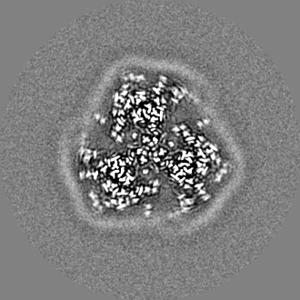
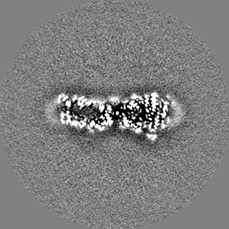
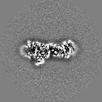
- EMDB-30420: Structure of the far-red light utilizing photosystem I of Acaryoc... -
+
データを開く
IDまたはキーワード:
読み込み中...
-
基本情報
登録情報
データベース: EMDB / ID: EMD-30420
タイトル
Structure of the far-red light utilizing photosystem I of Acaryochloris marina光化学系I
マップデータ
Postprocess_masked.mrc
試料
複合体: Photosystem I complex from Acaryochloris marina光化学系I
タンパク質・ペプチド: x 11種
リガンド: x 9種
機能・相同性
機能・相同性情報
photosystem I reaction center / 光化学系I / photosynthetic electron transport in photosystem I / 光化学系I / chlorophyll binding / plasma membrane-derived thylakoid membrane / 光合成 / 4 iron, 4 sulfur cluster binding / electron transfer activity / membrane => GO:0016020 ...photosystem I reaction center / 光化学系I / photosynthetic electron transport in photosystem I / 光化学系I / chlorophyll binding / plasma membrane-derived thylakoid membrane / 光合成 / 4 iron, 4 sulfur cluster binding / electron transfer activity / membrane => GO:0016020 / magnesium ion binding / metal ion binding 類似検索 - 分子機能
Photosystem I reaction center subunit PsaK / Photosystem I reaction centre subunit PsaK / Photosystem I reaction centre subunit PsaK superfamily / Photosystem I reaction center subunit V/PsaK / Photosystem I psaG / psaK / Photosystem I PsaL, reaction centre subunit XI / Photosystem I, reaction centre subunit XI / Photosystem I PsaL, reaction centre subunit XI superfamily / Photosystem I reaction centre subunit XI / Photosystem I PsaF, reaction centre subunit III ...Photosystem I reaction center subunit PsaK / Photosystem I reaction centre subunit PsaK / Photosystem I reaction centre subunit PsaK superfamily / Photosystem I reaction center subunit V/PsaK / Photosystem I psaG / psaK / Photosystem I PsaL, reaction centre subunit XI / Photosystem I, reaction centre subunit XI / Photosystem I PsaL, reaction centre subunit XI superfamily / Photosystem I reaction centre subunit XI / Photosystem I PsaF, reaction centre subunit III / Photosystem I PsaF, reaction centre subunit III superfamily / Photosystem I reaction centre subunit III / Photosystem I PsaJ, reaction centre subunit IX / Photosystem I PsaD / Photosystem I PsaJ, reaction centre subunit IX superfamily / Photosystem I, reaction centre subunit PsaD superfamily / Photosystem I reaction centre subunit IX / PsaJ / PsaD / Photosystem I PsaE, reaction centre subunit IV / Photosystem I reaction centre subunit IV / PsaE / Photosystem I protein PsaC / Photosystem I PsaA / Photosystem I PsaB / Photosystem I PsaA/PsaB, conserved site / Photosystem I psaA and psaB proteins signature. / Photosystem I PsaA/PsaB / Photosystem I PsaA/PsaB superfamily / Photosystem I psaA/psaB protein / Electron transport accessory-like domain superfamily / 4Fe-4S dicluster domain / 4Fe-4S ferredoxin, iron-sulphur binding, conserved site / 4Fe-4S ferredoxin-type iron-sulfur binding region signature. / 4Fe-4S ferredoxin-type iron-sulfur binding domain profile. / 4Fe-4S ferredoxin-type, iron-sulphur binding domain 類似検索 - ドメイン・相同性
Photosystem I P700 chlorophyll a apoprotein A1 / Photosystem I P700 chlorophyll a apoprotein A2 / Photosystem I reaction center subunit IV / Photosystem I reaction center subunit XI / Photosystem I reaction center subunit IX / Photosystem I reaction center subunit III / Photosystem I reaction center subunit II / Photosystem I reaction center subunit PsaK / Photosystem I iron-sulfur center 類似検索 - 構成要素
Japan Agency for Medical Research and Development (AMED)
日本
引用
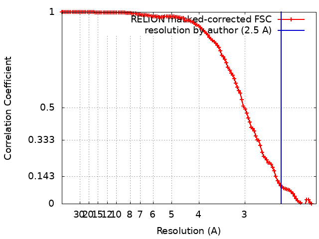
ジャーナル: Nat Commun / 年: 2021 タイトル: Structure of the far-red light utilizing photosystem I of Acaryochloris marina. 著者: Tasuku Hamaguchi / Keisuke Kawakami / Kyoko Shinzawa-Itoh / Natsuko Inoue-Kashino / Shigeru Itoh / Kentaro Ifuku / Eiki Yamashita / Kou Maeda / Koji Yonekura / Yasuhiro Kashino / 要旨: Acaryochloris marina is one of the cyanobacterial species that can use far-red light to drive photochemical reactions for oxygenic photosynthesis. Here, we report the structure of A. marina ...Acaryochloris marina is one of the cyanobacterial species that can use far-red light to drive photochemical reactions for oxygenic photosynthesis. Here, we report the structure of A. marina photosystem I (PSI) reaction center, determined by cryo-electron microscopy at 2.58 Å resolution. The structure reveals an arrangement of electron carriers and light-harvesting pigments distinct from other type I reaction centers. The paired chlorophyll, or special pair (also referred to as P740 in this case), is a dimer of chlorophyll d and its epimer chlorophyll d'. The primary electron acceptor is pheophytin a, a metal-less chlorin. We show the architecture of this PSI reaction center is composed of 11 subunits and we identify key components that help explain how the low energy yield from far-red light is efficiently utilized for driving oxygenic photosynthesis.
 ムービー
ムービー コントローラー


 データを開く
 基本情報
基本情報 光化学系I
光化学系I マップデータ
 試料
試料 機能・相同性情報
 データ登録者
 日本, 5件
  引用
引用 構造の表示
構造の表示
 ダウンロードとリンク
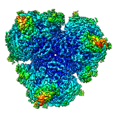 emd_30420.png
 http://ftp.pdbj.org/pub/emdb/structures/EMD-30420


 Z
Z Y
Y X
X


 試料の構成要素
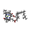


 解析
解析 電子顕微鏡法