 ムービー
ムービー コントローラー


+ データを開く

- 基本情報
基本情報
| 登録情報 | データベース: EMDB / ID: EMD-2886 | |||||||||
|---|---|---|---|---|---|---|---|---|---|---|
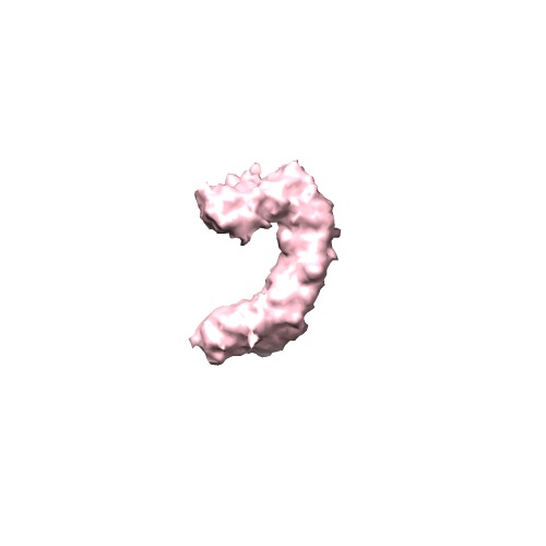
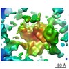
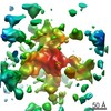
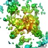
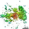
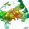
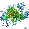
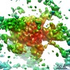
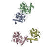
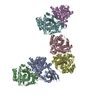
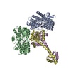
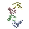
| タイトル | Cryo-Molecular electron tomography of PfEMP1-IT4Var60) | |||||||||
 マップデータ マップデータ | Subtomogram average of 10 PfEMP1s | |||||||||
 試料 試料 |
| |||||||||
 キーワード キーワード |  Malaria (マラリア) / Rosetting / PfEMP1 Malaria (マラリア) / Rosetting / PfEMP1 | |||||||||
| 生物種 |  Plasmodium falciparum (マラリア病原虫) Plasmodium falciparum (マラリア病原虫) | |||||||||
| 手法 | サブトモグラム平均法 / クライオ電子顕微鏡法 / 解像度: 20.0 Å | |||||||||
 データ登録者 データ登録者 | Akhouri RR / Goel S / Furusho H / Skoglund U / Wahlgren M | |||||||||
 引用 引用 | ジャーナル: Cell Rep / 年: 2016 タイトル: Architecture of Human IgM in Complex with P. falciparum Erythrocyte Membrane Protein 1. 著者: Reetesh Raj Akhouri / Suchi Goel / Hirotoshi Furusho / Ulf Skoglund / Mats Wahlgren /   要旨: Plasmodium falciparum virulence is associated with sequestration of infected erythrocytes. Microvascular binding mediated by PfEMP1 in complex with non-immune immunoglobulin M (IgM) is common among ...Plasmodium falciparum virulence is associated with sequestration of infected erythrocytes. Microvascular binding mediated by PfEMP1 in complex with non-immune immunoglobulin M (IgM) is common among parasites that cause both severe childhood malaria and pregnancy-associated malaria. Here, we present cryo-molecular electron tomography structures of human IgM, PfEMP1 and their complex. Three-dimensional reconstructions of IgM reveal that it has a dome-like core, randomly oriented Fab2s units, and the overall shape of a turtle. PfEMP1 is a C- shaped molecule with a flexible N terminus followed by an arc-shaped backbone and a bulky C terminus that interacts with IgM. Our data demonstrate that the PfEMP1 binding pockets on IgM overlap with those of C1q, and the bulkiness of PfEMP1 limits the capacity of IgM to interact with PfEMP1. We suggest that P. falciparum exploits IgM to cluster PfEMP1 into an organized matrix to augment its affinity to host cell receptors. | |||||||||
| 履歴 |
|
- 構造の表示
構造の表示
| ムービー |
 ムービービューア ムービービューア |
|---|---|
| 構造ビューア | EMマップ: SurfViewMolmilJmol/JSmol |
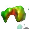
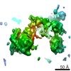
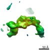
| 添付画像 |
- ダウンロードとリンク
-EMDBアーカイブ
| マップデータ | emd_2886.map.gz | 403.6 KB | EMDBマップデータ形式 | |
|---|---|---|---|---|
| ヘッダ (付随情報) | emd-2886-v30.xmlemd-2886.xml | 9.3 KB 9.3 KB | 表示 表示 | EMDBヘッダ |
| 画像 | emd_2886.tif | 47.1 KB | ||
| アーカイブディレクトリ |  http://ftp.pdbj.org/pub/emdb/structures/EMD-2886ftp://ftp.pdbj.org/pub/emdb/structures/EMD-2886 http://ftp.pdbj.org/pub/emdb/structures/EMD-2886ftp://ftp.pdbj.org/pub/emdb/structures/EMD-2886 | HTTPS FTP |
-関連構造データ


-リンク
| EMDBのページ | EMDB (EBI/PDBe) / EMDataResource |
|---|
-マップ
| ファイル | ダウンロード / ファイル: emd_2886.map.gz / 形式: CCP4 / 大きさ: 450.2 KB / タイプ: IMAGE STORED AS FLOATING POINT NUMBER (4 BYTES) | ||||||||||||||||||||||||||||||||||||||||||||||||||||||||||||
|---|---|---|---|---|---|---|---|---|---|---|---|---|---|---|---|---|---|---|---|---|---|---|---|---|---|---|---|---|---|---|---|---|---|---|---|---|---|---|---|---|---|---|---|---|---|---|---|---|---|---|---|---|---|---|---|---|---|---|---|---|---|
| 注釈 | Subtomogram average of 10 PfEMP1s | ||||||||||||||||||||||||||||||||||||||||||||||||||||||||||||
| ボクセルのサイズ | X=Y=Z: 4.534 Å | ||||||||||||||||||||||||||||||||||||||||||||||||||||||||||||
| 密度 |
| ||||||||||||||||||||||||||||||||||||||||||||||||||||||||||||
| 対称性 | 空間群: 1 | ||||||||||||||||||||||||||||||||||||||||||||||||||||||||||||
| 詳細 | EMDB XML:
CCP4マップ ヘッダ情報:
| ||||||||||||||||||||||||||||||||||||||||||||||||||||||||||||
-添付データ
- 試料の構成要素
-全体 : ectodomain of PfEMP1-IT4Var60
| 全体 | 名称: ectodomain of PfEMP1-IT4Var60 |
|---|---|
| 要素 |
|
-超分子 #1000: ectodomain of PfEMP1-IT4Var60
| 超分子 | 名称: ectodomain of PfEMP1-IT4Var60 / タイプ: sample / ID: 1000 / 詳細: the sample was monodisperse. / 集合状態: Monomer / Number unique components: 1 |
|---|---|
| 分子量 | 実験値: 320 KDa / 理論値: 280 KDa |
-分子 #1: PfEMP1
| 分子 | 名称: PfEMP1 / タイプ: protein_or_peptide / ID: 1 / コピー数: 1 / 集合状態: monomer / 組換発現: Yes |
|---|---|
| 由来(天然) | 生物種: Plasmodium falciparum (マラリア病原虫) / 株: FCR3S1.2 / 別称: Malaria Parasite / 細胞中の位置: Infected erythrocyte membrane |
| 分子量 | 実験値: 320 KDa / 理論値: 280 KDa |
| 組換発現 | 生物種:  Drosophila melanogaster (キイロショウジョウバエ) Drosophila melanogaster (キイロショウジョウバエ)組換株: S2 cells / 組換プラスミド: pMTBipV5HisA |
-実験情報
-構造解析
| 手法 | クライオ電子顕微鏡法 |
|---|---|
 解析 解析 | サブトモグラム平均法 |
| 試料の集合状態 | particle |
-試料調製
| 濃度 | 2 mg/mL |
|---|---|
| 緩衝液 | pH: 7.5 / 詳細: 20mM Hepes, 200mM NaCl |
| グリッド | 詳細: C-Flat (Copper grid with thin Carbon support) |
| 凍結 | 凍結剤: ETHANE / チャンバー内湿度: 100 % / チャンバー内温度: 78 K / 装置: FEI VITROBOT MARK IV / 手法: Blot for 4 seconds before plunging |
- 電子顕微鏡法
| 顕微鏡 | FEI TITAN KRIOS |
|---|---|
| 電子線 | 加速電圧: 300 kV / 電子線源: FIELD EMISSION GUN |
| 電子光学系 | 照射モード: OTHER / 撮影モード: BRIGHT FIELDBright-field microscopy / Cs: 2.7 mm / 最大 デフォーカス(公称値): 0.0015 µm / 最小 デフォーカス(公称値): 0.0007 µm / 倍率(公称値): 37000 |
| 特殊光学系 | エネルギーフィルター - 名称: FEI |
| 試料ステージ | 試料ホルダー: LN2 cooled 試料ホルダーモデル: FEI TITAN KRIOS AUTOGRID HOLDER Tilt series - Axis1 - Min angle: -70 ° / Tilt series - Axis1 - Max angle: 70 ° |
| 温度 | 最低: 78 K / 最高: 100 K |
| 日付 | 2013年10月25日 |
| 撮影 | カテゴリ: CCD フィルム・検出器のモデル: FEI FALCON II (4k x 4k) 実像数: 281 / 平均電子線量: 40 e/Å2 |
| 実験機器 |  モデル: Titan Krios / 画像提供: FEI Company |
-画像解析
| CTF補正 | 詳細: each frame |
|---|---|
| 最終 再構成 | 想定した対称性 - 点群: C1 (非対称) / 解像度のタイプ: BY AUTHOR / 解像度: 20.0 Å / ソフトウェア - 名称: COMET / 使用したサブトモグラム数: 10 |
