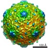
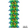
ジャーナル: J Virol / 年: 2014 タイトル: Cryo-electron microscopy structure of lactococcal siphophage 1358 virion. 著者: Silvia Spinelli / Cecilia Bebeacua / Igor Orlov / Denise Tremblay / Bruno P Klaholz / Sylvain Moineau / Christian Cambillau / 要旨: Lactococcus lactis, a Gram(+) lactic acid-producing bacterium used for the manufacture of several fermented dairy products, is subject to infection by diverse virulent tailed phages, leading to ...Lactococcus lactis, a Gram(+) lactic acid-producing bacterium used for the manufacture of several fermented dairy products, is subject to infection by diverse virulent tailed phages, leading to industrial fermentation failures. This constant viral risk has led to a sustained interest in the study of their biology, diversity, and evolution. Lactococcal phages now constitute a wide ensemble of at least 10 distinct genotypes within the Caudovirales order, many of them belonging to the Siphoviridae family. Lactococcal siphophage 1358, currently the only member of its group, displays a noticeably high genomic similarity to some Listeria phages as well as a host range limited to a few L. lactis strains. These genomic and functional characteristics stimulated our interest in this phage. Here, we report the cryo-electron microscopy structure of the complete 1358 virion. Phage 1358 exhibits noteworthy features, such as a capsid with dextro handedness and protruding decorations on its capsid and tail. Observations of the baseplate of virion particles revealed at least two conformations, a closed and an open, activated form. Functional assays uncovered that the adsorption of phage 1358 to its host is Ca(2+) independent, but this cation is necessary to complete its lytic cycle. Taken together, our results provide the complete structural picture of a unique lactococcal phage and expand our knowledge on the complex baseplate of phages of the Siphoviridae family. IMPORTANCE: Phages of Lactococcus lactis are investigated mainly because they are sources of milk fermentation failures in the dairy industry. Despite the availability of several antiphage measures, ...IMPORTANCE: Phages of Lactococcus lactis are investigated mainly because they are sources of milk fermentation failures in the dairy industry. Despite the availability of several antiphage measures, new phages keep emerging in this ecosystem. In this study, we provide the cryo-electron microscopy reconstruction of a unique lactococcal phage that possesses genomic similarity to particular Listeria phages and has a host range restricted to only a minority of L. lactis strains. The capsid of phage 1358 displays the almost unique characteristic of being dextro handed. Its capsid and tail exhibit decorations that we assigned to nonspecific sugar binding modules. We observed the baseplate of 1358 in two conformations, a closed and an open form. We also found that the adsorption to its host, but not infection, is Ca(2+) independent. Overall, this study advances our understanding of the adhesion mechanisms of siphophages.
 ムービー
ムービー コントローラー


 データを開く
 基本情報
基本情報 マップデータ
 試料
試料 キーワード
キーワード Lactococcus lactis /
  Lactococcus phage 1358 (ファージ)
 データ登録者
 引用
引用

 構造の表示
構造の表示 ムービービューア
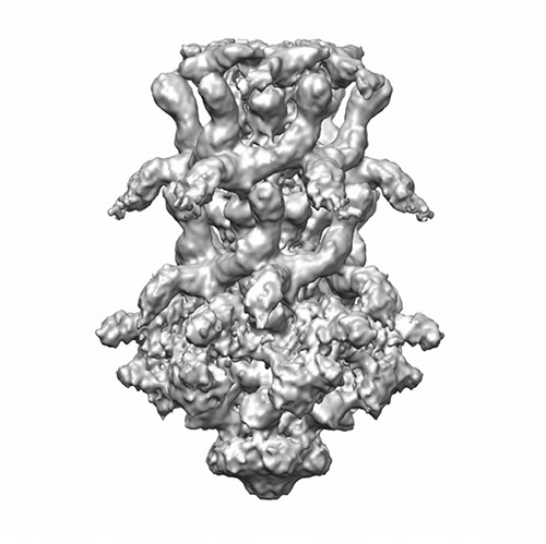
 ダウンロードとリンク
 http://ftp.pdbj.org/pub/emdb/structures/EMD-2817
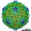


 試料の構成要素
 解析
解析 電子顕微鏡法
