 ムービー
ムービー コントローラー


+ データを開く

- 基本情報
基本情報
| 登録情報 | データベース: EMDB / ID: EMD-2136 | |||||||||
|---|---|---|---|---|---|---|---|---|---|---|
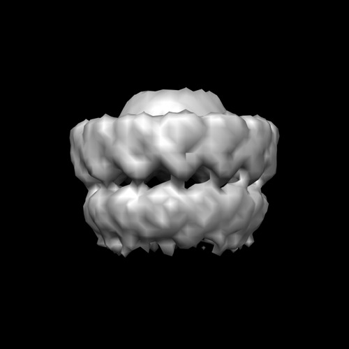
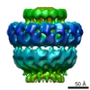
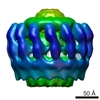
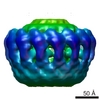
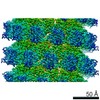
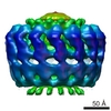
| タイトル | Negative stain EM composite structure (part 1) of the type IV secretion system subcomplex VirB4-VirB7-VirB9-VirB10 分泌 分泌 | |||||||||
 マップデータ マップデータ | Core complex part of the VirB4-core complex composite reconstruction | |||||||||
 試料 試料 |
| |||||||||
| 生物種 |  Escherichia coli (大腸菌) Escherichia coli (大腸菌) | |||||||||
| 手法 | 単粒子再構成法 / ネガティブ染色法 / 解像度: 20.0 Å | |||||||||
 データ登録者 データ登録者 | Williams R / Wallden K / Yan J / Lian PW / Wang L / Thalassinos K / Orlova EV / Waksman G | |||||||||
 引用 引用 | ジャーナル: Proc Natl Acad Sci U S A / 年: 2012 タイトル: Structure of the VirB4 ATPase, alone and bound to the core complex of a type IV secretion system. 著者: Karin Walldén / Robert Williams / Jun Yan / Pei W Lian / Luchun Wang / Konstantinos Thalassinos / Elena V Orlova / Gabriel Waksman /  要旨: Type IV secretion (T4S) systems mediate the transfer of proteins and DNA across the cell envelope of bacteria. These systems play important roles in bacterial pathogenesis and in horizontal transfer ...Type IV secretion (T4S) systems mediate the transfer of proteins and DNA across the cell envelope of bacteria. These systems play important roles in bacterial pathogenesis and in horizontal transfer of antibiotic resistance. The VirB4 ATPase of the T4S system is essential for both the assembly of the system and substrate transfer. In this article, we present the crystal structure of the C-terminal domain of Thermoanaerobacter pseudethanolicus VirB4. This structure is strikingly similar to that of another T4S ATPase, VirD4, a protein that shares only 12% sequence identity with VirB4. The VirB4 domain purifies as a monomer, but the full-length protein is observed in a monomer-dimer equilibrium, even in the presence of nucleotides and DNAs. We also report the negative stain electron microscopy structure of the core complex of the T4S system of the Escherichia coli pKM101 plasmid, with VirB4 bound. In this structure, VirB4 is also monomeric and bound through its N-terminal domain to the core's VirB9 protein. Remarkably, VirB4 is observed bound to the side of the complex where it is ideally placed to play its known regulatory role in substrate transfer. | |||||||||
| 履歴 |
|
- 構造の表示
構造の表示
| ムービー |
 ムービービューア ムービービューア |
|---|---|
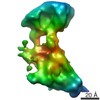
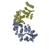
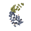
| 構造ビューア | EMマップ: SurfViewMolmilJmol/JSmol |
| 添付画像 |
- ダウンロードとリンク
-EMDBアーカイブ
| マップデータ | emd_2136.map.gz | 4.3 MB | EMDBマップデータ形式 | |
|---|---|---|---|---|
| ヘッダ (付随情報) | emd-2136-v30.xmlemd-2136.xml | 11.5 KB 11.5 KB | 表示 表示 | EMDBヘッダ |
| 画像 | EMD2136.tif | 74.2 KB | ||
| アーカイブディレクトリ |  http://ftp.pdbj.org/pub/emdb/structures/EMD-2136ftp://ftp.pdbj.org/pub/emdb/structures/EMD-2136 http://ftp.pdbj.org/pub/emdb/structures/EMD-2136ftp://ftp.pdbj.org/pub/emdb/structures/EMD-2136 | HTTPS FTP |
-関連構造データ
-リンク
| EMDBのページ | EMDB (EBI/PDBe) / EMDataResource |
|---|
-マップ
| ファイル | ダウンロード / ファイル: emd_2136.map.gz / 形式: CCP4 / 大きさ: 58.2 MB / タイプ: IMAGE STORED AS FLOATING POINT NUMBER (4 BYTES) | ||||||||||||||||||||||||||||||||||||||||||||||||||||||||||||||||||||
|---|---|---|---|---|---|---|---|---|---|---|---|---|---|---|---|---|---|---|---|---|---|---|---|---|---|---|---|---|---|---|---|---|---|---|---|---|---|---|---|---|---|---|---|---|---|---|---|---|---|---|---|---|---|---|---|---|---|---|---|---|---|---|---|---|---|---|---|---|---|
| 注釈 | Core complex part of the VirB4-core complex composite reconstruction | ||||||||||||||||||||||||||||||||||||||||||||||||||||||||||||||||||||
| ボクセルのサイズ | X=Y=Z: 1.67 Å | ||||||||||||||||||||||||||||||||||||||||||||||||||||||||||||||||||||
| 密度 |
| ||||||||||||||||||||||||||||||||||||||||||||||||||||||||||||||||||||
| 対称性 | 空間群: 1 | ||||||||||||||||||||||||||||||||||||||||||||||||||||||||||||||||||||
| 詳細 | EMDB XML:
CCP4マップ ヘッダ情報:
| ||||||||||||||||||||||||||||||||||||||||||||||||||||||||||||||||||||
-添付データ
- 試料の構成要素
-全体 : TraB/TraN/TraO/TraF complex encoded by pKM101
| 全体 | 名称: TraB/TraN/TraO/TraF complex encoded by pKM101 |
|---|---|
| 要素 |
|
-超分子 #1000: TraB/TraN/TraO/TraF complex encoded by pKM101
| 超分子 | 名称: TraB/TraN/TraO/TraF complex encoded by pKM101 / タイプ: sample / ID: 1000 集合状態: 14-mer of core complex (TraN/TraO/TraF) and monomer of TraB Number unique components: 4 |
|---|---|
| 分子量 | 実験値: 1.15 MDa / 理論値: 1.15 MDa |
-分子 #1: TraB
| 分子 | 名称: TraB / タイプ: protein_or_peptide / ID: 1 / Name.synonym: VirB4 詳細: The 1.05 MDa core complex (TraN/TraO/TraF) with TraB bound, see EMD-2137 コピー数: 1 / 組換発現: Yes |
|---|---|
| 由来(天然) | 生物種: Escherichia coli (大腸菌) / 株: pKM101 plasmid / 細胞中の位置: cell envelope |
| 組換発現 | 生物種: Escherichia coli (大腸菌) / 組換プラスミド: pASK IBA3C |
-分子 #2: TraN
| 分子 | 名称: TraN / タイプ: protein_or_peptide / ID: 2 / Name.synonym: VirB7 詳細: The 1.05 MDa core complex (TraN/TraO/TraF) with TraB bound, see EMD-2137 コピー数: 14 / 組換発現: Yes |
|---|---|
| 由来(天然) | 生物種: Escherichia coli (大腸菌) / 株: pKM101 plasmid / 細胞中の位置: cell envelope |
| 組換発現 | 生物種: Escherichia coli (大腸菌) / 組換プラスミド: pASK IBA3C |
-分子 #3: TraO
| 分子 | 名称: TraO / タイプ: protein_or_peptide / ID: 3 / Name.synonym: VirB9 詳細: The 1.05 MDa core complex (TraN/TraO/TraF) with TraB bound, see EMD-2137 コピー数: 14 / 組換発現: Yes |
|---|---|
| 由来(天然) | 生物種: Escherichia coli (大腸菌) / 株: pKM101 plasmid / 細胞中の位置: cell envelope |
| 組換発現 | 生物種: Escherichia coli (大腸菌) / 組換プラスミド: pASK IBA3C |
-分子 #4: TraF
| 分子 | 名称: TraF / タイプ: protein_or_peptide / ID: 4 / Name.synonym: VirB10 詳細: The 1.05 MDa core complex (TraN/TraO/TraF) with TraB bound, see EMD-2137 コピー数: 14 / 組換発現: Yes |
|---|---|
| 由来(天然) | 生物種: Escherichia coli (大腸菌) / 株: pKM101 plasmid / 細胞中の位置: cell envelope |
| 組換発現 | 生物種: Escherichia coli (大腸菌) / 組換プラスミド: pASK IBA3C |
-実験情報
-構造解析
| 手法 | ネガティブ染色法 |
|---|---|
 解析 解析 | 単粒子再構成法 |
| 試料の集合状態 | particle |
-試料調製
| 染色 | タイプ: NEGATIVE / 詳細: NanoW |
|---|---|
| グリッド | 詳細: Glow-discharged continuous carbon |
| 凍結 | 凍結剤: NONE / 装置: OTHER |
- 電子顕微鏡法
| 顕微鏡 | FEI TECNAI 12 |
|---|---|
| 電子線 | 加速電圧: 120 kV / 電子線源: OTHER |
| 電子光学系 | 照射モード: SPOT SCAN / 撮影モード: BRIGHT FIELDBright-field microscopy / 最大 デフォーカス(公称値): 2.0 µm / 最小 デフォーカス(公称値): 0.7 µm / 倍率(公称値): 42000 |
| 試料ステージ | 試料ホルダーモデル: SIDE ENTRY, EUCENTRIC |
| 日付 | 2009年12月22日 |
| 撮影 | カテゴリ: FILM / フィルム・検出器のモデル: GENERIC FILM / デジタル化 - スキャナー: ZEISS SCAI / デジタル化 - サンプリング間隔: 7 µm / 実像数: 12 / ビット/ピクセル: 8 |
| Tilt angle min | 0 |
| Tilt angle max | 0 |
-画像解析
| 最終 2次元分類 | クラス数: 330 |
|---|---|
| 最終 再構成 | 想定した対称性 - 点群: C1 (非対称) / アルゴリズム: OTHER / 解像度のタイプ: BY AUTHOR / 解像度: 20.0 Å / 解像度の算出法: FSC 0.5 CUT-OFF / ソフトウェア - 名称: IMAGIC 詳細: This map is part of a composite structure of two maps. 使用した粒子像数: 10000 |
