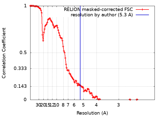
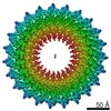
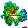
- EMDB-20831: Cryo-EM reconstruction shows that the needle complex's inner ring... -
+
データを開く
IDまたはキーワード:
読み込み中...
-
基本情報
登録情報
データベース: EMDB / ID: EMD-20831
タイトル
Cryo-EM reconstruction shows that the needle complex's inner rings from Salmonella assemble with 23-fold symmetry in the absence of the export apparatus.
マップデータ
試料
複合体: Complex of PrgH and PrgK, the protein components of the inner rings of Salmonella's needle complex, assembled in the absence of the export apparatus.
National Institutes of Health/National Institute Of Allergy and Infectious Diseases
米国
引用
ジャーナル: Proc Natl Acad Sci U S A / 年: 2019 タイトル: High-resolution view of the type III secretion export apparatus in situ reveals membrane remodeling and a secretion pathway. 著者: Carmen Butan / Maria Lara-Tejero / Wenwei Li / Jun Liu / Jorge E Galán / 要旨: Type III protein secretion systems are essential virulence factors for many important pathogenic bacteria. The entire protein secretion machine is composed of several substructures that organize into ...Type III protein secretion systems are essential virulence factors for many important pathogenic bacteria. The entire protein secretion machine is composed of several substructures that organize into a holostructure or injectisome. The core component of the injectisome is the needle complex, which houses the export apparatus that serves as a gate for the passage of the secreted proteins through the bacterial inner membrane. Here, we describe a high-resolution structure of the export apparatus of the type III secretion system in association with the needle complex and the underlying bacterial membrane, both in isolation and in situ. We show the precise location of the core export apparatus components within the injectisome and bacterial envelope and demonstrate that their deployment results in major membrane remodeling and thinning, which may be central for the protein translocation process. We also show that InvA, a critical export apparatus component, forms a multiring cytoplasmic conduit that provides a pathway for the type III secretion substrates to reach the entrance of the export gate. Combined with structure-guided mutagenesis, our studies provide major insight into potential mechanisms of protein translocation and injectisome assembly.
ダウンロード / ファイル: emd_20831.map.gz / 形式: CCP4 / 大きさ: 178 MB / タイプ: IMAGE STORED AS FLOATING POINT NUMBER (4 BYTES)
ボクセルのサイズ
X=Y=Z: 1.07 Å
密度
表面レベル
登録者による: 0.0098 / ムービー #1: 0.0108
最小 - 最大
-0.01748075 - 0.040073305
平均 (標準偏差)
0.00037343317 (±0.0028942786)
対称性
空間群: 1
詳細
EMDB XML:
マップ形状
Axis order
X
Y
Z
Origin
0
0
0
サイズ
360
360
360
Spacing
360
360
360
セル
A=B=C: 385.2 Å α=β=γ: 90.0 °
CCP4マップ ヘッダ情報:
mode
Image stored as Reals
Å/pix. X/Y/Z
1.07
1.07
1.07
M x/y/z
360
360
360
origin x/y/z
0.000
0.000
0.000
length x/y/z
385.200
385.200
385.200
α/β/γ
90.000
90.000
90.000
MAP C/R/S
1
2
3
start NC/NR/NS
0
0
0
NC/NR/NS
360
360
360
D min/max/mean
-0.017
0.040
0.000
-
添付データ
-
試料の構成要素
-
全体 : Complex of PrgH and PrgK, the protein components of the inner rin...
全体
名称: Complex of PrgH and PrgK, the protein components of the inner rings of Salmonella's needle complex, assembled in the absence of the export apparatus.
要素
複合体: Complex of PrgH and PrgK, the protein components of the inner rings of Salmonella's needle complex, assembled in the absence of the export apparatus.
-
超分子 #1: Complex of PrgH and PrgK, the protein components of the inner rin...
超分子
名称: Complex of PrgH and PrgK, the protein components of the inner rings of Salmonella's needle complex, assembled in the absence of the export apparatus. タイプ: complex / ID: 1 / 親要素: 0 / 含まれる分子: #1
 ムービー
ムービー コントローラー


 データを開く
 基本情報
基本情報 マップデータ
 試料
試料
 Salmonella enterica subsp. enterica serovar Typhimurium (サルモネラ菌)
 データ登録者
 米国, 1件
  引用
引用 構造の表示
構造の表示 ムービービューア
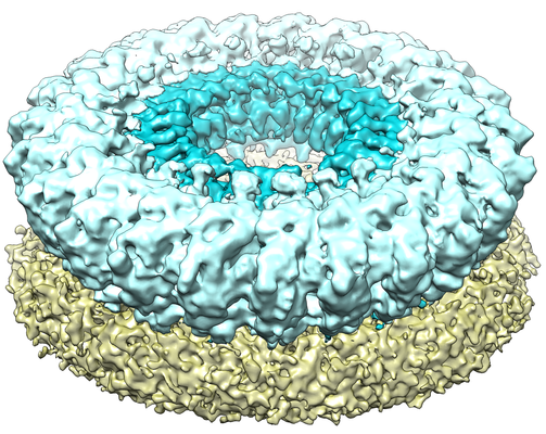
 ダウンロードとリンク
 emd_20831.png
 http://ftp.pdbj.org/pub/emdb/structures/EMD-20831


 試料の構成要素
 解析
解析 電子顕微鏡法