 ムービー
ムービー コントローラー


+ データを開く

- 基本情報
基本情報
| 登録情報 | データベース: EMDB / ID: EMD-2077 | |||||||||
|---|---|---|---|---|---|---|---|---|---|---|
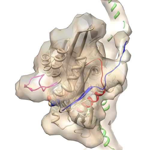
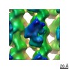
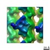
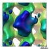
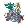
| タイトル | Electron cryo-microscopy of microtubule-bound human kinesin-5 motor domain in AMPPNP state. | |||||||||
 マップデータ マップデータ | 3D reconstruction of microtubule-bound human kinesin-5 motor domain with AMPPNP bound in the nucleotide-binding site | |||||||||
 試料 試料 |
| |||||||||
 キーワード キーワード |  cancer (悪性腫瘍) / electron cryo-microscopy (低温電子顕微鏡法) / kinesin (キネシン) / microtubule (微小管) / mitosis (有糸分裂) cancer (悪性腫瘍) / electron cryo-microscopy (低温電子顕微鏡法) / kinesin (キネシン) / microtubule (微小管) / mitosis (有糸分裂) | |||||||||
| 機能・相同性 |  機能・相同性情報 機能・相同性情報spindle elongation / Kinesins / plus-end-directed microtubule motor activity / regulation of mitotic centrosome separation / mitotic centrosome separation / positive regulation of axon guidance / COPI-dependent Golgi-to-ER retrograde traffic / kinesin complex / microtubule motor activity / spindle organization ...spindle elongation / Kinesins / plus-end-directed microtubule motor activity / regulation of mitotic centrosome separation / mitotic centrosome separation / positive regulation of axon guidance / COPI-dependent Golgi-to-ER retrograde traffic / kinesin complex / microtubule motor activity / spindle organization / microtubule-based movement / mitotic spindle assembly / microtubule-based process / MHC class II antigen presentation / mitotic spindle organization / 加水分解酵素; 酸無水物に作用; GTPに作用・細胞または細胞小器官の運動に関与 / structural constituent of cytoskeleton / spindle / 紡錘体 / microtubule cytoskeleton organization / 紡錘体 / microtubule cytoskeleton / mitotic cell cycle / nervous system development / microtubule binding / 微小管 / hydrolase activity / protein heterodimerization activity / 細胞分裂 / GTPase activity / GTP binding / protein kinase binding / protein-containing complex / ATP binding / 生体膜 / metal ion binding / 細胞核 / 細胞質基質 / 細胞質類似検索 - 分子機能 | |||||||||
| 生物種 |  Bos taurus (ウシ) / Bos taurus (ウシ) /  Homo sapiens (ヒト) Homo sapiens (ヒト) | |||||||||
| 手法 | 単粒子再構成法 / クライオ電子顕微鏡法 / 解像度: 9.7 Å | |||||||||
 データ登録者 データ登録者 | Goulet A / Behnke-Parks WM / Sindelar C / Rosenfeld S / Moores C | |||||||||
 引用 引用 | ジャーナル: J Biol Chem / 年: 2012 タイトル: The structural basis of force generation by the mitotic motor kinesin-5. 著者: Adeline Goulet / William M Behnke-Parks / Charles V Sindelar / Jennifer Major / Steven S Rosenfeld / Carolyn A Moores /  要旨: Kinesin-5 is required for forming the bipolar spindle during mitosis. Its motor domain, which contains nucleotide and microtubule binding sites and mechanical elements to generate force, has evolved ...Kinesin-5 is required for forming the bipolar spindle during mitosis. Its motor domain, which contains nucleotide and microtubule binding sites and mechanical elements to generate force, has evolved distinct properties for its spindle-based functions. In this study, we report subnanometer resolution cryoelectron microscopy reconstructions of microtubule-bound human kinesin-5 before and after nucleotide binding and combine this information with studies of the kinetics of nucleotide-induced neck linker and cover strand movement. These studies reveal coupled, nucleotide-dependent conformational changes that explain many of this motor's properties. We find that ATP binding induces a ratchet-like docking of the neck linker and simultaneous, parallel docking of the N-terminal cover strand. Loop L5, the binding site for allosteric inhibitors of kinesin-5, also undergoes a dramatic reorientation when ATP binds, suggesting that it is directly involved in controlling nucleotide binding. Our structures indicate that allosteric inhibitors of human kinesin-5, which are being developed as anti-cancer therapeutics, bind to a motor conformation that occurs in the course of normal function. However, due to evolutionarily defined sequence variations in L5, this conformation is not adopted by invertebrate kinesin-5s, explaining their resistance to drug inhibition. Together, our data reveal the precision with which the molecular mechanism of kinesin-5 motors has evolved for force generation. | |||||||||
| 履歴 |
|
- 構造の表示
構造の表示
| ムービー |
ムービービューア |
|---|---|
| 構造ビューア | EMマップ: SurfViewMolmilJmol/JSmol |
| 添付画像 |
- ダウンロードとリンク
-EMDBアーカイブ
| マップデータ | emd_2077.map.gz | 238.1 KB | EMDBマップデータ形式 | |
|---|---|---|---|---|
| ヘッダ (付随情報) | emd-2077-v30.xmlemd-2077.xml | 12.3 KB 12.3 KB | 表示 表示 | EMDBヘッダ |
| 画像 |  emd_2077.jpg emd_2077.jpg | 144.6 KB | ||
| アーカイブディレクトリ |  http://ftp.pdbj.org/pub/emdb/structures/EMD-2077ftp://ftp.pdbj.org/pub/emdb/structures/EMD-2077 http://ftp.pdbj.org/pub/emdb/structures/EMD-2077ftp://ftp.pdbj.org/pub/emdb/structures/EMD-2077 | HTTPS FTP |
-関連構造データ
-リンク
| EMDBのページ | EMDB (EBI/PDBe) / EMDataResource |
|---|---|
| 「今月の分子」の関連する項目 |


-マップ
| ファイル | ダウンロード / ファイル: emd_2077.map.gz / 形式: CCP4 / 大きさ: 348.6 KB / タイプ: IMAGE STORED AS FLOATING POINT NUMBER (4 BYTES) | ||||||||||||||||||||||||||||||||||||||||||||||||||||||||||||||||||||
|---|---|---|---|---|---|---|---|---|---|---|---|---|---|---|---|---|---|---|---|---|---|---|---|---|---|---|---|---|---|---|---|---|---|---|---|---|---|---|---|---|---|---|---|---|---|---|---|---|---|---|---|---|---|---|---|---|---|---|---|---|---|---|---|---|---|---|---|---|---|
| 注釈 | 3D reconstruction of microtubule-bound human kinesin-5 motor domain with AMPPNP bound in the nucleotide-binding site | ||||||||||||||||||||||||||||||||||||||||||||||||||||||||||||||||||||
| ボクセルのサイズ | X=Y=Z: 2.8 Å | ||||||||||||||||||||||||||||||||||||||||||||||||||||||||||||||||||||
| 密度 |
| ||||||||||||||||||||||||||||||||||||||||||||||||||||||||||||||||||||
| 対称性 | 空間群: 1 | ||||||||||||||||||||||||||||||||||||||||||||||||||||||||||||||||||||
| 詳細 | EMDB XML:
CCP4マップ ヘッダ情報:
| ||||||||||||||||||||||||||||||||||||||||||||||||||||||||||||||||||||
-添付データ
- 試料の構成要素
-全体 : 13-protofilament microtubule-bound human kinesin-5 motor domain w...
| 全体 | 名称: 13-protofilament microtubule-bound human kinesin-5 motor domain with AMPPNP |
|---|---|
| 要素 |
|
-超分子 #1000: 13-protofilament microtubule-bound human kinesin-5 motor domain w...
| 超分子 | 名称: 13-protofilament microtubule-bound human kinesin-5 motor domain with AMPPNP タイプ: sample / ID: 1000 集合状態: 13-protofilament microtubule with one kineisn-5 motor domain bound every tubulin heterodimers Number unique components: 2 |
|---|
-分子 #1: alpha-beta tubulin dimer
| 分子 | 名称: alpha-beta tubulin dimer / タイプ: protein_or_peptide / ID: 1 / 集合状態: heterodimer / 組換発現: No / データベース: NCBI |
|---|---|
| 由来(天然) | 生物種: Bos taurus (ウシ) / 別称: Cattle / 組織: brain |
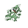
-分子 #2: Kinesin-5 motor domain
| 分子 | 名称: Kinesin-5 motor domain / タイプ: protein_or_peptide / ID: 2 / 集合状態: monomer / 組換発現: Yes |
|---|---|
| 由来(天然) | 生物種: Homo sapiens (ヒト) / 別称: Human |
| 組換発現 | 生物種:  Escherichia coli (大腸菌) / 組換プラスミド: pet21a Escherichia coli (大腸菌) / 組換プラスミド: pet21a |
-実験情報
-構造解析
| 手法 | クライオ電子顕微鏡法 |
|---|---|
 解析 解析 | 単粒子再構成法 |
| 試料の集合状態 | particle |
-試料調製
| 緩衝液 | pH: 6.8 / 詳細: 80 mM PIPES, 5 mM MgCl2, 1 mM EGTA, 5mM AMPPNP |
|---|---|
| グリッド | 詳細: 400 mesh holey carbon grids |
| 凍結 | 凍結剤: ETHANE / チャンバー内湿度: 100 % / 装置: FEI VITROBOT MARK I / 手法: chamber at 24 degrees C, blot 2.5 sec |
- 電子顕微鏡法
| 顕微鏡 | FEI TECNAI F20 |
|---|---|
| 電子線 | 加速電圧: 200 kV / 電子線源: FIELD EMISSION GUN |
| 電子光学系 | 照射モード: FLOOD BEAM / 撮影モード: BRIGHT FIELDBright-field microscopy / Cs: 2.0 mm / 最大 デフォーカス(公称値): 2.2 µm / 最小 デフォーカス(公称値): 0.7 µm / 倍率(公称値): 50000 |
| 試料ステージ | 試料ホルダーモデル: GATAN LIQUID NITROGEN |
| 温度 | 平均: 90 K |
| アライメント法 | Legacy - 非点収差: Objective lens astigmatism was corrected at 150,000 times magnification |
| 日付 | 2011年1月10日 |
| 撮影 | カテゴリ: FILM / フィルム・検出器のモデル: KODAK SO-163 FILM / デジタル化 - スキャナー: ZEISS SCAI / デジタル化 - サンプリング間隔: 7 µm / 実像数: 46 / 平均電子線量: 18 e/Å2 / ビット/ピクセル: 8 |
| 実験機器 |  モデル: Tecnai F20 / 画像提供: FEI Company |
-画像解析
| CTF補正 | 詳細: FREALIGN |
|---|---|
| 最終 再構成 | 解像度のタイプ: BY AUTHOR / 解像度: 9.7 Å / 解像度の算出法: FSC 0.5 CUT-OFF / ソフトウェア - 名称: SPIDER, FREALIGN 詳細: Approximately 50,000 asymmetric units were averaged in the final reconstruction. 使用した粒子像数: 3587 |
| 詳細 | The particles were selected along individual microtubules. |
-原子モデル構築 1
| 初期モデル | PDB ID: Chain - Chain ID: A |
|---|---|
| ソフトウェア | 名称: Chimera, FlexEM |
| 詳細 | Protocol: rigid body then flexible fitting. The domain was fitted as a rigid body. The N-terminal residues 6 to 16 were built in the EM map and the final model was refined by flexible fitting. |
| 精密化 | 空間: REAL / プロトコル: FLEXIBLE FIT / 当てはまり具合の基準: cross-correlation |
| 得られたモデル | 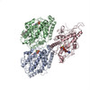 PDB-4aqv: |
-原子モデル構築 2
| 初期モデル | PDB ID: Chain - #0 - Chain ID: A / Chain - #1 - Chain ID: B |
|---|---|
| ソフトウェア | 名称: Chimera |
| 詳細 | Protocol: rigid body. alpha- and b-tubulin were separately fitted. |
| 精密化 | 空間: REAL / プロトコル: RIGID BODY FIT / 当てはまり具合の基準: cross-correlation |
| 得られたモデル | PDB-4aqv: |

