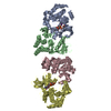
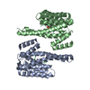
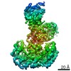
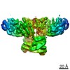
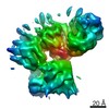
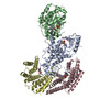
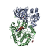
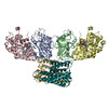
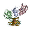
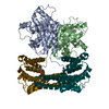
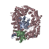
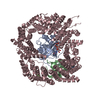
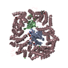
登録情報 データベース : EMDB / ID : EMD-20551タイトル Structure of a MAPK pathway complex Structure of a MAPK pathway complex 複合体 : ERK pathway complexタンパク質・ペプチド : Serine/threonine-protein kinase B-rafタンパク質・ペプチド : 14-3-3 protein zeta/delta機能・相同性 分子機能 ドメイン・相同性 構成要素
/ / / / / / / / / / / / / / / / / / / / / / / / / / / / / / / / / / / / / / / / / / / / / / / / / / / / / / / / / / / / / / / / / / / / / / / / / / / / / / / / / / / / / / / / / / / / / / / / / / / / / / / / / / / / / / / / / / / / / / / / / / / / / / / / / / / / / / / / / / / / / / / / / / / 生物種 Homo sapiens (ヒト) / Human (ヒト)手法 / / 解像度 : 6.8 Å Park E / Rawson S / Jeon H / Eck MJ 資金援助 Organization Grant number 国 National Institutes of Health/National Human Genome Research Institute (NIH/NHGRI) P50CA165962 National Institutes of Health/National Cancer Institute (NIH/NCI) R50CA221830
ジャーナル : Nature / 年 : 2019タイトル : Architecture of autoinhibited and active BRAF-MEK1-14-3-3 complexes.著者 : Eunyoung Park / Shaun Rawson / Kunhua Li / Byeong-Won Kim / Scott B Ficarro / Gonzalo Gonzalez-Del Pino / Humayun Sharif / Jarrod A Marto / Hyesung Jeon / Michael J Eck / 要旨 : RAF family kinases are RAS-activated switches that initiate signalling through the MAP kinase cascade to control cellular proliferation, differentiation and survival. RAF activity is tightly ... RAF family kinases are RAS-activated switches that initiate signalling through the MAP kinase cascade to control cellular proliferation, differentiation and survival. RAF activity is tightly regulated and inappropriate activation is a frequent cause of cancer; however, the structural basis for RAF regulation is poorly understood at present. Here we use cryo-electron microscopy to determine autoinhibited and active-state structures of full-length BRAF in complexes with MEK1 and a 14-3-3 dimer. The reconstruction reveals an inactive BRAF-MEK1 complex restrained in a cradle formed by the 14-3-3 dimer, which binds the phosphorylated S365 and S729 sites that flank the BRAF kinase domain. The BRAF cysteine-rich domain occupies a central position that stabilizes this assembly, but the adjacent RAS-binding domain is poorly ordered and peripheral. The 14-3-3 cradle maintains autoinhibition by sequestering the membrane-binding cysteine-rich domain and blocking dimerization of the BRAF kinase domain. In the active state, these inhibitory interactions are released and a single 14-3-3 dimer rearranges to bridge the C-terminal pS729 binding sites of two BRAFs, which drives the formation of an active, back-to-back BRAF dimer. Our structural snapshots provide a foundation for understanding normal RAF regulation and its mutational disruption in cancer and developmental syndromes. 履歴 登録 2019年8月1日 - ヘッダ(付随情報) 公開 2019年10月2日 - マップ公開 2019年10月9日 - 更新 2020年4月22日 - 現状 2020年4月22日 処理サイト : RCSB / 状態 : 公開
すべて表示 表示を減らす
 ムービー
ムービー コントローラー


 データを開く
 基本情報
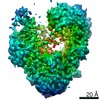
基本情報 マップデータ
 試料
試料 機能・相同性情報
 stress fiber assembly / positive regulation of axon regeneration / face development / synaptic vesicle exocytosis / somatic stem cell population maintenance /
 データ登録者
 米国, 2件
  引用
引用 構造の表示
構造の表示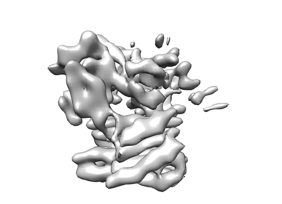
 ダウンロードとリンク
 emd_20551.png
 http://ftp.pdbj.org/pub/emdb/structures/EMD-20551
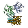


 試料の構成要素
 解析
解析 電子顕微鏡法