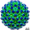
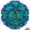
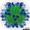
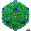
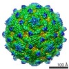
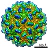
ジャーナル: J Virol / 年: 2012 タイトル: Structural analysis of coxsackievirus A7 reveals conformational changes associated with uncoating. 著者: Jani J T Seitsonen / Shabih Shakeel / Petri Susi / Arun P Pandurangan / Robert S Sinkovits / Heini Hyvönen / Pasi Laurinmäki / Jani Ylä-Pelto / Maya Topf / Timo Hyypiä / Sarah J Butcher / 要旨: Coxsackievirus A7 (CAV7) is a rarely detected and poorly characterized serotype of the Enterovirus species Human enterovirus A (HEV-A) within the Picornaviridae family. The CAV7-USSR strain has ...Coxsackievirus A7 (CAV7) is a rarely detected and poorly characterized serotype of the Enterovirus species Human enterovirus A (HEV-A) within the Picornaviridae family. The CAV7-USSR strain has caused polio-like epidemics and was originally thought to represent the fourth poliovirus type, but later evidence linked this strain to the CAV7-Parker prototype. Another isolate, CAV7-275/58, was also serologically similar to Parker but was noninfectious in a mouse model. Sequencing of the genomic region encoding the capsid proteins of the USSR and 275/58 strains and subsequent comparison with the corresponding amino acid sequences of the Parker strain revealed that the Parker and USSR strains are nearly identical, while the 275/58 strain is more distant. Using electron cryomicroscopy and three-dimensional image reconstruction, the structures of the CAV7-USSR virion and empty capsid were resolved to 8.2-Å and 6.1-Å resolutions, respectively. This is one of the first detailed structural analyses of the HEV-A species. Using homology modeling, reconstruction segmentation, and flexible fitting, we constructed a pseudoatomic T = 1 (pseudo T = 3) model incorporating the three major capsid proteins (VP1 to VP3), addressed the conformational changes of the capsid and its constituent viral proteins occurring during RNA release, and mapped the capsid proteins' variable regions to the structure. During uncoating, VP4 and RNA are released analogously to poliovirus 1, the interfaces of VP2 and VP3 are rearranged, and VP1 rotates. Variable regions in the capsid proteins were predicted to map mainly to the surface of VP1 and are thus likely to affect the tropism and pathogenicity of CAV7.
 ムービー
ムービー コントローラー


 データを開く
 基本情報
基本情報 マップデータ
 試料
試料 キーワード
キーワード coxsackievirus (コクサッキーウイルス) / CAV7 /
  機能・相同性情報
 データ登録者
 引用
引用
 構造の表示
構造の表示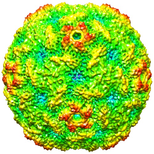
 ダウンロードとリンク
 http://ftp.pdbj.org/pub/emdb/structures/EMD-2027


 試料の構成要素
 解析
解析 電子顕微鏡法
