 ムービー
ムービー コントローラー


+ データを開く

- 基本情報
基本情報
| 登録情報 | データベース: EMDB / ID: EMD-20091 | |||||||||
|---|---|---|---|---|---|---|---|---|---|---|
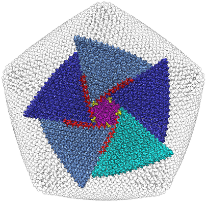
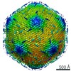
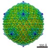
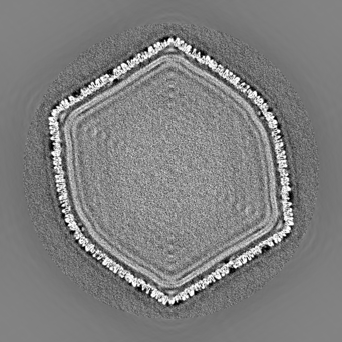
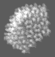
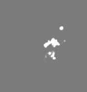
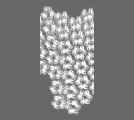
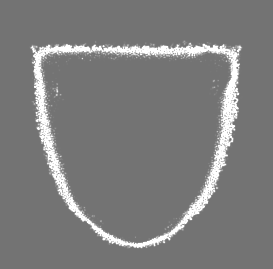
| タイトル | Singapore Grouper Iridovirus (SGIV) at 8.6 Angstrom Resolution | |||||||||
 マップデータ マップデータ | Independent reconstruction half of the micrographs | |||||||||
 試料 試料 |
| |||||||||
 キーワード キーワード |  icosahedral (二十面体) / trimers / outer coat / anchor proteins (錨) / zip proteins / VIRAL PROTEIN (ウイルスタンパク質) icosahedral (二十面体) / trimers / outer coat / anchor proteins (錨) / zip proteins / VIRAL PROTEIN (ウイルスタンパク質) | |||||||||
| 機能・相同性 | Major capsid protein, N-terminal / Major capsid protein N-terminus / Major capsid protein, C-terminal / Major capsid protein, C-terminal domain superfamily / Large eukaryotic DNA virus major capsid protein / Group II dsDNA virus coat/capsid protein / カプシド / structural molecule activity / Major capsid protein 機能・相同性情報 機能・相同性情報 | |||||||||
| 生物種 |  Singapore grouper iridovirus (ウイルス) Singapore grouper iridovirus (ウイルス) | |||||||||
| 手法 | 単粒子再構成法 / クライオ電子顕微鏡法 / 解像度: 8.6 Å | |||||||||
 データ登録者 データ登録者 | Pintilie G / Chen D-H | |||||||||
| 資金援助 |  米国, 2件 米国, 2件
| |||||||||
 引用 引用 | ジャーナル: Structure / 年: 2019 タイトル: Segmentation and Comparative Modeling in an 8.6-Å Cryo-EM Map of the Singapore Grouper Iridovirus. 著者: Grigore Pintilie / Dong-Hua Chen / Bich Ngoc Tran / Joanita Jakana / Jinlu Wu / Choy Leong Hew / Wah Chiu /  要旨: SGIV, or Singapore grouper iridovirus, is a large double-stranded DNA virus, reaching a diameter of 220 nm and packaging a genome of 140 kb. We present a 3D cryoelectron microscopy (cryo-EM) ...SGIV, or Singapore grouper iridovirus, is a large double-stranded DNA virus, reaching a diameter of 220 nm and packaging a genome of 140 kb. We present a 3D cryoelectron microscopy (cryo-EM) icosahedral reconstruction of SGIV determined at 8.6-Å resolution. It reveals several layers including a T = 247 icosahedral outer coat, anchor proteins, a lipid bilayer, and the encapsidated DNA. A new segmentation tool, iSeg, was applied to extract these layers from the reconstructed map. The outer coat was further segmented into major and minor capsid proteins. None of the proteins extracted by segmentation have known atomic structures. We generated models for the major coat protein using three comparative modeling tools, and evaluated each model using the cryo-EM map. Our analysis reveals a new architecture in the Iridoviridae family of viruses. It shares similarities with others in the same family, e.g., Chilo iridescent virus, but also shows new features of the major and minor capsid proteins. | |||||||||
| 履歴 |
|
- 構造の表示
構造の表示
| ムービー |
ムービービューア |
|---|---|
| 構造ビューア | EMマップ: SurfViewMolmilJmol/JSmol |
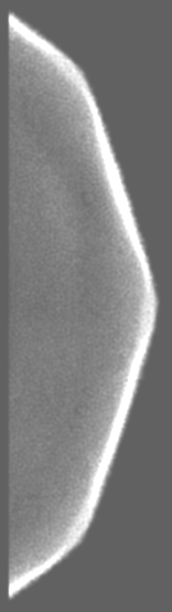
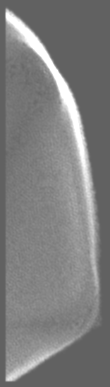
| 添付画像 |
- ダウンロードとリンク
-EMDBアーカイブ
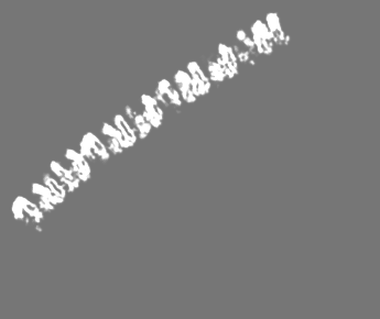
 emd_20091.png
-関連構造データ


- リンク
リンク
| EMDBのページ | EMDB (EBI/PDBe) / EMDataResource |
|---|---|
| 「今月の分子」の関連する項目 |


-マップ
| ファイル | ダウンロード / ファイル: emd_20091.map.gz / 形式: CCP4 / 大きさ: 6.4 GB / タイプ: IMAGE STORED AS FLOATING POINT NUMBER (4 BYTES) | ||||||||||||||||||||||||||||||||||||||||||||||||||||||||||||
|---|---|---|---|---|---|---|---|---|---|---|---|---|---|---|---|---|---|---|---|---|---|---|---|---|---|---|---|---|---|---|---|---|---|---|---|---|---|---|---|---|---|---|---|---|---|---|---|---|---|---|---|---|---|---|---|---|---|---|---|---|---|
| 注釈 | Independent reconstruction half of the micrographs | ||||||||||||||||||||||||||||||||||||||||||||||||||||||||||||
| ボクセルのサイズ | X=Y=Z: 2.37 Å | ||||||||||||||||||||||||||||||||||||||||||||||||||||||||||||
| 密度 |
| ||||||||||||||||||||||||||||||||||||||||||||||||||||||||||||
| 対称性 | 空間群: 1 | ||||||||||||||||||||||||||||||||||||||||||||||||||||||||||||
| 詳細 | EMDB XML:
CCP4マップ ヘッダ情報:
| ||||||||||||||||||||||||||||||||||||||||||||||||||||||||||||
-添付データ
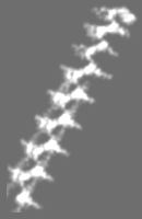
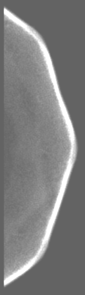
+追加マップ: Independent reconstruction other half of the micrographs
 Z
Z Y
Y X
X


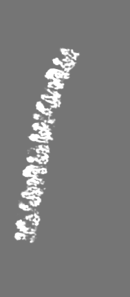
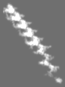
+追加マップ: A pentasymmetron
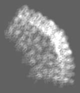
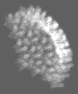

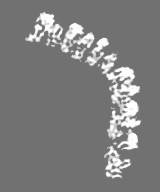
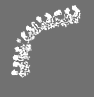


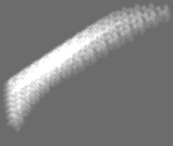
+追加マップ: Averaged densities from trimers from a trisymmetron within...


+追加マップ: Trisymmetron 1 of 5 around a pentasymmetron


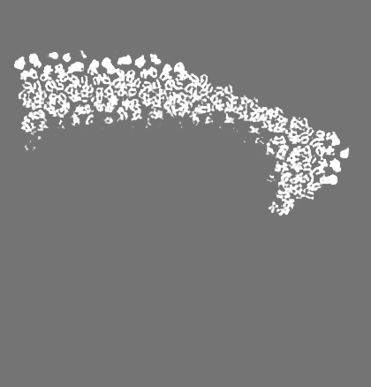


+追加マップ: Trisymmetron 2 of 5 around a pentasymmetron


+追加マップ: Trisymmetron 3 of 5 around a pentasymmetron


+添付マップデータ: emd 20091 additional 15.map


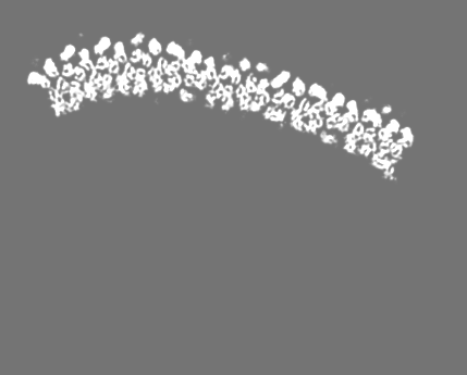

+追加マップ: Trisymmetron 5 of 5 around a pentasymmetron


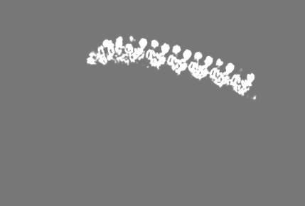
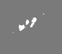
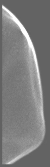
+追加マップ: Zip proteins 1


+追加マップ: Zip proteins 2


+追加マップ: Zip proteins 3


+追加マップ: Anchor protein 1


+追加マップ: Zip proteins 4


+追加マップ: Zip proteins 5


+追加マップ: Anchor protein 2


+追加マップ: Anchor protein 3


+追加マップ: Anchor protein 4


+追加マップ: Anchor protein 5


+追加マップ: An asymmetric unit from the icosahedrally-symmetric map


+追加マップ: Inner half of the bilayer lipid membrane


+追加マップ: Outer half of the bilayer lipid membrane


- 試料の構成要素
-全体 : Singapore grouper iridovirus
| 全体 | 名称: Singapore grouper iridovirus (ウイルス) |
|---|---|
| 要素 |
|
-超分子 #1: Singapore grouper iridovirus
| 超分子 | 名称: Singapore grouper iridovirus / タイプ: virus / ID: 1 / 親要素: 0 / 含まれる分子: all / NCBI-ID: 262968 / 生物種: Singapore grouper iridovirus / ウイルスタイプ: VIRION / ウイルス・単離状態: SPECIES / ウイルス・エンベロープ: Yes / ウイルス・中空状態: No |
|---|---|
| 宿主 | 生物種:  Epinephelus tauvina (ヒトミハタ) Epinephelus tauvina (ヒトミハタ) |
| ウイルス殻 | Shell ID: 1 / 名称: Outer coat / 直径: 2200.0 Å / T番号(三角分割数): 247 |
-分子 #1: Major capsid protein
| 分子 | 名称: Major capsid protein / タイプ: protein_or_peptide / ID: 1 / コピー数: 3 / 光学異性体: LEVO |
|---|---|
| 由来(天然) | 生物種: Singapore grouper iridovirus (ウイルス) |
| 分子量 | 理論値: 50.573141 KDa |
| 配列 | 文字列: MTCTTGAGVT SGFIDLATYD NLDRALYGGK DATTYFIKEH YPVGWFTKLP TMATRVSGNP AFGQEFSVGV PRSGDYVLNA WLTLKTPEI KLLETNRLGA NGTVRWTKNL MHNAVEHASL TFNDICAQQF NTAYLDAWTQ FNMCEGKRIG YDNMIGNTSD M TNPTPAQG ...文字列: MTCTTGAGVT SGFIDLATYD NLDRALYGGK DATTYFIKEH YPVGWFTKLP TMATRVSGNP AFGQEFSVGV PRSGDYVLNA WLTLKTPEI KLLETNRLGA NGTVRWTKNL MHNAVEHASL TFNDICAQQF NTAYLDAWTQ FNMCEGKRIG YDNMIGNTSD M TNPTPAQG QDGARTLPSK NLVLPLPFFF SRDCGLALPT VVLPYNEIRI NIKLRSLQEL LVFQNKDTGN VIPISATDIA GG LADTVEA YVYMTVGLVS NVERCAMAGT VRDMVVEQMQ AAPTHIVNPQ NTNNVHVDMR FSHAVKALFF MVQNVTYKSV GSN YTCVTP VNGPGNTVME PAMSVDPIKS ASLTYENTTR LANMGVEYYS LVQPWYFSAS IPVYTGYHMY SYALNVGSVH PSGS TNYGR LTNASITVTM SPESVVAAAG GGNNNSGYNE PQRFALVVIA VNHNVIRIMN GSMGFPIL UniProtKB: Major capsid protein |
-実験情報
-構造解析
| 手法 | クライオ電子顕微鏡法 |
|---|---|
 解析 解析 | 単粒子再構成法 |
| 試料の集合状態 | particle |
-試料調製
| 緩衝液 | pH: 7.4 |
|---|---|
| グリッド | モデル: Quantifoil R2/1 |
| 凍結 | 凍結剤: ETHANE / チャンバー内湿度: 100 % / 装置: FEI VITROBOT MARK IV / 詳細: Blotted for 1 second before plunging.. |
- 電子顕微鏡法
| 顕微鏡 | FEI TITAN KRIOS |
|---|---|
| 電子線 | 加速電圧: 300 kV / 電子線源: FIELD EMISSION GUN |
| 電子光学系 | C2レンズ絞り径: 70.0 µm / 倍率(補正後): 63290 / 照射モード: FLOOD BEAM / 撮影モード: BRIGHT FIELDBright-field microscopy / Cs: 2.7 mm / 最大 デフォーカス(公称値): 3.0 µm / 最小 デフォーカス(公称値): 1.5 µm |
| 試料ステージ | 試料ホルダーモデル: FEI TITAN KRIOS AUTOGRID HOLDER ホルダー冷却材: NITROGEN |
| 撮影 | フィルム・検出器のモデル: GATAN ULTRASCAN 4000 (4k x 4k) 平均露光時間: 1.0 sec. / 平均電子線量: 25.0 e/Å2 |
| 実験機器 |  モデル: Titan Krios / 画像提供: FEI Company |
-画像解析
| 粒子像選択 | 選択した数: 19628 |
|---|---|
| 初期モデル | モデルのタイプ: OTHER / 詳細: EMAN1 starticos was used. |
| 初期 角度割当 | タイプ: COMMON LINE / ソフトウェア - 名称: MPSA |
| 最終 3次元分類 | クラス数: 1 |
| 最終 角度割当 | タイプ: COMMON LINE / ソフトウェア - 名称: MPSA |
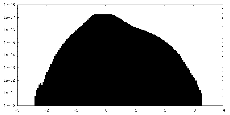
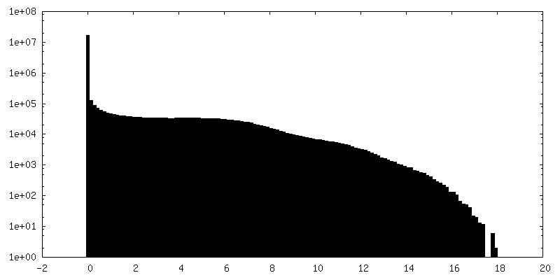
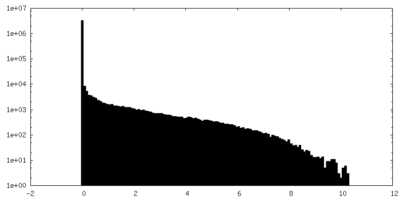
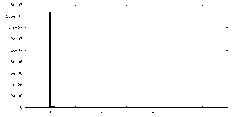
| 最終 再構成 | 想定した対称性 - 点群: I (正20面体型対称) / アルゴリズム: FOURIER SPACE / 解像度のタイプ: BY AUTHOR / 解像度: 8.6 Å / 解像度の算出法: FSC 0.143 CUT-OFF / ソフトウェア - 名称: EMAN / 使用した粒子像数: 9422 |
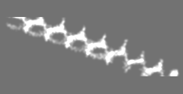
| FSC曲線 (解像度の算出) | 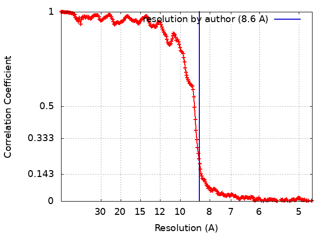 |

