 ムービー
ムービー コントローラー


+ データを開く

- 基本情報
基本情報
| 登録情報 | データベース: EMDB / ID: EMD-1508 | |||||||||
|---|---|---|---|---|---|---|---|---|---|---|
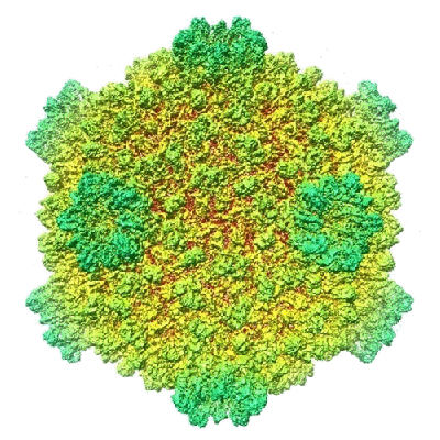
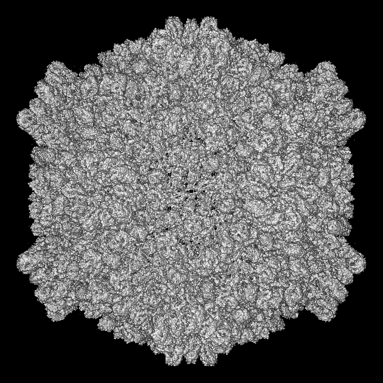
| タイトル | 3.88 Angstrom structure of cytoplasmic polyhedrosis virus by single-particle cryo-electron microscopy | |||||||||
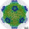
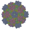
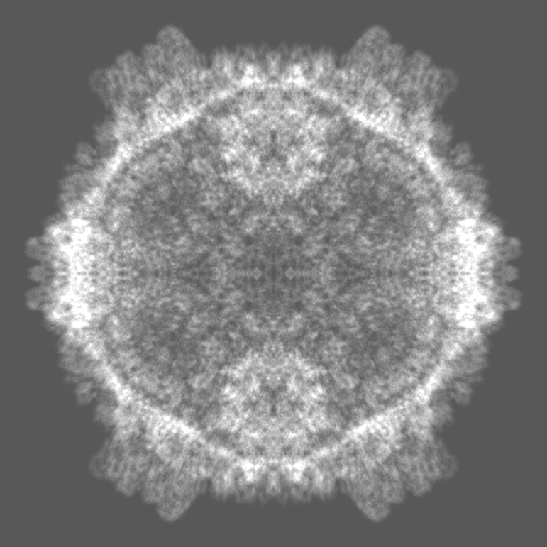
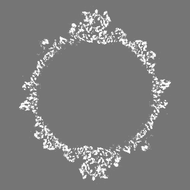
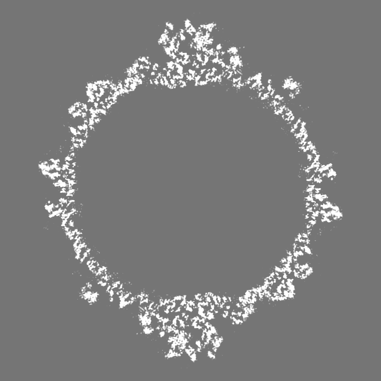
 マップデータ マップデータ | This is the whole cryoEM 2f map for the icosahedral Cytoplasmic Polyhedrosis Virus (CPV). | |||||||||
 試料 試料 |
| |||||||||
 キーワード キーワード |  virus (ウイルス) / strcuture / CPV / cryo-electron microscopy (低温電子顕微鏡法) virus (ウイルス) / strcuture / CPV / cryo-electron microscopy (低温電子顕微鏡法) | |||||||||
| 機能・相同性 | : / : / CPV Capsid shell protein VP1, small protrusion domain / Inner layer core protein VP1-like, C-terminal / T=2 icosahedral viral capsid / viral inner capsid / Capsid protein VP1 / VP3 機能・相同性情報 機能・相同性情報 | |||||||||
| 生物種 |   Bombyx mori cypovirus 1 (ウイルス) Bombyx mori cypovirus 1 (ウイルス) | |||||||||
| 手法 | 単粒子再構成法 / クライオ電子顕微鏡法 / 解像度: 3.88 Å | |||||||||
 データ登録者 データ登録者 | YU X / Jin L / Zhou ZH | |||||||||
 引用 引用 | ジャーナル: Nature / 年: 2008 タイトル: 3.88 A structure of cytoplasmic polyhedrosis virus by cryo-electron microscopy. 著者: Xuekui Yu / Lei Jin / Z Hong Zhou /  要旨: Cytoplasmic polyhedrosis virus (CPV) is unique within the Reoviridae family in having a turreted single-layer capsid contained within polyhedrin inclusion bodies, yet being fully capable of cell ...Cytoplasmic polyhedrosis virus (CPV) is unique within the Reoviridae family in having a turreted single-layer capsid contained within polyhedrin inclusion bodies, yet being fully capable of cell entry and endogenous RNA transcription. Biochemical data have shown that the amino-terminal 79 residues of the CPV turret protein (TP) is sufficient to bring CPV or engineered proteins into the polyhedrin matrix for micro-encapsulation. Here we report the three-dimensional structure of CPV at 3.88 A resolution using single-particle cryo-electron microscopy. Our map clearly shows the turns and deep grooves of alpha-helices, the strand separation in beta-sheets, and densities for loops and many bulky side chains; thus permitting atomic model-building effort from cryo-electron microscopy maps. We observed a helix-to-beta-hairpin conformational change between the two conformational states of the capsid shell protein in the region directly interacting with genomic RNA. We have also discovered a messenger RNA release hole coupled with the mRNA capping machinery unique to CPV. Furthermore, we have identified the polyhedrin-binding domain, a structure that has potential in nanobiotechnology applications. | |||||||||
| 履歴 |
|
- 構造の表示
構造の表示
| ムービー |
ムービービューア |
|---|---|
| 構造ビューア | EMマップ: SurfViewMolmilJmol/JSmol |
| 添付画像 |
- ダウンロードとリンク
-EMDBアーカイブ
| マップデータ | emd_1508.map.gz | 187.6 MB | EMDBマップデータ形式 | |
|---|---|---|---|---|
| ヘッダ (付随情報) | emd-1508-v30.xmlemd-1508.xml | 11.2 KB 11.2 KB | 表示 表示 | EMDBヘッダ |
| 画像 |  1508.gif 1508.gif | 103.8 KB | ||
| マスクデータ | emd_1508_msk_1.map | 14.9 MB | マスクマップ | |
| アーカイブディレクトリ |  http://ftp.pdbj.org/pub/emdb/structures/EMD-1508ftp://ftp.pdbj.org/pub/emdb/structures/EMD-1508 http://ftp.pdbj.org/pub/emdb/structures/EMD-1508ftp://ftp.pdbj.org/pub/emdb/structures/EMD-1508 | HTTPS FTP |
-関連構造データ


-リンク
| EMDBのページ | EMDB (EBI/PDBe) / EMDataResource |
|---|---|
| 「今月の分子」の関連する項目 |

-マップ
| ファイル | ダウンロード / ファイル: emd_1508.map.gz / 形式: CCP4 / 大きさ: 1.7 GB / タイプ: IMAGE STORED AS FLOATING POINT NUMBER (4 BYTES) | ||||||||||||||||||||||||||||||||||||||||||||||||||||||||||||||||||||
|---|---|---|---|---|---|---|---|---|---|---|---|---|---|---|---|---|---|---|---|---|---|---|---|---|---|---|---|---|---|---|---|---|---|---|---|---|---|---|---|---|---|---|---|---|---|---|---|---|---|---|---|---|---|---|---|---|---|---|---|---|---|---|---|---|---|---|---|---|---|
| 注釈 | This is the whole cryoEM 2f map for the icosahedral Cytoplasmic Polyhedrosis Virus (CPV). | ||||||||||||||||||||||||||||||||||||||||||||||||||||||||||||||||||||
| 投影像・断面図 | 画像のコントロール
画像は Spider により作成 | ||||||||||||||||||||||||||||||||||||||||||||||||||||||||||||||||||||
| ボクセルのサイズ | X=Y=Z: 0.97 Å | ||||||||||||||||||||||||||||||||||||||||||||||||||||||||||||||||||||
| 密度 |
| ||||||||||||||||||||||||||||||||||||||||||||||||||||||||||||||||||||
| 対称性 | 空間群: 1 | ||||||||||||||||||||||||||||||||||||||||||||||||||||||||||||||||||||
| 詳細 | EMDB XML:
CCP4マップ ヘッダ情報:
| ||||||||||||||||||||||||||||||||||||||||||||||||||||||||||||||||||||
 Z (Sec.)
 Y (Row.)
 X (Col.)


-添付データ
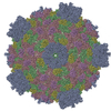
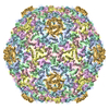
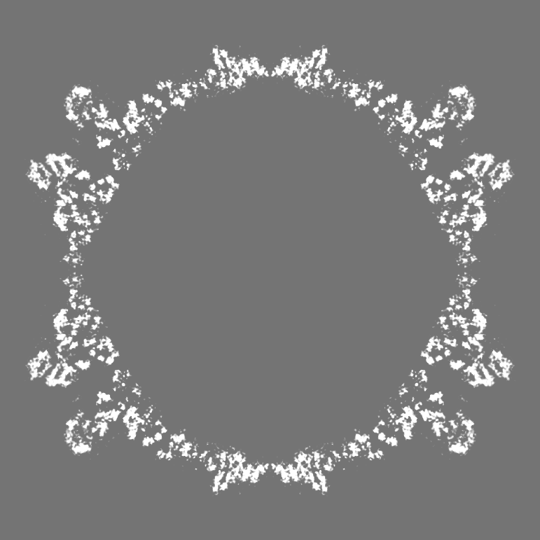
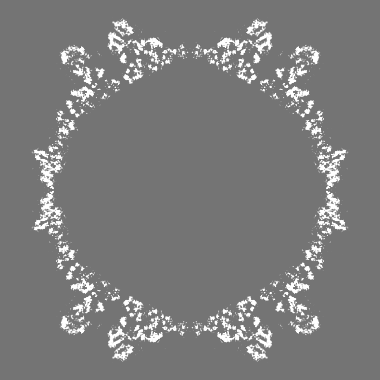
-セグメンテーションマップ: This is a segment of the asymmetric unit
| 注釈 | This is a segment of the asymmetric unit | ||||||||||||
|---|---|---|---|---|---|---|---|---|---|---|---|---|---|
| ファイル | emd_1508_msk_1.map | ||||||||||||
| 投影像・断面図 |
| ||||||||||||
| 密度ヒストグラム |


- 試料の構成要素
-全体 : cytoplasmic polyhedrosis virus
| 全体 | 名称: cytoplasmic polyhedrosis virusCypovirus |
|---|---|
| 要素 |
|
-超分子 #1000: cytoplasmic polyhedrosis virus
| 超分子 | 名称: cytoplasmic polyhedrosis virus / タイプ: sample / ID: 1000 / 詳細: whole infectious virus / 集合状態: icosahedral particle of whole virus / Number unique components: 5 |
|---|
-超分子 #1: Bombyx mori cypovirus 1
| 超分子 | 名称: Bombyx mori cypovirus 1 / タイプ: virus / ID: 1 / Name.synonym: CPV 詳細: CPV is an unenveloped virus wiht a single-shell capsid and diameter of 750 Angstroms. NCBI-ID: 110829 / 生物種: Bombyx mori cypovirus 1 / ウイルスタイプ: VIRION / ウイルス・単離状態: STRAIN / ウイルス・エンベロープ: No / ウイルス・中空状態: No / Syn species name: CPV |
|---|---|
| 宿主 | 生物種:  Bombyx mori (カイコ) / 別称: INVERTEBRATES Bombyx mori (カイコ) / 別称: INVERTEBRATES |
| ウイルス殻 | Shell ID: 1 / 直径: 750 Å / T番号(三角分割数): 1 |
-実験情報
-構造解析
| 手法 | クライオ電子顕微鏡法 |
|---|---|
 解析 解析 | 単粒子再構成法 |
| 試料の集合状態 | particle |
-試料調製
| 緩衝液 | pH: 7.4 / 詳細: 10mM PBS |
|---|---|
| グリッド | 詳細: the holes of holey carbon films |
| 凍結 | 凍結剤: ETHANE / チャンバー内温度: 100 K / 装置: HOMEMADE PLUNGER 詳細: Vitrification instrument: lab-made plunger. Vitrification was carried out at room temperature. CPV were embedded in a thin layer of vitreous ice suspended across the holes of holey carbon ...詳細: Vitrification instrument: lab-made plunger. Vitrification was carried out at room temperature. CPV were embedded in a thin layer of vitreous ice suspended across the holes of holey carbon films for cryoEM imaging. 手法: blot for 3 seconds wiht filter paper before plunging |
- 電子顕微鏡法
| 顕微鏡 | FEI POLARA 300 |
|---|---|
| 電子線 | 加速電圧: 300 kV / 電子線源: FIELD EMISSION GUN |
| 電子光学系 | 倍率(補正後): 154380 / 照射モード: FLOOD BEAM / 撮影モード: BRIGHT FIELDBright-field microscopy / Cs: 2 mm / 最大 デフォーカス(公称値): 1.3 µm / 最小 デフォーカス(公称値): 0.15 µm / 倍率(公称値): 154380 |
| 試料ステージ | 試料ホルダー: Eucentric / 試料ホルダーモデル: GATAN LIQUID NITROGEN |
| 温度 | 平均: 100 K |
| 撮影 | カテゴリ: CCD / フィルム・検出器のモデル: GENERIC TVIPS / 平均電子線量: 20 e/Å2 |
| 実験機器 |  モデル: Tecnai Polara / 画像提供: FEI Company |
-画像解析
| CTF補正 | 詳細: each particle |
|---|---|
| 最終 再構成 | 想定した対称性 - 点群: I (正20面体型対称) / アルゴリズム: OTHER / 解像度のタイプ: BY AUTHOR / 解像度: 3.88 Å / 解像度の算出法: FSC 0.5 CUT-OFF / ソフトウェア - 名称: IMIRS 詳細: Determination of particle orientation and center parameters and subsequent 3D reconstruction were carried out using programs in the IMIRS software package, which are based on Fourier common ...詳細: Determination of particle orientation and center parameters and subsequent 3D reconstruction were carried out using programs in the IMIRS software package, which are based on Fourier common lines and Fourier-Bessel synthesis methods. Prior to the merging of particles for 3D reconstruction, the Fourier transform values of individual images were corrected for the CTF with 15 percent amplitude contrast and a decay factor of 35 sq. Angstroms. 使用した粒子像数: 12814 |
| 詳細 | Focal pairs of micrographs were recorded on 4KX4K charge-coupled device (CCD) camera. |
