 ムービー
ムービー コントローラー


+ データを開く

- 基本情報
基本情報
| 登録情報 | データベース: EMDB / ID: EMD-11030 | |||||||||
|---|---|---|---|---|---|---|---|---|---|---|
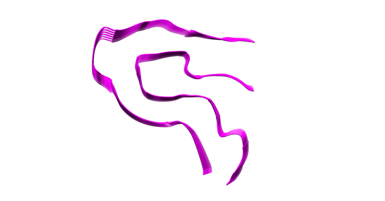
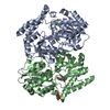
| タイトル | AL amyloid fibril from a lambda 3 light chain in conformation B | |||||||||
 マップデータ マップデータ | EM map of amyloid fibril from a patient-derived lambda 3 immunoglobulin light chain conformation B | |||||||||
 試料 試料 |
| |||||||||
| 機能・相同性 |  機能・相同性情報 機能・相同性情報CD22 mediated BCR regulation / Fc epsilon receptor (FCERI) signaling / Classical antibody-mediated complement activation /  immunoglobulin complex / Initial triggering of complement / FCGR activation / Role of phospholipids in phagocytosis / Role of LAT2/NTAL/LAB on calcium mobilization / Scavenging of heme from plasma / antigen binding ...CD22 mediated BCR regulation / Fc epsilon receptor (FCERI) signaling / Classical antibody-mediated complement activation / immunoglobulin complex / Initial triggering of complement / FCGR activation / Role of phospholipids in phagocytosis / Role of LAT2/NTAL/LAB on calcium mobilization / Scavenging of heme from plasma / antigen binding / FCERI mediated Ca+2 mobilization / FCGR3A-mediated IL10 synthesis / Antigen activates B Cell Receptor (BCR) leading to generation of second messengers / Regulation of Complement cascade / Cell surface interactions at the vascular wall / FCGR3A-mediated phagocytosis / FCERI mediated MAPK activation / Regulation of actin dynamics for phagocytic cup formation / FCERI mediated NF-kB activation / Immunoregulatory interactions between a Lymphoid and a non-Lymphoid cell / Potential therapeutics for SARS / 獲得免疫系 / 免疫応答 / extracellular space / extracellular exosome / extracellular region / 細胞膜 immunoglobulin complex / Initial triggering of complement / FCGR activation / Role of phospholipids in phagocytosis / Role of LAT2/NTAL/LAB on calcium mobilization / Scavenging of heme from plasma / antigen binding ...CD22 mediated BCR regulation / Fc epsilon receptor (FCERI) signaling / Classical antibody-mediated complement activation / immunoglobulin complex / Initial triggering of complement / FCGR activation / Role of phospholipids in phagocytosis / Role of LAT2/NTAL/LAB on calcium mobilization / Scavenging of heme from plasma / antigen binding / FCERI mediated Ca+2 mobilization / FCGR3A-mediated IL10 synthesis / Antigen activates B Cell Receptor (BCR) leading to generation of second messengers / Regulation of Complement cascade / Cell surface interactions at the vascular wall / FCGR3A-mediated phagocytosis / FCERI mediated MAPK activation / Regulation of actin dynamics for phagocytic cup formation / FCERI mediated NF-kB activation / Immunoregulatory interactions between a Lymphoid and a non-Lymphoid cell / Potential therapeutics for SARS / 獲得免疫系 / 免疫応答 / extracellular space / extracellular exosome / extracellular region / 細胞膜類似検索 - 分子機能 | |||||||||
| 生物種 |  Homo sapiens (ヒト) / Human (ヒト) Homo sapiens (ヒト) / Human (ヒト) | |||||||||
| 手法 | らせん対称体再構成法 / クライオ電子顕微鏡法 / 解像度: 3.4 Å | |||||||||
 データ登録者 データ登録者 | Radamaker L / Fandrich M | |||||||||
| 資金援助 |  ドイツ, 1件 ドイツ, 1件
| |||||||||
 引用 引用 | ジャーナル: Nat Commun / 年: 2021 タイトル: Cryo-EM reveals structural breaks in a patient-derived amyloid fibril from systemic AL amyloidosis. 著者: Lynn Radamaker / Julian Baur / Stefanie Huhn / Christian Haupt / Ute Hegenbart / Stefan Schönland / Akanksha Bansal / Matthias Schmidt / Marcus Fändrich / 要旨: Systemic AL amyloidosis is a debilitating and potentially fatal disease that arises from the misfolding and fibrillation of immunoglobulin light chains (LCs). The disease is patient-specific with ...Systemic AL amyloidosis is a debilitating and potentially fatal disease that arises from the misfolding and fibrillation of immunoglobulin light chains (LCs). The disease is patient-specific with essentially each patient possessing a unique LC sequence. In this study, we present two ex vivo fibril structures of a λ3 LC. The fibrils were extracted from the explanted heart of a patient (FOR005) and consist of 115-residue fibril proteins, mainly from the LC variable domain. The fibril structures imply that a 180° rotation around the disulfide bond and a major unfolding step are necessary for fibrils to form. The two fibril structures show highly similar fibril protein folds, differing in only a 12-residue segment. Remarkably, the two structures do not represent separate fibril morphologies, as they can co-exist at different z-axial positions within the same fibril. Our data imply the presence of structural breaks at the interface of the two structural forms. | |||||||||
| 履歴 |
|
- 構造の表示
構造の表示
| ムービー |
ムービービューア |
|---|---|
| 構造ビューア | EMマップ: SurfViewMolmilJmol/JSmol |
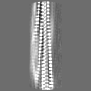
| 添付画像 |
- ダウンロードとリンク
-EMDBアーカイブ
| マップデータ | emd_11030.map.gz | 91.9 MB | EMDBマップデータ形式 | |
|---|---|---|---|---|
| ヘッダ (付随情報) | emd-11030-v30.xmlemd-11030.xml | 17.8 KB 17.8 KB | 表示 表示 | EMDBヘッダ |
| FSC (解像度算出) | emd_11030_fsc.xml | 10.7 KB | 表示 | FSCデータファイル |
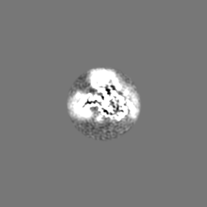
| 画像 |  emd_11030.png emd_11030.png | 50.4 KB | ||
| その他 | emd_11030_half_map_1.map.gzemd_11030_half_map_2.map.gz | 10.3 MB 10.2 MB | ||
| アーカイブディレクトリ |  http://ftp.pdbj.org/pub/emdb/structures/EMD-11030ftp://ftp.pdbj.org/pub/emdb/structures/EMD-11030 http://ftp.pdbj.org/pub/emdb/structures/EMD-11030ftp://ftp.pdbj.org/pub/emdb/structures/EMD-11030 | HTTPS FTP |
-関連構造データ
| 関連構造データ |  6z1iMC  6z1oC M: このマップから作成された原子モデル C: 同じ文献を引用 ( |
|---|---|
| 類似構造データ | |
| 電子顕微鏡画像生データ | EMPIAR-10457 (タイトル: AL amyloid fibril from a lambda 3 light chain / Data size: 297.8 Data #1: Raw cryo-EM movies of AL fibrils extracted from human heart tissue [micrographs - multiframe]) |


-リンク
| EMDBのページ | EMDB (EBI/PDBe) / EMDataResource |
|---|---|
| 「今月の分子」の関連する項目 |


-マップ
| ファイル | ダウンロード / ファイル: emd_11030.map.gz / 形式: CCP4 / 大きさ: 103 MB / タイプ: IMAGE STORED AS FLOATING POINT NUMBER (4 BYTES) | ||||||||||||||||||||||||||||||||||||||||||||||||||||||||||||||||||||
|---|---|---|---|---|---|---|---|---|---|---|---|---|---|---|---|---|---|---|---|---|---|---|---|---|---|---|---|---|---|---|---|---|---|---|---|---|---|---|---|---|---|---|---|---|---|---|---|---|---|---|---|---|---|---|---|---|---|---|---|---|---|---|---|---|---|---|---|---|---|
| 注釈 | EM map of amyloid fibril from a patient-derived lambda 3 immunoglobulin light chain conformation B | ||||||||||||||||||||||||||||||||||||||||||||||||||||||||||||||||||||
| ボクセルのサイズ | X=Y=Z: 1.04 Å | ||||||||||||||||||||||||||||||||||||||||||||||||||||||||||||||||||||
| 密度 |
| ||||||||||||||||||||||||||||||||||||||||||||||||||||||||||||||||||||
| 対称性 | 空間群: 1 | ||||||||||||||||||||||||||||||||||||||||||||||||||||||||||||||||||||
| 詳細 | EMDB XML:
CCP4マップ ヘッダ情報:
| ||||||||||||||||||||||||||||||||||||||||||||||||||||||||||||||||||||
-添付データ
-ハーフマップ: #2
| ファイル | emd_11030_half_map_1.map | ||||||||||||
|---|---|---|---|---|---|---|---|---|---|---|---|---|---|
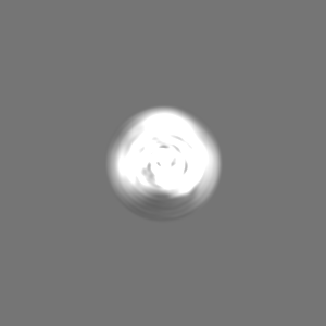
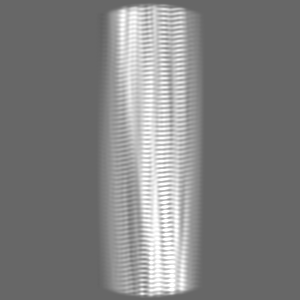
| 投影像・断面図 |
| ||||||||||||
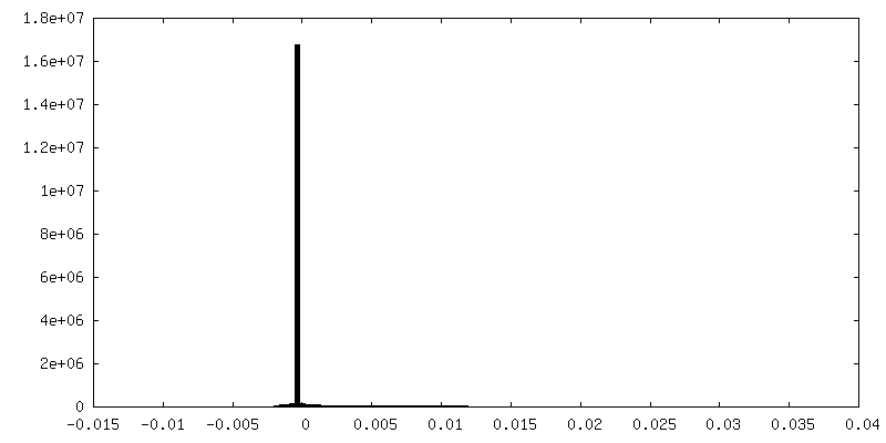
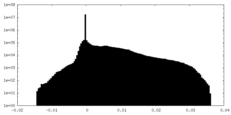
| 密度ヒストグラム |
 Z
Z Y
Y X
X


-ハーフマップ: #1
| ファイル | emd_11030_half_map_2.map | ||||||||||||
|---|---|---|---|---|---|---|---|---|---|---|---|---|---|
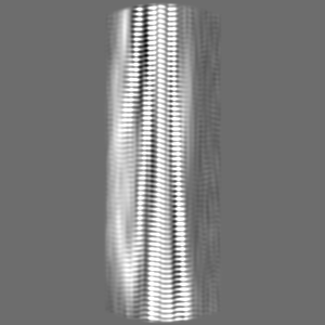
| 投影像・断面図 |
| ||||||||||||
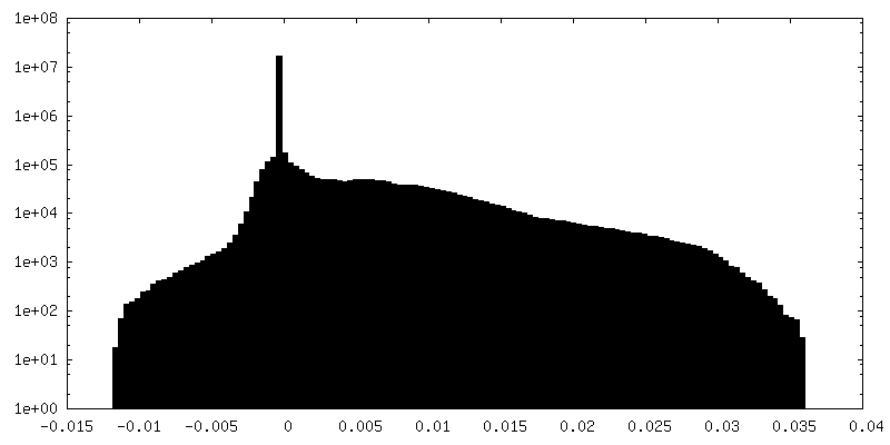
| 密度ヒストグラム |


- 試料の構成要素
-全体 : Amyloid fibril of an antibody lambda 3 immunoglobulin light chain
| 全体 | 名称: Amyloid fibril of an antibody lambda 3 immunoglobulin light chain |
|---|---|
| 要素 |
|
-超分子 #1: Amyloid fibril of an antibody lambda 3 immunoglobulin light chain
| 超分子 | 名称: Amyloid fibril of an antibody lambda 3 immunoglobulin light chain タイプ: complex / ID: 1 / 親要素: 0 / 含まれる分子: all 詳細: Extracted fibrils from the explanted heart of a systemic AL amyloidosis patient |
|---|---|
| 由来(天然) | 生物種: Homo sapiens (ヒト) / 器官: Heart / 組織: Heart muscle |
-分子 #1: lambda 3 light chain fragment, residues 2-116
| 分子 | 名称: lambda 3 light chain fragment, residues 2-116 / タイプ: protein_or_peptide / ID: 1 / コピー数: 6 / 光学異性体: LEVO |
|---|---|
| 由来(天然) | 生物種: Human (ヒト) / 器官: Heart / 組織: heart muscle |
| 分子量 | 理論値: 8.664521 KDa |
| 配列 | 文字列: AVSVALGQTV RITCQGDSLR SYSASWYQQK PGQAPVLVIF RNTASLTITG AQAEDEADYY CNSRDSSANH QVFGGGTKLT V |
-実験情報
-構造解析
| 手法 | クライオ電子顕微鏡法 |
|---|---|
 解析 解析 | らせん対称体再構成法 |
| 試料の集合状態 | filament |
-試料調製
| 緩衝液 | pH: 7 / 構成要素 - 式: H2O / 構成要素 - 名称: Distilled water |
|---|---|
| グリッド | モデル: C-flat-1.2/1.3 / 材質: COPPER / メッシュ: 400 / 支持フィルム - 材質: CARBON / 支持フィルム - トポロジー: HOLEY / 前処理 - タイプ: GLOW DISCHARGE / 前処理 - 雰囲気: OTHER / 詳細: 40 mA |
| 凍結 | 凍結剤: ETHANE / チャンバー内湿度: 95 % / チャンバー内温度: 295 K / 装置: FEI VITROBOT MARK III / 詳細: blot for 9s before plunging. |
| 詳細 | Sample in pure water, pH not determined |
- 電子顕微鏡法
| 顕微鏡 | FEI TITAN KRIOS |
|---|---|
| 電子線 | 加速電圧: 300 kV / 電子線源: FIELD EMISSION GUN |
| 電子光学系 | 照射モード: FLOOD BEAM / 撮影モード: BRIGHT FIELDBright-field microscopy / Cs: 2.7 mm |
| 特殊光学系 | エネルギーフィルター - スリット幅: 20 eV |
| 試料ステージ | 試料ホルダーモデル: FEI TITAN KRIOS AUTOGRID HOLDER ホルダー冷却材: NITROGEN |
| 撮影 | フィルム・検出器のモデル: GATAN K2 SUMMIT (4k x 4k) 検出モード: COUNTING / デジタル化 - サイズ - 横: 3838 pixel / デジタル化 - サイズ - 縦: 3710 pixel / 撮影したグリッド数: 1 / 実像数: 1964 / 平均電子線量: 40.0 e/Å2 |
| 実験機器 |  モデル: Titan Krios / 画像提供: FEI Company |
-画像解析
| Segment selection | 選択した数: 194502 / ソフトウェア - 名称: RELION (ver. 2.1.0) 詳細: manual particle picking helical start-end coordinates |
|---|---|
| CTF補正 | ソフトウェア - 名称: Gctf (ver. 1.06) 詳細: CTF was estimated from the non-dose-weighted micrographs |
| 初期モデル | モデルのタイプ: NONE 詳細: Initial model generation in RELION, followed by a 3D classification of particles picked from only 9 micrographs to generate rough 3D model which was used as a reference |
| 最終 角度割当 | タイプ: NOT APPLICABLE |
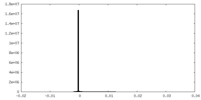
| 最終 再構成 | 使用したクラス数: 1 想定した対称性 - らせんパラメータ - Δz: 4.8 Å 想定した対称性 - らせんパラメータ - ΔΦ: -1.1 ° 想定した対称性 - らせんパラメータ - 軸対称性: C1 (非対称) アルゴリズム: FOURIER SPACE / 解像度のタイプ: BY AUTHOR / 解像度: 3.4 Å / 解像度の算出法: FSC 0.143 CUT-OFF / ソフトウェア - 名称: RELION (ver. 2.1.0) / 使用した粒子像数: 12122 |
| 詳細 | Motion-corrected and dose-weighted movie frames |
| FSC曲線 (解像度の算出) | 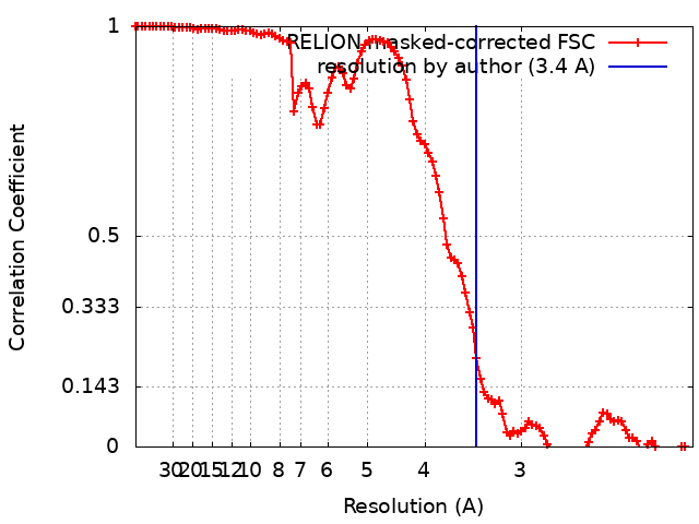 |
-原子モデル構築 1
| 詳細 | Secondary structure restraints and NCS were applied during refinement |
|---|---|
| 精密化 | 空間: REAL / プロトコル: OTHER / 温度因子: 73.24 当てはまり具合の基準: REAL-SPACE (WEIGHTED MAP SUM AT ATOM CENTERS) |
| 得られたモデル | PDB-6z1i: |
