 ムービー
ムービー コントローラー


+ データを開く

- 基本情報
基本情報
| 登録情報 | データベース: EMDB / ID: EMD-10064 | |||||||||
|---|---|---|---|---|---|---|---|---|---|---|
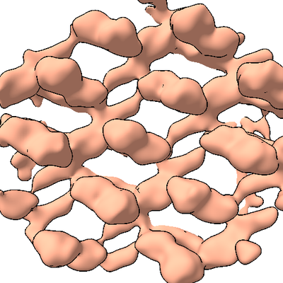
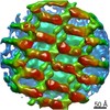
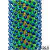
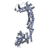
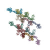
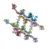
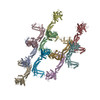
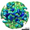
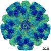
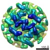
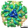
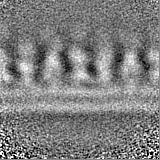
| タイトル | Structure of s-Mgm1 decorating the inner surface of tubulated lipid membranes | |||||||||
 マップデータ マップデータ | ||||||||||
 試料 試料 |
| |||||||||
| 機能・相同性 |  機能・相同性情報 機能・相同性情報 | |||||||||
| 生物種 |  Chaetomium thermophilum var. thermophilum DSM 1495 (菌類) Chaetomium thermophilum var. thermophilum DSM 1495 (菌類) | |||||||||
| 手法 | サブトモグラム平均法 /  クライオ電子顕微鏡法 / 解像度: 20.6 Å クライオ電子顕微鏡法 / 解像度: 20.6 Å | |||||||||
 データ登録者 データ登録者 | Faelber K / Dietrich L / Noel JK / Sanchez R / Kudryashev M / Kuelbrandt W / Daumke O | |||||||||
| 資金援助 |  ドイツ, 1件 ドイツ, 1件
| |||||||||
 引用 引用 | ジャーナル: Nature / 年: 2019 タイトル: Structure and assembly of the mitochondrial membrane remodelling GTPase Mgm1. 著者: Katja Faelber / Lea Dietrich / Jeffrey K Noel / Florian Wollweber / Anna-Katharina Pfitzner / Alexander Mühleip / Ricardo Sánchez / Misha Kudryashev / Nicolas Chiaruttini / Hauke Lilie / ...著者: Katja Faelber / Lea Dietrich / Jeffrey K Noel / Florian Wollweber / Anna-Katharina Pfitzner / Alexander Mühleip / Ricardo Sánchez / Misha Kudryashev / Nicolas Chiaruttini / Hauke Lilie / Jeanette Schlegel / Eva Rosenbaum / Manuel Hessenberger / Claudia Matthaeus / Séverine Kunz / Alexander von der Malsburg / Frank Noé / Aurélien Roux / Martin van der Laan / Werner Kühlbrandt / Oliver Daumke /  要旨: Balanced fusion and fission are key for the proper function and physiology of mitochondria. Remodelling of the mitochondrial inner membrane is mediated by the dynamin-like protein mitochondrial ...Balanced fusion and fission are key for the proper function and physiology of mitochondria. Remodelling of the mitochondrial inner membrane is mediated by the dynamin-like protein mitochondrial genome maintenance 1 (Mgm1) in fungi or the related protein optic atrophy 1 (OPA1) in animals. Mgm1 is required for the preservation of mitochondrial DNA in yeast, whereas mutations in the OPA1 gene in humans are a common cause of autosomal dominant optic atrophy-a genetic disorder that affects the optic nerve. Mgm1 and OPA1 are present in mitochondria as a membrane-integral long form and a short form that is soluble in the intermembrane space. Yeast strains that express temperature-sensitive mutants of Mgm1 or mammalian cells that lack OPA1 display fragmented mitochondria, which suggests that Mgm1 and OPA1 have an important role in inner-membrane fusion. Consistently, only the mitochondrial outer membrane-not the inner membrane-fuses in the absence of functional Mgm1. Mgm1 and OPA1 have also been shown to maintain proper cristae architecture; for example, OPA1 prevents the release of pro-apoptotic factors by tightening crista junctions. Finally, the short form of OPA1 localizes to mitochondrial constriction sites, where it presumably promotes mitochondrial fission. How Mgm1 and OPA1 perform their diverse functions in membrane fusion, scission and cristae organization is at present unknown. Here we present crystal and electron cryo-tomography structures of Mgm1 from Chaetomium thermophilum. Mgm1 consists of a GTPase (G) domain, a bundle signalling element domain, a stalk, and a paddle domain that contains a membrane-binding site. Biochemical and cell-based experiments demonstrate that the Mgm1 stalk mediates the assembly of bent tetramers into helical filaments. Electron cryo-tomography studies of Mgm1-decorated lipid tubes and fluorescence microscopy experiments on reconstituted membrane tubes indicate how the tetramers assemble on positively or negatively curved membranes. Our findings convey how Mgm1 and OPA1 filaments dynamically remodel the mitochondrial inner membrane. | |||||||||
| 履歴 |
|
- 構造の表示
構造の表示
| ムービー |
ムービービューア |
|---|---|
| 構造ビューア | EMマップ: SurfViewMolmilJmol/JSmol |
| 添付画像 |
- ダウンロードとリンク
-EMDBアーカイブ
| マップデータ | emd_10064.map.gz | 4.1 MB | EMDBマップデータ形式 | |
|---|---|---|---|---|
| ヘッダ (付随情報) | emd-10064-v30.xmlemd-10064.xml | 19.8 KB 19.8 KB | 表示 表示 | EMDBヘッダ |
| FSC (解像度算出) | emd_10064_fsc.xml | 5.8 KB | 表示 | FSCデータファイル |
| 画像 |  emd_10064.png emd_10064.png | 161.5 KB | ||
| マスクデータ | emd_10064_msk_1.map | 15.6 MB | マスクマップ | |
| その他 | emd_10064_half_map_1.map.gzemd_10064_half_map_2.map.gz | 14.5 MB 14.5 MB | ||
| アーカイブディレクトリ |  http://ftp.pdbj.org/pub/emdb/structures/EMD-10064ftp://ftp.pdbj.org/pub/emdb/structures/EMD-10064 http://ftp.pdbj.org/pub/emdb/structures/EMD-10064ftp://ftp.pdbj.org/pub/emdb/structures/EMD-10064 | HTTPS FTP |
-関連構造データ


-リンク
| EMDBのページ | EMDB (EBI/PDBe) / EMDataResource |
|---|
-マップ
| ファイル | ダウンロード / ファイル: emd_10064.map.gz / 形式: CCP4 / 大きさ: 15.6 MB / タイプ: IMAGE STORED AS FLOATING POINT NUMBER (4 BYTES) | ||||||||||||||||||||||||||||||||||||||||||||||||||||||||||||
|---|---|---|---|---|---|---|---|---|---|---|---|---|---|---|---|---|---|---|---|---|---|---|---|---|---|---|---|---|---|---|---|---|---|---|---|---|---|---|---|---|---|---|---|---|---|---|---|---|---|---|---|---|---|---|---|---|---|---|---|---|---|
| ボクセルのサイズ | X=Y=Z: 2.7 Å | ||||||||||||||||||||||||||||||||||||||||||||||||||||||||||||
| 密度 |
| ||||||||||||||||||||||||||||||||||||||||||||||||||||||||||||
| 対称性 | 空間群: 1 | ||||||||||||||||||||||||||||||||||||||||||||||||||||||||||||
| 詳細 | EMDB XML:
CCP4マップ ヘッダ情報:
| ||||||||||||||||||||||||||||||||||||||||||||||||||||||||||||
-添付データ
-マスク #1
| ファイル | emd_10064_msk_1.map | ||||||||||||
|---|---|---|---|---|---|---|---|---|---|---|---|---|---|
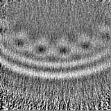
| 投影像・断面図 |
| ||||||||||||
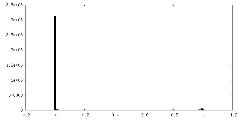
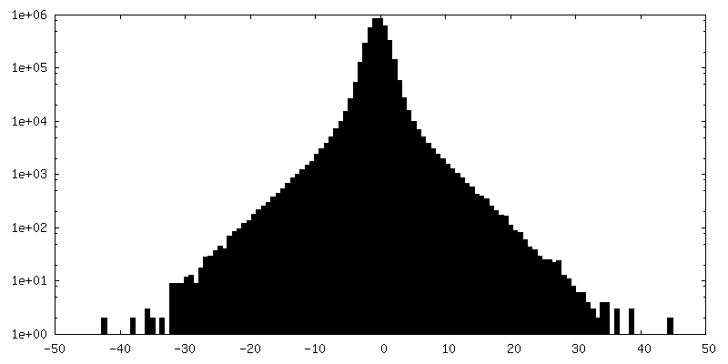
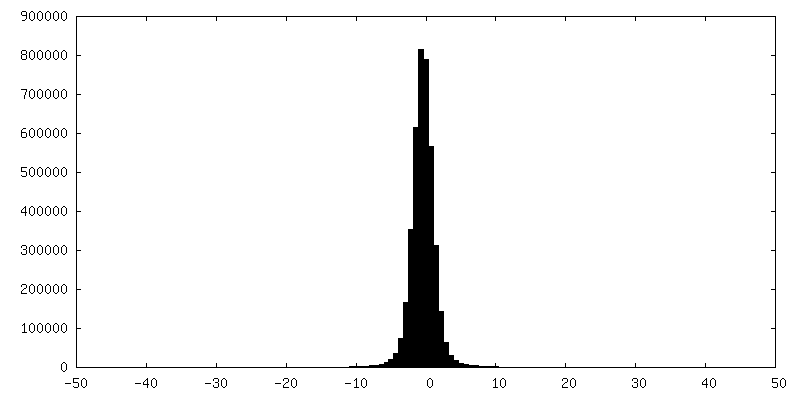
| 密度ヒストグラム |
 Z
Z Y
Y X
X


-ハーフマップ: Unfiltered non-masked half-map #1
| ファイル | emd_10064_half_map_1.map | ||||||||||||
|---|---|---|---|---|---|---|---|---|---|---|---|---|---|
| 注釈 | Unfiltered non-masked half-map #1 | ||||||||||||
| 投影像・断面図 |
| ||||||||||||
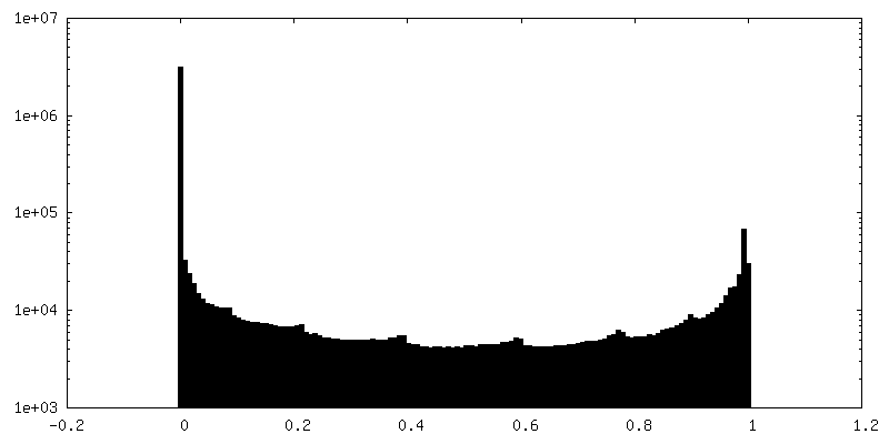
| 密度ヒストグラム |


-ハーフマップ: Unfiltered non-masked half-map #2
| ファイル | emd_10064_half_map_2.map | ||||||||||||
|---|---|---|---|---|---|---|---|---|---|---|---|---|---|
| 注釈 | Unfiltered non-masked half-map #2 | ||||||||||||
| 投影像・断面図 |
| ||||||||||||
| 密度ヒストグラム |

- 試料の構成要素
-全体 : Mgm1
| 全体 | 名称: Mgm1 |
|---|---|
| 要素 |
|
-超分子 #1: Mgm1
| 超分子 | 名称: Mgm1 / タイプ: complex / ID: 1 / 親要素: 0 / 含まれる分子: all / 詳細: short isoform with C- and N-terminal truncations |
|---|---|
| 由来(天然) | 生物種: Chaetomium thermophilum var. thermophilum DSM 1495 (菌類) |
| 組換発現 | 生物種:  Escherichia coli (大腸菌) Escherichia coli (大腸菌) |
-分子 #1: Putative mitochondrial dynamin protein
| 分子 | 名称: Putative mitochondrial dynamin protein / タイプ: protein_or_peptide / ID: 1 / コピー数: 16 / 光学異性体: LEVO |
|---|---|
| 由来(天然) | 生物種: Chaetomium thermophilum var. thermophilum DSM 1495 (菌類) |
| 分子量 | 理論値: 77.360172 KDa |
| 組換発現 | 生物種: Escherichia coli (大腸菌) |
| 配列 | 文字列: EEIMRDDNMM FITKKMIEIR NLLQKVGQGS TVTLPSIVVI GSQSSGKSSV LEAIVGHEFL PKGSNMITRR PIELTLVNDP EAKVDYGEF PDLGLARVTD FSLIQKTLTE LNQSVPESEC VTDDPIRLTI HSPNIPDLSL IDLPGYIQVA GENQPRELKR K ITELCDKY ...文字列: EEIMRDDNMM FITKKMIEIR NLLQKVGQGS TVTLPSIVVI GSQSSGKSSV LEAIVGHEFL PKGSNMITRR PIELTLVNDP EAKVDYGEF PDLGLARVTD FSLIQKTLTE LNQSVPESEC VTDDPIRLTI HSPNIPDLSL IDLPGYIQVA GENQPRELKR K ITELCDKY IRGPNIILAI SAADTDLANS TALQASRRVD PRGERTIGVI TKMDLVEPEK GAAILSDRQY PLKLGYVGVI SK LPPQSGL FRRDTGNLLA SINRNEKNYF GSHPTEFGPD SGVSTGVMTL RKKLLQVLEQ QMSSKLNETT EAIQRELEET TYQ FKVQYN EQPMSAESYL AASLDDFKHQ FHEFASSFGR PQLQTLLKDA LDQKVLDQLA ARYWNRPIED LSPAPREPDN IIDL PKADP DSPYWHRQLD TACSGLTRLG VGRLAATVAA SAIQQHVEKL LDKSSFAKHP SARKVISDAA ATVLADRSYA TSDGI EISL KPYKFDPDIQ PNEWAQGREH VVGVLQAELE QCQAAMKALE NSVGGRKKLK EVMSFVDKAR KGEIIVEGDH PSGAGG FSA ALLARGREAV FLRDRADILS LRIQAAKSRQ CKTLTNKYYC PEVFLDAVAT KLAQTAVLFL NVEMLNDFYV RFPREVE AK LHEHMHAGGG LEKFAREDPK VRRHLDLIRR KELLETVLGK IEELHRISSG TAG |
-実験情報
-構造解析
| 手法 | クライオ電子顕微鏡法 |
|---|---|
 解析 解析 | サブトモグラム平均法 |
| 試料の集合状態 | particle |
-試料調製
| 濃度 | 5 mg/mL |
|---|---|
| 緩衝液 | pH: 7.4 / 詳細: 20 mM HEPES, 200 mM NaCl, residual MgCl2, 9mM KCl. |
| 凍結 | 凍結剤: ETHANE / チャンバー内湿度: 70 % / チャンバー内温度: 277.15 K / 装置: FEI VITROBOT MARK IV |
| 詳細 | The sample was prepared in the described buffer. Just before freezing 6 nm colloidal gold fiducial marker were added in a 1:1 ratio |
- 電子顕微鏡法
| 顕微鏡 | FEI TITAN KRIOS |
|---|---|
| 電子線 | 加速電圧: 300 kV / 電子線源: FIELD EMISSION GUN |
| 電子光学系 | C2レンズ絞り径: 70.0 µm / 照射モード: FLOOD BEAM / 撮影モード: BRIGHT FIELDBright-field microscopy / Cs: 2.7 mm / 最大 デフォーカス(公称値): 4.0 µm / 最小 デフォーカス(公称値): 2.0 µm / 倍率(公称値): 53000 |
| 特殊光学系 | エネルギーフィルター - スリット幅: 20 eV |
| 試料ステージ | 試料ホルダーモデル: FEI TITAN KRIOS AUTOGRID HOLDER ホルダー冷却材: NITROGEN |
| 撮影 | フィルム・検出器のモデル: GATAN K2 SUMMIT (4k x 4k) 検出モード: COUNTING / 平均電子線量: 2.0 e/Å2 |
| 実験機器 |  モデル: Titan Krios / 画像提供: FEI Company |
-画像解析
| 抽出 | トモグラム数: 1 / 使用した粒子像数: 1874 参照モデル: global average of the particles rotated to the known initial orientations 手法: Geometry-assisted particle picking form tube surfaces ソフトウェア - 名称: Dynamo (ver. 1.1.266) / 詳細: with the use of Dynamo Catalogue system | ||||||
|---|---|---|---|---|---|---|---|
| CTF補正 | ソフトウェア:
詳細: CTF determination was done by Gctf and correction was performed by ctfphaseflip from Imod | ||||||
| 最終 3次元分類 | クラス数: 1 / 平均メンバー数/クラス: 1792 / ソフトウェア - 名称: Dynamo (ver. 1.3) | ||||||
| 最終 角度割当 | タイプ: OTHER / ソフトウェア - 名称: Dynamo (ver. 1.1.266) ソフトウェア - 詳細: independent half-set refinement 詳細: subtomogram averaging by cross correlation maximization | ||||||
| 最終 再構成 | 使用したクラス数: 1 / 想定した対称性 - 点群: C1 (非対称) / アルゴリズム: BACK PROJECTION / 解像度のタイプ: BY AUTHOR / 解像度: 20.6 Å / 解像度の算出法: FSC 0.143 CUT-OFF / ソフトウェア - 名称: Dynamo (ver. 1.1.266) 詳細: Half sets were generated not even-odd but as upper and lower-halves of particles in given tomogram 使用したサブトモグラム数: 1792 | ||||||
| FSC曲線 (解像度の算出) | 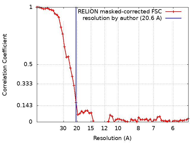 |
