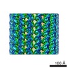
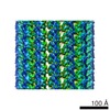
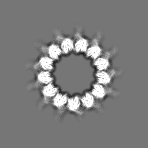
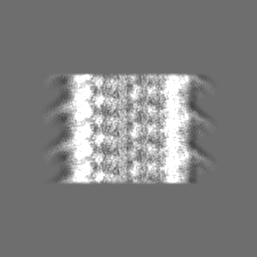
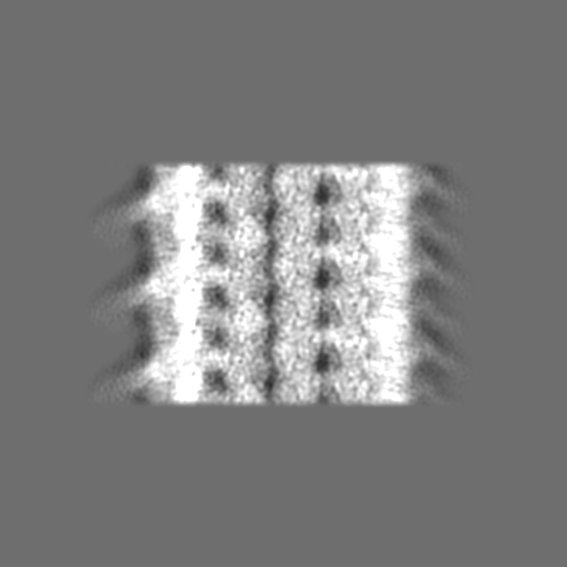
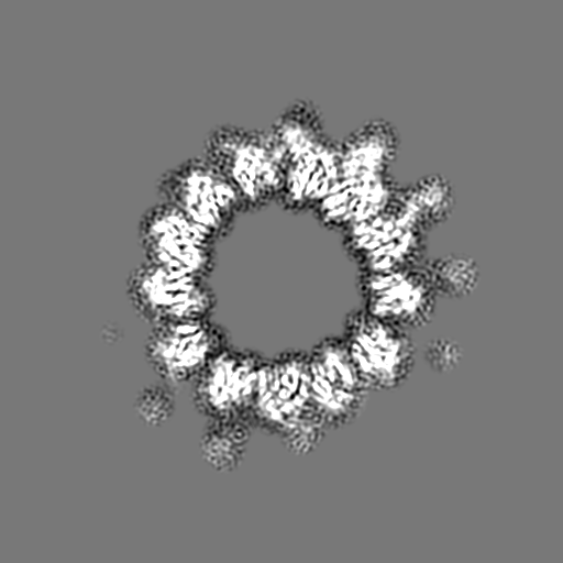
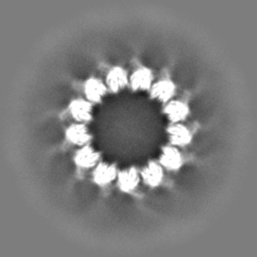
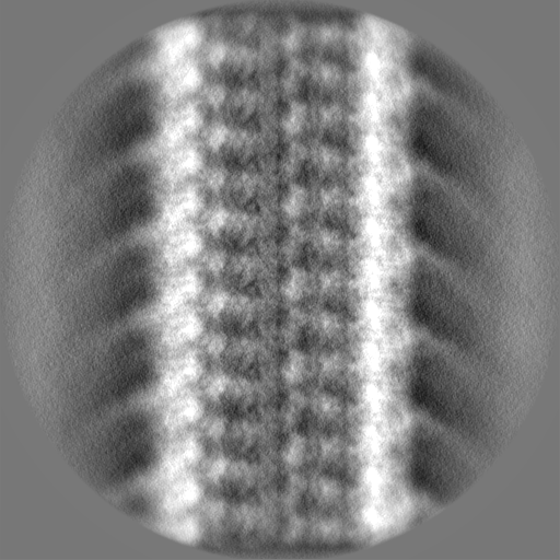
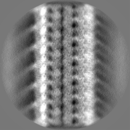
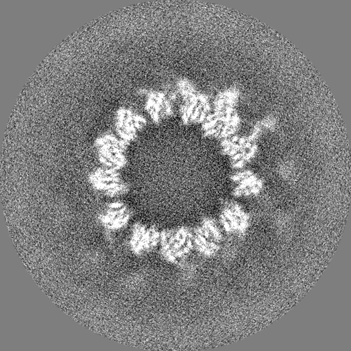
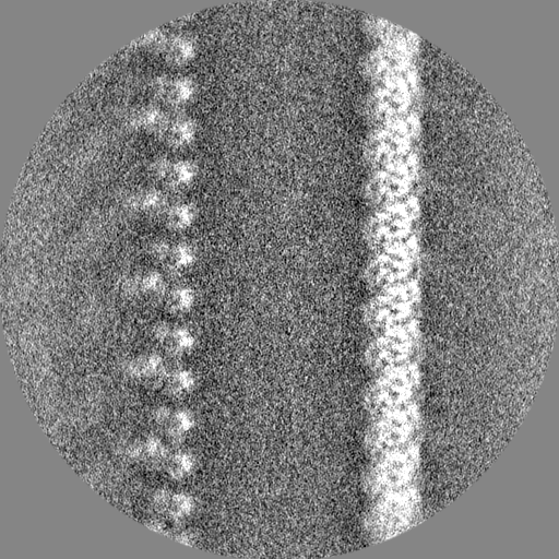
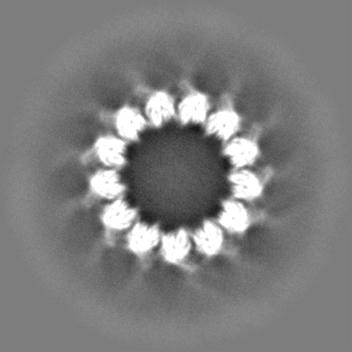
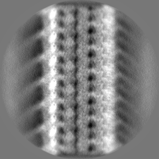
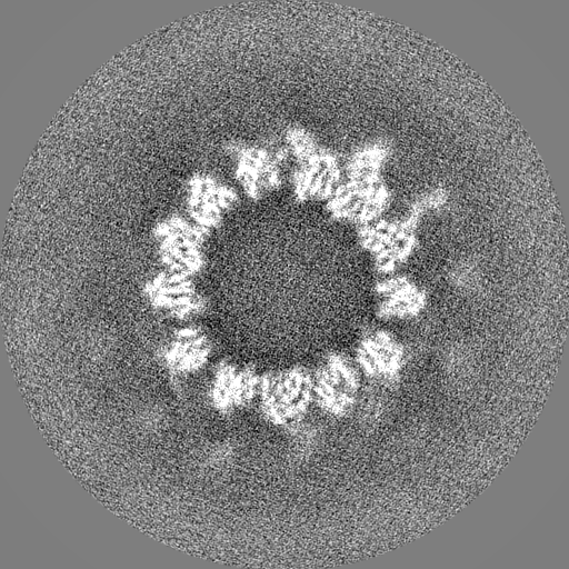
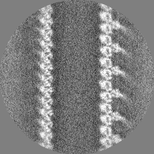
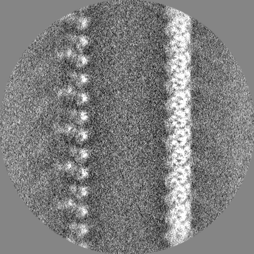
- EMDB-10060: Cryo-EM structure of the human inner arm dynein DNAH7 microtubule... -
+
データを開く
IDまたはキーワード:
読み込み中...
-
基本情報
登録情報
データベース: EMDB / ID: EMD-10060
タイトル
Cryo-EM structure of the human inner arm dynein DNAH7 microtubule binding domain bound to microtubules
マップデータ
None
試料
複合体: DNAH7 microtubule binding domain bound to microtubules
複合体: DNAH7
タンパク質・ペプチド: Cytoplasmic dynein 1 heavy chain 1,Dynein heavy chain 7, axonemal,Cytoplasmic dynein 1 heavy chain 1
複合体: tubulinチューブリン
タンパク質・ペプチド: Tubulin alpha-1B chain
タンパク質・ペプチド: Tubulin beta chain
リガンド: GUANOSINE-5'-TRIPHOSPHATEグアノシン三リン酸
リガンド: MAGNESIUM ION
リガンド: GUANOSINE-5'-DIPHOSPHATE
リガンド: TAXOLパクリタキセル
機能・相同性
機能・相同性情報
inner dynein arm / axonemal dynein complex / inner dynein arm assembly / cilium-dependent cell motility / COPI-independent Golgi-to-ER retrograde traffic / HSP90 chaperone cycle for steroid hormone receptors (SHR) in the presence of ligand / Amplification of signal from unattached kinetochores via a MAD2 inhibitory signal / cilium movement involved in cell motility / 9+2 motile cilium / COPI-mediated anterograde transport ...inner dynein arm / axonemal dynein complex / inner dynein arm assembly / cilium-dependent cell motility / COPI-independent Golgi-to-ER retrograde traffic / HSP90 chaperone cycle for steroid hormone receptors (SHR) in the presence of ligand / Amplification of signal from unattached kinetochores via a MAD2 inhibitory signal / cilium movement involved in cell motility / 9+2 motile cilium / COPI-mediated anterograde transport / cilium movement / Aggrephagy / positive regulation of intracellular transport / Mitotic Prometaphase / EML4 and NUDC in mitotic spindle formation / Resolution of Sister Chromatid Cohesion / regulation of metaphase plate congression / establishment of spindle localization / RHO GTPases Activate Formins / positive regulation of spindle assembly / Separation of Sister Chromatids / Loss of Nlp from mitotic centrosomes / Recruitment of mitotic centrosome proteins and complexes / Loss of proteins required for interphase microtubule organization from the centrosome / Recruitment of NuMA to mitotic centrosomes / Anchoring of the basal body to the plasma membrane / AURKA Activation by TPX2 / manchette / Regulation of PLK1 Activity at G2/M Transition / P-body assembly / dynein complex / MHC class II antigen presentation / minus-end-directed microtubule motor activity / cytoplasmic dynein complex / retrograde axonal transport / dynein light intermediate chain binding / Microtubule-dependent trafficking of connexons from Golgi to the plasma membrane / Hedgehog 'off' state / Cilium Assembly / Intraflagellar transport / COPI-dependent Golgi-to-ER retrograde traffic / Carboxyterminal post-translational modifications of tubulin / RHOH GTPase cycle / Sealing of the nuclear envelope (NE) by ESCRT-III / Kinesins / PKR-mediated signaling / Resolution of Sister Chromatid Cohesion / Mitotic Prometaphase / EML4 and NUDC in mitotic spindle formation / Separation of Sister Chromatids / The role of GTSE1 in G2/M progression after G2 checkpoint / Aggrephagy / Recruitment of NuMA to mitotic centrosomes / RHO GTPases activate IQGAPs / RHO GTPases Activate Formins / HSP90 chaperone cycle for steroid hormone receptors (SHR) in the presence of ligand / COPI-independent Golgi-to-ER retrograde traffic / MHC class II antigen presentation / nuclear migration / COPI-mediated anterograde transport / microtubule motor activity / dynein intermediate chain binding / cytoplasmic microtubule / cytoplasmic microtubule organization / stress granule assembly / regulation of mitotic spindle organization / axon cytoplasm / Neutrophil degranulation / mitotic spindle organization / filopodium / 加水分解酵素; 酸無水物に作用; GTPに作用・細胞または細胞小器官の運動に関与 / 繊毛 / structural constituent of cytoskeleton / microtubule cytoskeleton organization / microtubule cytoskeleton / mitotic cell cycle / 核膜 / positive regulation of cold-induced thermogenesis / 細胞皮質 / 微小管 / 細胞分裂 / 神経繊維 / GTPase activity / 中心体 / neuronal cell body / calcium ion binding / GTP binding / ATP hydrolysis activity / ATP binding / metal ion binding / 細胞質基質 / 細胞質 類似検索 - 分子機能
Dynein heavy chain, AAA 5 extension domain / Dynein heavy chain AAA lid domain / Dynein heavy chain 3, AAA+ lid domain / AAA+ lid domain / Dynein heavy chain, C-terminal domain / Dynein heavy chain, C-terminal domain, barrel region / Dynein heavy chain C-terminal domain / P-loop containing dynein motor region / Dynein heavy chain, tail / Dynein heavy chain, N-terminal region 1 ...Dynein heavy chain, AAA 5 extension domain / Dynein heavy chain AAA lid domain / Dynein heavy chain 3, AAA+ lid domain / AAA+ lid domain / Dynein heavy chain, C-terminal domain / Dynein heavy chain, C-terminal domain, barrel region / Dynein heavy chain C-terminal domain / P-loop containing dynein motor region / Dynein heavy chain, tail / Dynein heavy chain, N-terminal region 1 / Dynein heavy chain / Dynein heavy chain region D6 P-loop domain / Dynein heavy chain, linker / Dynein heavy chain, AAA module D4 / Dynein heavy chain, coiled coil stalk / Dynein heavy chain, hydrolytic ATP-binding dynein motor region / Dynein heavy chain, ATP-binding dynein motor region / Dynein heavy chain AAA lid domain / Dynein heavy chain AAA lid domain superfamily / Dynein heavy chain, domain 2, N-terminal / Dynein heavy chain, linker, subdomain 3 / Dynein heavy chain, AAA1 domain, small subdomain / Dynein heavy chain region D6 P-loop domain / Dynein heavy chain, N-terminal region 2 / Hydrolytic ATP binding site of dynein motor region / Microtubule-binding stalk of dynein motor / P-loop containing dynein motor region D4 / ATP-binding dynein motor region / Dynein heavy chain AAA lid domain / Tubulin-beta mRNA autoregulation signal. / Alpha tubulin / Beta tubulin, autoregulation binding site / Beta tubulin / チューブリン / Tubulin, C-terminal / Tubulin C-terminal domain / Tubulin, conserved site / Tubulin subunits alpha, beta, and gamma signature. / Tubulin/FtsZ family, C-terminal domain / Tubulin/FtsZ-like, C-terminal domain / Tubulin/FtsZ, C-terminal / Tubulin/FtsZ, 2-layer sandwich domain / Tubulin/FtsZ family, GTPase domain / Tubulin/FtsZ family, GTPase domain / Tubulin/FtsZ, GTPase domain / Tubulin/FtsZ, GTPase domain superfamily / EF-Hand 1, calcium-binding site / EF-hand domain / ATPases associated with a variety of cellular activities / AAA+ ATPase domain / P-loop containing nucleoside triphosphate hydrolase 類似検索 - ドメイン・相同性
ジャーナル: Elife / 年: 2019 タイトル: Cryo-EM of dynein microtubule-binding domains shows how an axonemal dynein distorts the microtubule. 著者: Samuel E Lacey / Shaoda He / Sjors Hw Scheres / Andrew P Carter / 要旨: Dyneins are motor proteins responsible for transport in the cytoplasm and the beating of axonemes in cilia and flagella. They bind and release microtubules via a compact microtubule-binding domain ...Dyneins are motor proteins responsible for transport in the cytoplasm and the beating of axonemes in cilia and flagella. They bind and release microtubules via a compact microtubule-binding domain (MTBD) at the end of a coiled-coil stalk. We address how cytoplasmic and axonemal dynein MTBDs bind microtubules at near atomic resolution. We decorated microtubules with MTBDs of cytoplasmic dynein-1 and axonemal dynein DNAH7 and determined their cryo-EM structures using helical Relion. The majority of the MTBD is rigid upon binding, with the transition to the high-affinity state controlled by the movement of a single helix at the MTBD interface. DNAH7 contains an 18-residue insertion, found in many axonemal dyneins, that contacts the adjacent protofilament. Unexpectedly, we observe that DNAH7, but not dynein-1, induces large distortions in the microtubule cross-sectional curvature. This raises the possibility that dynein coordination in axonemes is mediated via conformational changes in the microtubule.
分子 #1: Cytoplasmic dynein 1 heavy chain 1,Dynein heavy chain 7, axonemal...
分子
名称: Cytoplasmic dynein 1 heavy chain 1,Dynein heavy chain 7, axonemal,Cytoplasmic dynein 1 heavy chain 1 タイプ: protein_or_peptide / ID: 1 詳細: Fusion between two proteins. Cytoplasmic dynein 1 sequence from position 1 to 15 DNAH7 sequence from proline at position 16 to proline at position 154 Cytoplasmic dynein 1 sequence again from ...詳細: Fusion between two proteins. Cytoplasmic dynein 1 sequence from position 1 to 15 DNAH7 sequence from proline at position 16 to proline at position 154 Cytoplasmic dynein 1 sequence again from from 155 to end,Fusion between two proteins. Cytoplasmic dynein 1 sequence from position 1 to 15 DNAH7 sequence from proline at position 16 to proline at position 154 Cytoplasmic dynein 1 sequence again from from 155 to end,Fusion between two proteins. Cytoplasmic dynein 1 sequence from position 1 to 15 DNAH7 sequence from proline at position 16 to proline at position 154 Cytoplasmic dynein 1 sequence again from from 155 to end,Fusion between two proteins. Cytoplasmic dynein 1 sequence from position 1 to 15 DNAH7 sequence from proline at position 16 to proline at position 154 Cytoplasmic dynein 1 sequence again from from 155 to end,Fusion between two proteins. Cytoplasmic dynein 1 sequence from position 1 to 15 DNAH7 sequence from proline at position 16 to proline at position 154 Cytoplasmic dynein 1 sequence again from from 155 to end,Fusion between two proteins. Cytoplasmic dynein 1 sequence from position 1 to 15 DNAH7 sequence from proline at position 16 to proline at position 154 Cytoplasmic dynein 1 sequence again from from 155 to end,Fusion between two proteins. Cytoplasmic dynein 1 sequence from position 1 to 15 DNAH7 sequence from proline at position 16 to proline at position 154 Cytoplasmic dynein 1 sequence again from from 155 to end,Fusion between two proteins. Cytoplasmic dynein 1 sequence from position 1 to 15 DNAH7 sequence from proline at position 16 to proline at position 154 Cytoplasmic dynein 1 sequence again from from 155 to end,Fusion between two proteins. Cytoplasmic dynein 1 sequence from position 1 to 15 DNAH7 sequence from proline at position 16 to proline at position 154 Cytoplasmic dynein 1 sequence again from from 155 to end,Fusion between two proteins. Cytoplasmic dynein 1 sequence from position 1 to 15 DNAH7 sequence from proline at position 16 to proline at position 154 Cytoplasmic dynein 1 sequence again from from 155 to end,Fusion between two proteins. Cytoplasmic dynein 1 sequence from position 1 to 15 DNAH7 sequence from proline at position 16 to proline at position 154 Cytoplasmic dynein 1 sequence again from from 155 to end,Fusion between two proteins. Cytoplasmic dynein 1 sequence from position 1 to 15 DNAH7 sequence from proline at position 16 to proline at position 154 Cytoplasmic dynein 1 sequence again from from 155 to end,Fusion between two proteins. Cytoplasmic dynein 1 sequence from position 1 to 15 DNAH7 sequence from proline at position 16 to proline at position 154 Cytoplasmic dynein 1 sequence again from from 155 to end,Fusion between two proteins. Cytoplasmic dynein 1 sequence from position 1 to 15 DNAH7 sequence from proline at position 16 to proline at position 154 Cytoplasmic dynein 1 sequence again from from 155 to end,Fusion between two proteins. Cytoplasmic dynein 1 sequence from position 1 to 15 DNAH7 sequence from proline at position 16 to proline at position 154 Cytoplasmic dynein 1 sequence again from from 155 to end,Fusion between two proteins. Cytoplasmic dynein 1 sequence from position 1 to 15 DNAH7 sequence from proline at position 16 to proline at position 154 Cytoplasmic dynein 1 sequence again from from 155 to end,Fusion between two proteins. Cytoplasmic dynein 1 sequence from position 1 to 15 DNAH7 sequence from proline at position 16 to proline at position 154 Cytoplasmic dynein 1 sequence again from from 155 to end,Fusion between two proteins. Cytoplasmic dynein 1 sequence from position 1 to 15 DNAH7 sequence from proline at position 16 to proline at position 154 Cytoplasmic dynein 1 sequence again from from 155 to end,Fusion between two proteins. Cytoplasmic dynein 1 sequence from position 1 to 15 DNAH7 sequence from proline at position 16 to proline at position 154 Cytoplasmic dynein 1 sequence again from from 155 to end,Fusion between two proteins. Cytoplasmic dynein 1 sequence from position 1 to 15 DNAH7 sequence from proline at position 16 to proline at position 154 Cytoplasmic dynein 1 sequence again from from 155 to end,Fusion between two proteins. Cytoplasmic dynein 1 sequence from position 1 to 15 DNAH7 sequence from proline at position 16 to proline at position 154 Cytoplasmic dynein 1 sequence again from from 155 to end,Fusion between two proteins. Cytoplasmic dynein 1 sequence from position 1 to 15 DNAH7 sequence from proline at position 16 to proline at position 154 Cytoplasmic dynein 1 sequence again from from 155 to end,Fusion between two proteins. Cytoplasmic dynein 1 sequence from position 1 to 15 DNAH7 sequence from proline at position 16 to proline at position 154 Cytoplasmic dynein 1 sequence again from from 155 to end,Fusion between two proteins. Cytoplasmic dynein 1 sequence from position 1 to 15 DNAH7 sequence from proline at position 16 to proline at position 154 Cytoplasmic dynein 1 sequence again from from 155 to end,Fusion between two proteins. Cytoplasmic dynein 1 sequence from position 1 to 15 DNAH7 sequence from proline at position 16 to proline at position 154 Cytoplasmic dynein 1 sequence again from from 155 to end,Fusion between two proteins. Cytoplasmic dynein 1 sequence from position 1 to 15 DNAH7 sequence from proline at position 16 to proline at position 154 Cytoplasmic dynein 1 sequence again from from 155 to end,Fusion between two proteins. Cytoplasmic dynein 1 sequence from position 1 to 15 DNAH7 sequence from proline at position 16 to proline at position 154 Cytoplasmic dynein 1 sequence again from from 155 to end コピー数: 1 / 光学異性体: LEVO
 ムービー
ムービー コントローラー


 データを開く
 基本情報
基本情報 マップデータ
 試料
試料 機能・相同性情報
 axonemal dynein complex / inner dynein arm assembly / cilium-dependent cell motility / COPI-independent Golgi-to-ER retrograde traffic / HSP90 chaperone cycle for steroid hormone receptors (SHR) in the presence of ligand / Amplification of signal from unattached kinetochores via a MAD2 inhibitory signal / cilium movement involved in cell motility / 9+2 motile cilium / COPI-mediated anterograde transport ...inner dynein arm /

 データ登録者
 英国, 2件
  引用
引用 構造の表示
構造の表示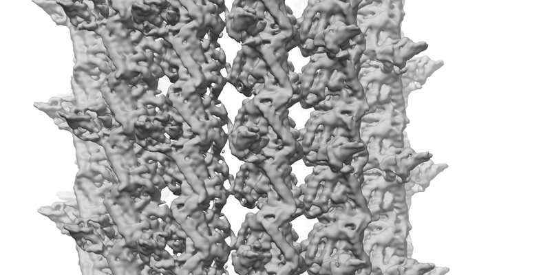
 ダウンロードとリンク
 emd_10060.png
 http://ftp.pdbj.org/pub/emdb/structures/EMD-10060
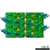


 Z
Z Y
Y X
X


 試料の構成要素


 解析
解析 電子顕微鏡法

