ムービー
ムービー コントローラー


+ データを開く

- 基本情報
基本情報
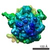
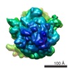
| 登録情報 | データベース: EMDB / ID: EMD-1006 | |||||||||
|---|---|---|---|---|---|---|---|---|---|---|
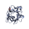
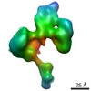
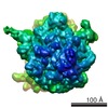
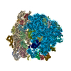
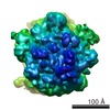
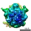
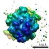
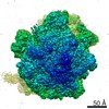
| タイトル | A cryo-electron microscopic study of ribosome-bound termination factor RF2. | |||||||||
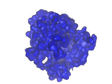
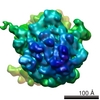
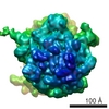
 マップデータ マップデータ | Ribosome-bound termination factor RF2 | |||||||||
 試料 試料 |
| |||||||||
| 機能・相同性 |  機能・相同性情報 機能・相同性情報translation release factor activity, codon specific / translational termination / large ribosomal subunit rRNA binding / small ribosomal subunit / cytosolic large ribosomal subunit /  tRNA binding / rRNA binding / structural constituent of ribosome / 翻訳 (生物学) / 細胞質基質 tRNA binding / rRNA binding / structural constituent of ribosome / 翻訳 (生物学) / 細胞質基質類似検索 - 分子機能 | |||||||||
| 生物種 |  Escherichia coli (大腸菌) Escherichia coli (大腸菌) | |||||||||
| 手法 | 単粒子再構成法 / クライオ電子顕微鏡法 / 解像度: 11.3 Å | |||||||||
 データ登録者 データ登録者 | Rawat U / Gao H / Zavialov A / Gursky R / Ehrenberg M / Frank J | |||||||||
 引用 引用 | ジャーナル: Nature / 年: 2003 タイトル: A cryo-electron microscopic study of ribosome-bound termination factor RF2. 著者: Urmila B S Rawat / Andrey V Zavialov / Jayati Sengupta / Mikel Valle / Robert A Grassucci / Jamie Linde / Bente Vestergaard / Måns Ehrenberg / Joachim Frank /  要旨: Protein synthesis takes place on the ribosome, where genetic information carried by messenger RNA is translated into a sequence of amino acids. This process is terminated when a stop codon moves into ...Protein synthesis takes place on the ribosome, where genetic information carried by messenger RNA is translated into a sequence of amino acids. This process is terminated when a stop codon moves into the ribosomal decoding centre (DC) and is recognized by a class-1 release factor (RF). RFs have a conserved GGQ amino-acid motif, which is crucial for peptide release and is believed to interact directly with the peptidyl-transferase centre (PTC) of the 50S ribosomal subunit. Another conserved motif of RFs (SPF in RF2) has been proposed to interact directly with stop codons in the DC of the 30S subunit. The distance between the DC and PTC is approximately 73 A. However, in the X-ray structure of RF2, SPF and GGQ are only 23 A apart, indicating that they cannot be at DC and PTC simultaneously. Here we show that RF2 is in an open conformation when bound to the ribosome, allowing GGQ to reach the PTC while still allowing SPF-stop-codon interaction. The results indicate new interpretations of accuracy in termination, and have implications for how the presence of a stop codon in the DC is signalled to PTC. | |||||||||
| 履歴 |
|
- 構造の表示
構造の表示
| ムービー |
ムービービューア |
|---|---|
| 構造ビューア | EMマップ: SurfViewMolmilJmol/JSmol |
| 添付画像 |
- ダウンロードとリンク
-EMDBアーカイブ
| マップデータ | emd_1006.map.gz | 7.9 MB | EMDBマップデータ形式 | |
|---|---|---|---|---|
| ヘッダ (付随情報) | emd-1006-v30.xmlemd-1006.xml | 11.4 KB 11.4 KB | 表示 表示 | EMDBヘッダ |
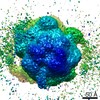
| 画像 |  1006.gif 1006.gif | 29.8 KB | ||
| アーカイブディレクトリ |  http://ftp.pdbj.org/pub/emdb/structures/EMD-1006ftp://ftp.pdbj.org/pub/emdb/structures/EMD-1006 http://ftp.pdbj.org/pub/emdb/structures/EMD-1006ftp://ftp.pdbj.org/pub/emdb/structures/EMD-1006 | HTTPS FTP |
-関連構造データ

-リンク
| EMDBのページ | EMDB (EBI/PDBe) / EMDataResource |
|---|---|
| 「今月の分子」の関連する項目 |


-マップ
| ファイル | ダウンロード / ファイル: emd_1006.map.gz / 形式: CCP4 / 大きさ: 8.2 MB / タイプ: IMAGE STORED AS FLOATING POINT NUMBER (4 BYTES) | ||||||||||||||||||||||||||||||||||||||||||||||||||||||||||||
|---|---|---|---|---|---|---|---|---|---|---|---|---|---|---|---|---|---|---|---|---|---|---|---|---|---|---|---|---|---|---|---|---|---|---|---|---|---|---|---|---|---|---|---|---|---|---|---|---|---|---|---|---|---|---|---|---|---|---|---|---|---|
| 注釈 | Ribosome-bound termination factor RF2 | ||||||||||||||||||||||||||||||||||||||||||||||||||||||||||||
| ボクセルのサイズ | X=Y=Z: 2.82 Å | ||||||||||||||||||||||||||||||||||||||||||||||||||||||||||||
| 密度 |
| ||||||||||||||||||||||||||||||||||||||||||||||||||||||||||||
| 対称性 | 空間群: 1 | ||||||||||||||||||||||||||||||||||||||||||||||||||||||||||||
| 詳細 | EMDB XML:
CCP4マップ ヘッダ情報:
| ||||||||||||||||||||||||||||||||||||||||||||||||||||||||||||
-添付データ
- 試料の構成要素
-全体 : E.coli 70s ribosome
| 全体 | 名称: E.coli 70s ribosome |
|---|---|
| 要素 |
|
-超分子 #1000: E.coli 70s ribosome
| 超分子 | 名称: E.coli 70s ribosome / タイプ: sample / ID: 1000 / Number unique components: 4 |
|---|---|
| 分子量 | 理論値: 2.5 MDa |
-超分子 #1: E.coli 70s ribosome
| 超分子 | 名称: E.coli 70s ribosome / タイプ: complex / ID: 1 / 組換発現: No / Ribosome-details: ribosome-prokaryote: LSU 50S |
|---|---|
| 由来(天然) | 生物種: Escherichia coli (大腸菌) |
| 分子量 | 理論値: 2.5 MDa |
-分子 #1: peptidyl P-tRNA
| 分子 | 名称: peptidyl P-tRNA / タイプ: ligand / ID: 1 / 組換発現: No |
|---|---|
| 由来(天然) | 生物種: Escherichia coli (大腸菌) |
-分子 #2: E-tRNA
| 分子 | 名称: E-tRNA / タイプ: ligand / ID: 2 / コピー数: 1 / 組換発現: No |
|---|---|
| 由来(天然) | 生物種: Escherichia coli (大腸菌) |
-分子 #3: MFTI-mRNA
| 分子 | 名称: MFTI-mRNA / タイプ: ligand / ID: 3 詳細: Zavialov et al., A posttermination ribosomal complex is the guanine nucleotide exchange factor for peptide release factor RF3. Cell. 107,1-20 (2001). コピー数: 1 / 組換発現: Yes |
|---|---|
| 由来(天然) | 生物種: Escherichia coli (大腸菌) |
-実験情報
-構造解析
| 手法 | クライオ電子顕微鏡法 |
|---|---|
 解析 解析 | 単粒子再構成法 |
| 試料の集合状態 | particle |
-試料調製
| 濃度 | 0.09 mg/mL |
|---|---|
| 緩衝液 | pH: 7.5 詳細: Polymix buffer, containing at final concentration 5 mM potassium phosphate, 5 mM magnesium acetate, 5mM ammonium chloride, 95 mM potassium chloride, 0.5 mM calcium chloride, 8mM putrescine, 1mM dithioerythritol |
| 凍結 | 凍結剤: ETHANE / チャンバー内湿度: 58 % / チャンバー内温度: 36 K / 装置: HOMEMADE PLUNGER 詳細: Vitrification instrument: Two side blotting plunger. Rapid-freezing in liquid ethane 手法: Blot and Plunge |
- 電子顕微鏡法
| 顕微鏡 | FEI TECNAI F20 |
|---|---|
| 電子線 | 加速電圧: 200 kV / 電子線源: FIELD EMISSION GUN |
| 電子光学系 | 倍率(補正後): 49696 / 照射モード: FLOOD BEAM / 撮影モード: BRIGHT FIELDBright-field microscopy / Cs: 2.0 mm / 最大 デフォーカス(公称値): 5.975 µm / 最小 デフォーカス(公称値): 1.39 µm / 倍率(公称値): 50000 |
| 試料ステージ | 試料ホルダー: Oxford,cryo-transfer 3500 / 試料ホルダーモデル: GATAN LIQUID NITROGEN |
| 温度 | 平均: 93 K |
| 日付 | 2001年8月9日 |
| 撮影 | カテゴリ: FILM / フィルム・検出器のモデル: KODAK SO-163 FILM / デジタル化 - スキャナー: ZEISS SCAI / デジタル化 - サンプリング間隔: 14 µm / 実像数: 34 / 平均電子線量: 20 e/Å2 / ビット/ピクセル: 12 |
| Tilt angle min | 0 |
| Tilt angle max | 0 |
| 実験機器 |  モデル: Tecnai F20 / 画像提供: FEI Company |
-画像解析
| CTF補正 | 詳細: Wiener filtration of defocus groups |
|---|---|
| 最終 再構成 | 想定した対称性 - 点群: C1 (非対称) / アルゴリズム: OTHER / 解像度のタイプ: BY AUTHOR / 解像度: 11.3 Å / 解像度の算出法: FSC 0.5 CUT-OFF / ソフトウェア - 名称: SPIDER/WEB 詳細: Frank, J. (2000) Three-Dimensional Cryoelectron Microscopy of Ribosomes, Methods of Enzymology (Ch.18) 317, 276-291. 使用した粒子像数: 14965 |
-原子モデル構築 1
| 初期モデル | (PDB ID: , ) |
|---|---|
| 詳細 | manual fitting using O |
| 精密化 | プロトコル: RIGID BODY FIT |
| 得られたモデル | 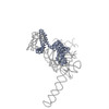 PDB-1mi6: 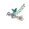 PDB-1mvr: |