 ムービー
ムービー コントローラー


+ データを開く

- 基本情報
基本情報
| 登録情報 | データベース: EMDB / ID: EMD-0622 | |||||||||
|---|---|---|---|---|---|---|---|---|---|---|
| タイトル | Structure of the AAV2 with its Cell Receptor, AAVR | |||||||||
 マップデータ マップデータ | AAV2 bound with AAVR (class2) | |||||||||
 試料 試料 |
| |||||||||
| 生物種 |    Adeno-associated virus (アデノ随伴ウイルス) Adeno-associated virus (アデノ随伴ウイルス) | |||||||||
| 手法 | サブトモグラム平均法 / クライオ電子顕微鏡法 / 解像度: 20.0 Å | |||||||||
 データ登録者 データ登録者 | Hu GQ / Meyer NL / Stagg SM / Chapman MS / Davulcu O / Xie Q / Noble AJ / Yoshioka C / Gingerich D / Trzynka A / David L | |||||||||
 引用 引用 | ジャーナル: Elife / 年: 2019 タイトル: Structure of the gene therapy vector, adeno-associated virus with its cell receptor, AAVR. 著者: Nancy L Meyer / Guiqing Hu / Omar Davulcu / Qing Xie / Alex J Noble / Craig Yoshioka / Drew S Gingerich / Andrew Trzynka / Larry David / Scott M Stagg / Michael Stewart Chapman /  要旨: Adeno-associated virus (AAV) vectors are preeminent in emerging clinical gene therapies. Generalizing beyond the most tractable genetic diseases will require modulation of cell specificity and immune ...Adeno-associated virus (AAV) vectors are preeminent in emerging clinical gene therapies. Generalizing beyond the most tractable genetic diseases will require modulation of cell specificity and immune neutralization. Interactions of AAV with its cellular receptor, AAVR, are key to understanding cell-entry and trafficking with the rigor needed to engineer tissue-specific vectors. -electron tomography shows ordered binding of part of the flexible receptor to the viral surface, with distal domains in multiple conformations. Regions of the virus and receptor in close physical proximity can be identified by cross-linking/mass spectrometry. -electron microscopy with a two-domain receptor fragment reveals the interactions at 2.4 Å resolution. AAVR binds between AAV's spikes on a plateau that is conserved, except in one clade whose structure is AAVR-incompatible. AAVR's footprint overlaps the epitopes of several neutralizing antibodies, prompting a re-evaluation of neutralization mechanisms. The structure provides a roadmap for experimental probing and manipulation of viral-receptor interactions. | |||||||||
| 履歴 |
|
- 構造の表示
構造の表示
| ムービー |
 ムービービューア ムービービューア |
|---|---|
| 構造ビューア | EMマップ: SurfViewMolmilJmol/JSmol |
| 添付画像 |

- ダウンロードとリンク
-EMDBアーカイブ
| マップデータ | emd_0622.map.gz | 136.4 KB | EMDBマップデータ形式 | |
|---|---|---|---|---|
| ヘッダ (付随情報) | emd-0622-v30.xmlemd-0622.xml | 14.5 KB 14.5 KB | 表示 表示 | EMDBヘッダ |
| 画像 | 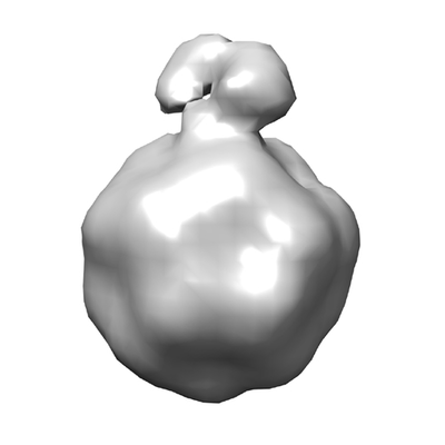 emd_0622.png emd_0622.png | 46 KB | ||
| アーカイブディレクトリ |  http://ftp.pdbj.org/pub/emdb/structures/EMD-0622ftp://ftp.pdbj.org/pub/emdb/structures/EMD-0622 http://ftp.pdbj.org/pub/emdb/structures/EMD-0622ftp://ftp.pdbj.org/pub/emdb/structures/EMD-0622 | HTTPS FTP |
-関連構造データ


-リンク
| EMDBのページ | EMDB (EBI/PDBe) / EMDataResource |
|---|
-マップ
| ファイル | ダウンロード / ファイル: emd_0622.map.gz / 形式: CCP4 / 大きさ: 182.6 KB / タイプ: IMAGE STORED AS FLOATING POINT NUMBER (4 BYTES) | ||||||||||||||||||||||||||||||||||||||||||||||||||||||||||||
|---|---|---|---|---|---|---|---|---|---|---|---|---|---|---|---|---|---|---|---|---|---|---|---|---|---|---|---|---|---|---|---|---|---|---|---|---|---|---|---|---|---|---|---|---|---|---|---|---|---|---|---|---|---|---|---|---|---|---|---|---|---|
| 注釈 | AAV2 bound with AAVR (class2) | ||||||||||||||||||||||||||||||||||||||||||||||||||||||||||||
| ボクセルのサイズ | X=Y=Z: 15.6 Å | ||||||||||||||||||||||||||||||||||||||||||||||||||||||||||||
| 密度 |
| ||||||||||||||||||||||||||||||||||||||||||||||||||||||||||||
| 対称性 | 空間群: 1 | ||||||||||||||||||||||||||||||||||||||||||||||||||||||||||||
| 詳細 | EMDB XML:
CCP4マップ ヘッダ情報:
| ||||||||||||||||||||||||||||||||||||||||||||||||||||||||||||
-添付データ
- 試料の構成要素
-全体 : AAV2 complex with AAVR receptor
| 全体 | 名称: AAV2 complex with AAVR receptor |
|---|---|
| 要素 |
|
-超分子 #1: AAV2 complex with AAVR receptor
| 超分子 | 名称: AAV2 complex with AAVR receptor / タイプ: complex / ID: 1 / 親要素: 0 / 詳細: class 1 |
|---|---|
| 由来(天然) | 生物種: Adeno-associated virus (アデノ随伴ウイルス) 株: hybrid of serotypes 2, 8, and 9 |
| 組換発現 | 生物種:   Spodoptera frugiperda (ツマジロクサヨトウ) Spodoptera frugiperda (ツマジロクサヨトウ)組換株: SF9 / 組換細胞: Sf9 / 組換プラスミド: pFBDDJM11 |
| 分子量 | 理論値: 3.75 MDa |
-実験情報
-構造解析
| 手法 | クライオ電子顕微鏡法 |
|---|---|
 解析 解析 | サブトモグラム平均法 |
| 試料の集合状態 | particle |
-試料調製
| 濃度 | 0.6 mg/mL | ||||||||||||
|---|---|---|---|---|---|---|---|---|---|---|---|---|---|
| 緩衝液 | pH: 7.4 構成要素:
詳細: blot force = 1, blot time = 3 seconds, total blots = 1 | ||||||||||||
| 凍結 | 凍結剤: ETHANE / チャンバー内湿度: 100 % / チャンバー内温度: 4 K / 装置: FEI VITROBOT MARK IV |
- 電子顕微鏡法
| 顕微鏡 | FEI TITAN KRIOS |
|---|---|
| 電子線 | 加速電圧: 300 kV / 電子線源: FIELD EMISSION GUN |
| 電子光学系 | C2レンズ絞り径: 70.0 µm / 最大 デフォーカス(補正後): 0.011 µm / 最小 デフォーカス(補正後): 9.0 µm / 倍率(補正後): 18000 / 照射モード: FLOOD BEAM / 撮影モード: BRIGHT FIELDBright-field microscopy / Cs: 2.7 mm / 最大 デフォーカス(公称値): 0.011 µm / 最小 デフォーカス(公称値): 9.0 µm / 倍率(公称値): 18000 |
| 試料ステージ | 試料ホルダーモデル: FEI TITAN KRIOS AUTOGRID HOLDER ホルダー冷却材: NITROGEN |
| 温度 | 最低: 290.0 K / 最高: 300.0 K |
| 詳細 | preliminary grid screening was performed manually |
| 撮影 | フィルム・検出器のモデル: DIRECT ELECTRON DE-20 (5k x 3k) 検出モード: INTEGRATING / デジタル化 - サイズ - 横: 5000 pixel / デジタル化 - サイズ - 縦: 3000 pixel / デジタル化 - サンプリング間隔: 10.0 µm / デジタル化 - 画像ごとのフレーム数: 1-7 / 撮影したグリッド数: 1 / 平均露光時間: 1.5 sec. / 平均電子線量: 1.42 e/Å2 / 詳細: none |
| 実験機器 |  モデル: Titan Krios / 画像提供: FEI Company |
-画像解析
| 抽出 | トモグラム数: 8 / 使用した粒子像数: 1321 / 参照モデル: AAVDJ low passfilter to 50 agntrom / 手法: automatic / ソフトウェア - 名称: Dynamo (ver. 1) / 詳細: none |
|---|---|
| CTF補正 | ソフトウェア - 名称: TOMOCTF (ver. 1.0) / ソフトウェア - 詳細: none / 詳細: tomoctf |
| 最終 3次元分類 | クラス数: 20 / 平均メンバー数/クラス: 150 / 詳細: none |
| 最終 角度割当 | タイプ: COMMON LINE / ソフトウェア - 名称: Dynamo (ver. 1) / ソフトウェア - 詳細: none / 詳細: none |
| 最終 再構成 | 使用したクラス数: 20 / 想定した対称性 - 点群: I (正20面体型対称) / アルゴリズム: BACK PROJECTION / 解像度のタイプ: BY AUTHOR / 解像度: 20.0 Å / 解像度の算出法: OTHER / ソフトウェア - 名称: Dynamo (ver. 1.0) / ソフトウェア - 詳細: none / 詳細: none / 使用したサブトモグラム数: 8 |
| 詳細 | nond |
-原子モデル構築 1
| 初期モデル | PDB ID: |
|---|---|
| 精密化 | 空間: REAL / プロトコル: RIGID BODY FIT / 当てはまり具合の基準: correlation coeeficient |
