ジャーナル: EMBO J / 年: 2019 タイトル: Frozen-hydrated chromatin from metaphase chromosomes has an interdigitated multilayer structure. 著者: Andrea Chicano / Eva Crosas / Joaquín Otón / Roberto Melero / Benjamin D Engel / Joan-Ramon Daban / 要旨: Cryo-electron tomography and small-angle X-ray scattering were used to investigate the chromatin folding in metaphase chromosomes. The tomographic 3D reconstructions show that frozen-hydrated ...Cryo-electron tomography and small-angle X-ray scattering were used to investigate the chromatin folding in metaphase chromosomes. The tomographic 3D reconstructions show that frozen-hydrated chromatin emanated from chromosomes is planar and forms multilayered plates. The layer thickness was measured accounting for the contrast transfer function fringes at the plate edges, yielding a width of ~ 7.5 nm, which is compatible with the dimensions of a monolayer of nucleosomes slightly tilted with respect to the layer surface. Individual nucleosomes are visible decorating distorted plates, but typical plates are very dense and nucleosomes are not identifiable as individual units, indicating that they are tightly packed. Two layers in contact are ~ 13 nm thick, which is thinner than the sum of two independent layers, suggesting that nucleosomes in the layers interdigitate. X-ray scattering of whole chromosomes shows a main scattering peak at ~ 6 nm, which can be correlated with the distance between layers and between interdigitating nucleosomes interacting through their faces. These observations support a model where compact chromosomes are composed of many chromatin layers stacked along the chromosome axis.
 ムービー
ムービー コントローラー


 データを開く
 基本情報
基本情報 マップデータ
 試料
試料
 Homo sapiens (ヒト)
 データ登録者
 スペイン, 1件
  引用
引用
 構造の表示
構造の表示 ムービービューア
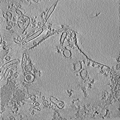
 ダウンロードとリンク
 emd_0119.png
 http://ftp.pdbj.org/pub/emdb/structures/EMD-0119
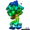
 試料の構成要素
 解析
解析 電子顕微鏡法
