 ムービー
ムービー コントローラー


+ データを開く

- 基本情報
基本情報
| 登録情報 | データベース: EMDB / ID: EMD-2132 | |||||||||
|---|---|---|---|---|---|---|---|---|---|---|
| タイトル | Cryo-electron tomography averaged map of microtubule doublet 2-8 in the distal/central region of Chlamydomonas flagella | |||||||||
 マップデータ マップデータ | Reconstruction of the average of outer doublets 2-8 from Chlamydomonas flagella in the distal/central region. | |||||||||
 試料 試料 |
| |||||||||
 キーワード キーワード |  axoneme / dynein (ダイニン) / Chlamydomonas (クラミドモナス) / microtubule doublet / flagella (鞭毛) axoneme / dynein (ダイニン) / Chlamydomonas (クラミドモナス) / microtubule doublet / flagella (鞭毛) | |||||||||
| 生物種 |   Chlamydomonas reinhardtii (クラミドモナス) Chlamydomonas reinhardtii (クラミドモナス) | |||||||||
| 手法 | サブトモグラム平均法 / クライオ電子顕微鏡法 / 解像度: 36.8 Å | |||||||||
 データ登録者 データ登録者 | Bui KH / Yagi T / Yamamoto R / Kamiya R / Ishikawa T | |||||||||
 引用 引用 | ジャーナル: J Cell Biol / 年: 2012 タイトル: Polarity and asymmetry in the arrangement of dynein and related structures in the Chlamydomonas axoneme. 著者: Khanh Huy Bui / Toshiki Yagi / Ryosuke Yamamoto / Ritsu Kamiya / Takashi Ishikawa /  要旨: Understanding the molecular architecture of the flagellum is crucial to elucidate the bending mechanism produced by this complex organelle. The current known structure of the flagellum has not yet ...Understanding the molecular architecture of the flagellum is crucial to elucidate the bending mechanism produced by this complex organelle. The current known structure of the flagellum has not yet been fully correlated with the complex composition and localization of flagellar components. Using cryoelectron tomography and subtomogram averaging while distinguishing each one of the nine outer doublet microtubules, we systematically collected and reconstructed the three-dimensional structures in different regions of the Chlamydomonas flagellum. We visualized the radial and longitudinal differences in the flagellum. One doublet showed a distinct structure, whereas the other eight were similar but not identical to each other. In the proximal region, some dyneins were missing or replaced by minor dyneins, and outer-inner arm dynein links were variable among different microtubule doublets. These findings shed light on the intricate organization of Chlamydomonas flagella, provide clues to the mechanism that produces asymmetric flagellar beating, and pose a new challenge for the functional study of the flagella. | |||||||||
| 履歴 |
|
- 構造の表示
構造の表示
| ムービー |
 ムービービューア ムービービューア |
|---|---|
| 構造ビューア | EMマップ: SurfViewMolmilJmol/JSmol |
| 添付画像 |

- ダウンロードとリンク
-EMDBアーカイブ
| マップデータ | emd_2132.map.gz | 25.1 MB | EMDBマップデータ形式 | |
|---|---|---|---|---|
| ヘッダ (付随情報) | emd-2132-v30.xmlemd-2132.xml | 10.1 KB 10.1 KB | 表示 表示 | EMDBヘッダ |
| 画像 |  emd_2132.png emd_2132.png | 185.3 KB | ||
| アーカイブディレクトリ |  http://ftp.pdbj.org/pub/emdb/structures/EMD-2132ftp://ftp.pdbj.org/pub/emdb/structures/EMD-2132 http://ftp.pdbj.org/pub/emdb/structures/EMD-2132ftp://ftp.pdbj.org/pub/emdb/structures/EMD-2132 | HTTPS FTP |
-関連構造データ
| 関連構造データ | 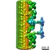 2113C 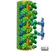 2114C  2115C 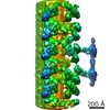 2116C  2117C  2118C 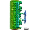 2119C 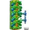 2120C 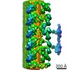 2121C 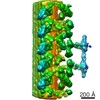 2122C  2123C 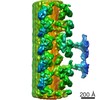 2124C 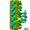 2125C 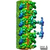 2126C 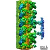 2127C 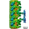 2128C 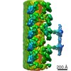 2129C 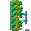 2130C 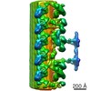 2131C C: 同じ文献を引用 ( |
|---|---|
| 類似構造データ |
-リンク
| EMDBのページ | EMDB (EBI/PDBe) / EMDataResource |
|---|
-マップ
| ファイル | ダウンロード / ファイル: emd_2132.map.gz / 形式: CCP4 / 大きさ: 29.8 MB / タイプ: IMAGE STORED AS FLOATING POINT NUMBER (4 BYTES) | ||||||||||||||||||||||||||||||||||||||||||||||||||||||||||||||||||||
|---|---|---|---|---|---|---|---|---|---|---|---|---|---|---|---|---|---|---|---|---|---|---|---|---|---|---|---|---|---|---|---|---|---|---|---|---|---|---|---|---|---|---|---|---|---|---|---|---|---|---|---|---|---|---|---|---|---|---|---|---|---|---|---|---|---|---|---|---|---|
| 注釈 | Reconstruction of the average of outer doublets 2-8 from Chlamydomonas flagella in the distal/central region. | ||||||||||||||||||||||||||||||||||||||||||||||||||||||||||||||||||||
| ボクセルのサイズ | X=Y=Z: 7.25 Å | ||||||||||||||||||||||||||||||||||||||||||||||||||||||||||||||||||||
| 密度 |
| ||||||||||||||||||||||||||||||||||||||||||||||||||||||||||||||||||||
| 対称性 | 空間群: 1 | ||||||||||||||||||||||||||||||||||||||||||||||||||||||||||||||||||||
| 詳細 | EMDB XML:
CCP4マップ ヘッダ情報:
| ||||||||||||||||||||||||||||||||||||||||||||||||||||||||||||||||||||
-添付データ
- 試料の構成要素
-全体 : Outer doublets 2-8 of Chlamydomonas flagella in the distal/centra...
| 全体 | 名称: Outer doublets 2-8 of Chlamydomonas flagella in the distal/central region |
|---|---|
| 要素 |
|
-超分子 #1000: Outer doublets 2-8 of Chlamydomonas flagella in the distal/centra...
| 超分子 | 名称: Outer doublets 2-8 of Chlamydomonas flagella in the distal/central region タイプ: sample / ID: 1000 / 詳細: Flagella were purified from Chlamydomonas / Number unique components: 1 |
|---|
-超分子 #1: flagellum
| 超分子 | 名称: flagellum / タイプ: organelle_or_cellular_component / ID: 1 / Name.synonym: axoneme, cilia / 組換発現: No / データベース: NCBI |
|---|---|
| 由来(天然) | 生物種: Chlamydomonas reinhardtii (クラミドモナス) 株: c137 (mt+) / 別称: green algae / Organelle: flagella / 細胞中の位置: distal part of the flagellum |
-実験情報
-構造解析
| 手法 | クライオ電子顕微鏡法 |
|---|---|
 解析 解析 | サブトモグラム平均法 |
-試料調製
| 濃度 | 2 mg/mL |
|---|---|
| 緩衝液 | pH: 7.4 詳細: 30 mM Hepes, pH 7.4, 5 mM MgSO4, 1 mM DTT, 0.5 mM EDTA, 25 mM KCl and 0.5% (wt/vol) polyethylene glycol (MW 20,000) |
| グリッド | 詳細: 300 mesh Quantifoil Holey Carbon copper grid R2/1 |
| 凍結 | 凍結剤: ETHANE / チャンバー内湿度: 90 % / チャンバー内温度: 110 K / 装置: FEI VITROBOT MARK II / 手法: Offset -3, Blot 3s, Drain time 0s |
- 電子顕微鏡法
| 顕微鏡 | FEI TECNAI F20 |
|---|---|
| 電子線 | 加速電圧: 200 kV / 電子線源: FIELD EMISSION GUN |
| 電子光学系 | 倍率(補正後): 19303 / 照射モード: FLOOD BEAM / 撮影モード: BRIGHT FIELDBright-field microscopy / Cs: 2 mm / 最大 デフォーカス(公称値): 6.0 µm / 最小 デフォーカス(公称値): 4.0 µm / 倍率(公称値): 27500 |
| 特殊光学系 | エネルギーフィルター - 名称: Gatan Tridiem エネルギーフィルター - エネルギー下限: 0.0 eV エネルギーフィルター - エネルギー上限: 20.0 eV |
| 試料ステージ | 試料ホルダー: Gatan 626 / 試料ホルダーモデル: GATAN LIQUID NITROGEN / Tilt series - Axis1 - Min angle: -60 ° / Tilt series - Axis1 - Max angle: 60 ° |
| 温度 | 最低: 93 K / 最高: 118 K / 平均: 98 K |
| 日付 | 2010年11月26日 |
| 撮影 | カテゴリ: CCD フィルム・検出器のモデル: GATAN ULTRASCAN 1000 (2k x 2k) 実像数: 3770 / 平均電子線量: 60 e/Å2 / ビット/ピクセル: 16 |
| 実験機器 |  モデル: Tecnai F20 / 画像提供: FEI Company |
-画像解析
| 最終 再構成 | アルゴリズム: OTHER / 解像度のタイプ: BY AUTHOR / 解像度: 36.8 Å / 解像度の算出法: FSC 0.5 CUT-OFF ソフトウェア - 名称: IMOD, BSOFT, SPIDER, TOM, package, Matlab |
|---|---|
| 詳細 | R-weighted backprojection using IMOD with fiducial markers. Average number of tilts used in the 3D reconstructions: 61. Average tomographic tilt angle increment: 2. |
