 Movie
Movie Controller

[English] 日本語
 Yorodumi
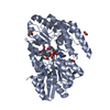
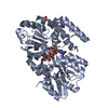
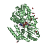
- PDB-7c0u: Crystal structure of a dinucleotide-binding protein (S127A) of AB... -
+ Open data

- Basic information
| Entry | Database: PDB / ID: 7c0u | ||||||
|---|---|---|---|---|---|---|---|
| Title | Crystal structure of a dinucleotide-binding protein (S127A) of ABC transporter endogenously bound to uridylyl-3'-5'-phospho-guanosine | ||||||
 Components Components | Sugar ABC transporter, periplasmic sugar-binding protein | ||||||
 Keywords Keywords |  TRANSPORT PROTEIN / c-di-GMP/AMP / Substrate-binding protein / Thermus thermophilus / tRNA synthesis and/or modification / Venus Fly-trap mechanism / UgpB TRANSPORT PROTEIN / c-di-GMP/AMP / Substrate-binding protein / Thermus thermophilus / tRNA synthesis and/or modification / Venus Fly-trap mechanism / UgpB | ||||||
| Function / homology | Bacterial extracellular solute-binding protein / Solute-binding family 1, conserved site / Bacterial extracellular solute-binding proteins, family 1 signature. / Bacterial extracellular solute-binding protein / transmembrane transport / Chem-FGO / DI(HYDROXYETHYL)ETHER / Sugar ABC transporter, periplasmic sugar-binding protein Function and homology information Function and homology information | ||||||
| Biological species |   Thermus thermophilus HB8 (bacteria) Thermus thermophilus HB8 (bacteria) | ||||||
| Method | X-RAY DIFFRACTION / MOLECULAR REPLACEMENT / molecular replacement / Resolution: 1.8 Å | ||||||
 Authors Authors | Kanaujia, S.P. / Chandravanshi, M. / Samanta, R. | ||||||
| Funding support |  India, 1items India, 1items
| ||||||
 Citation Citation | Journal: Febs J. / Year: 2021 Title: Structural and thermodynamic insights into the novel dinucleotide-binding protein of ABC transporter unveils its moonlighting function. Authors: Chandravanshi, M. / Samanta, R. / Kanaujia, S.P. | ||||||
| History |
|
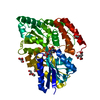
- Structure visualization
| Structure viewer | Molecule: MolmilJmol/JSmol |
|---|
- Downloads & links
-Download
| PDBx/mmCIF format | 7c0u.cif.gz | 330.7 KB | Display | PDBx/mmCIF format |
|---|---|---|---|---|
| PDB format | pdb7c0u.ent.gz | 266.1 KB | Display | PDB format |
| PDBx/mmJSON format | 7c0u.json.gz | Tree view | PDBx/mmJSON format | |
| Others |  Other downloads Other downloads |
-Validation report
| Arichive directory | https://data.pdbj.org/pub/pdb/validation_reports/c0/7c0uftp://data.pdbj.org/pub/pdb/validation_reports/c0/7c0u | HTTPS FTP |
|---|
-Related structure data
| Related structure data | 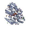 7c0fSC 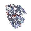 7c0kC  7c0lC  7c0oC 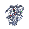 7c0rC  7c0sC 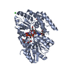 7c0tC 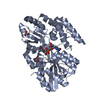 7c0vC 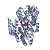 7c0wC 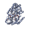 7c0xC 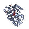 7c0yC 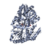 7c0zC 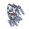 7c14C  7c15C 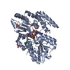 7c16C 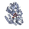 7c19C 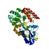 7c1bC S: Starting model for refinement C: citing same article ( |
|---|---|
| Similar structure data |


-Links
 PDBj
PDBj


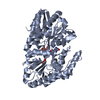
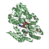
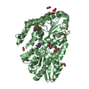
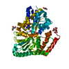
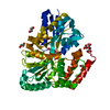
- Assembly
| Deposited unit | 
| ||||||||||||||||||
|---|---|---|---|---|---|---|---|---|---|---|---|---|---|---|---|---|---|---|---|
| 1 | 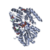
| ||||||||||||||||||
| 2 | 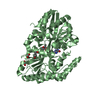
| ||||||||||||||||||
| Unit cell |
| ||||||||||||||||||
| Noncrystallographic symmetry (NCS) | NCS domain:
NCS domain segments: Component-ID: 0 / Ens-ID: 1 / Beg auth comp-ID: MET / Beg label comp-ID: MET / End auth comp-ID: SER / End label comp-ID: SER / Refine code: 0 / Auth seq-ID: -1 - 395 / Label seq-ID: 1 - 397
|
-Components
-Protein , 1 types, 2 molecules AB
| #1: Protein | Mass: 44488.785 Da / Num. of mol.: 2 / Mutation: S127A Source method: isolated from a genetically manipulated source Source: (gene. exp.) Thermus thermophilus HB8 (bacteria) / Gene: TTHA0379 / Plasmid: pET22b / Production host: Escherichia coli BL21(DE3) (bacteria) / Variant (production host): Rosetta / References: UniProt: Q5SLB4 |
|---|
-Non-polymers , 6 types, 726 molecules 


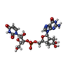

| #2: Chemical | Chloride Mass: 35.453 Da / Num. of mol.: 3 / Source method: obtained synthetically / Formula: Cl Mass: 35.453 Da / Num. of mol.: 3 / Source method: obtained synthetically / Formula: Cl#3: Chemical | ChemComp-EDO / Ethylene glycol Mass: 62.068 Da / Num. of mol.: 4 / Source method: obtained synthetically / Formula: C2H6O2 Mass: 62.068 Da / Num. of mol.: 4 / Source method: obtained synthetically / Formula: C2H6O2#4: Chemical | Glycerol Mass: 92.094 Da / Num. of mol.: 2 / Source method: obtained synthetically / Formula: C3H8O3 Mass: 92.094 Da / Num. of mol.: 2 / Source method: obtained synthetically / Formula: C3H8O3#5: Chemical | ChemComp-PEG / Diethylene glycol Mass: 106.120 Da / Num. of mol.: 4 / Source method: obtained synthetically / Formula: C4H10O3 Mass: 106.120 Da / Num. of mol.: 4 / Source method: obtained synthetically / Formula: C4H10O3#6: Chemical |  Mass: 629.471 Da / Num. of mol.: 2 / Source method: obtained synthetically / Formula: C22H28N7O13P / Feature type: SUBJECT OF INVESTIGATION Mass: 629.471 Da / Num. of mol.: 2 / Source method: obtained synthetically / Formula: C22H28N7O13P / Feature type: SUBJECT OF INVESTIGATION#7: Water | ChemComp-HOH / | WaterMass: 18.015 Da / Num. of mol.: 711 / Source method: isolated from a natural source / Formula: H2O |
|---|
-Details
| Has ligand of interest | Y |
|---|
-Experimental details
-Experiment
| Experiment | Method: X-RAY DIFFRACTION / Number of used crystals: 1 |
|---|
- Sample preparation
| Crystal | Density Matthews: 2.63 Å3/Da / Density % sol: 53.26 % / Description: Monoclinic |
|---|---|
| Crystal grow | Temperature: 277 K / Method: vapor diffusion, hanging drop / pH: 6.5 Details: 0.2M ammonium phosphate, 0.1M sodium cacodylate pH 6.5, 30% PEG 8000 |
-Data collection
| Diffraction | Mean temperature: 100 K / Serial crystal experiment: N | ||||||||||||||||||||||||||||||
|---|---|---|---|---|---|---|---|---|---|---|---|---|---|---|---|---|---|---|---|---|---|---|---|---|---|---|---|---|---|---|---|
| Diffraction source | Source: ROTATING ANODE / Type: RIGAKU MICROMAX-007 HF / Wavelength: 1.5418 Å | ||||||||||||||||||||||||||||||
| Detector | Type: RIGAKU RAXIS IV++ / Detector: IMAGE PLATE / Date: Apr 14, 2019 / Details: VariMax HF | ||||||||||||||||||||||||||||||
| Radiation | Protocol: SINGLE WAVELENGTH / Monochromatic (M) / Laue (L): M / Scattering type: x-ray | ||||||||||||||||||||||||||||||
| Radiation wavelength | Wavelength: 1.5418 Å / Relative weight: 1 | ||||||||||||||||||||||||||||||
| Reflection | Resolution: 1.8→46.95 Å / Num. obs: 82790 / % possible obs: 97.1 % / Redundancy: 3.5 % / CC1/2: 0.985 / Rmerge(I) obs: 0.118 / Rpim(I) all: 0.072 / Rrim(I) all: 0.139 / Net I/σ(I): 7 | ||||||||||||||||||||||||||||||
| Reflection shell | Diffraction-ID: 1
|
-Phasing
| Phasing | Method: molecular replacement | |||||||||
|---|---|---|---|---|---|---|---|---|---|---|
| Phasing MR | Model details: Phaser MODE: MR_AUTO
|
- Processing
| Software |
| |||||||||||||||||||||||||||||||||||||||||||||||||||||||||||||||||||||||||||
|---|---|---|---|---|---|---|---|---|---|---|---|---|---|---|---|---|---|---|---|---|---|---|---|---|---|---|---|---|---|---|---|---|---|---|---|---|---|---|---|---|---|---|---|---|---|---|---|---|---|---|---|---|---|---|---|---|---|---|---|---|---|---|---|---|---|---|---|---|---|---|---|---|---|---|---|---|
| Refinement | Method to determine structure: MOLECULAR REPLACEMENT Starting model: 7C0F Resolution: 1.8→46.95 Å / Cor.coef. Fo:Fc: 0.907 / Cor.coef. Fo:Fc free: 0.868 / SU B: 7.163 / SU ML: 0.119 / SU R Cruickshank DPI: 0.156 / Cross valid method: THROUGHOUT / σ(F): 0 / ESU R: 0.156 / ESU R Free: 0.152 Details: U VALUES : WITH TLS ADDED HYDROGENS HAVE BEEN ADDED IN THE RIDING POSITIONS
| |||||||||||||||||||||||||||||||||||||||||||||||||||||||||||||||||||||||||||
| Solvent computation | Ion probe radii: 0.8 Å / Shrinkage radii: 0.8 Å / VDW probe radii: 1.2 Å | |||||||||||||||||||||||||||||||||||||||||||||||||||||||||||||||||||||||||||
| Displacement parameters | Biso max: 49.54 Å2 / Biso mean: 17.567 Å2 / Biso min: 6.16 Å2
| |||||||||||||||||||||||||||||||||||||||||||||||||||||||||||||||||||||||||||
| Refinement step | Cycle: final / Resolution: 1.8→46.95 Å
| |||||||||||||||||||||||||||||||||||||||||||||||||||||||||||||||||||||||||||
| Refine LS restraints |
| |||||||||||||||||||||||||||||||||||||||||||||||||||||||||||||||||||||||||||
| Refine LS restraints NCS | Ens-ID: 1 / Number: 12908 / Refine-ID: X-RAY DIFFRACTION / Type: interatomic distance / Rms dev position: 0.06 Å / Weight position: 0.05
| |||||||||||||||||||||||||||||||||||||||||||||||||||||||||||||||||||||||||||
| LS refinement shell | Resolution: 1.8→1.83 Å / Rfactor Rfree error: 0
| |||||||||||||||||||||||||||||||||||||||||||||||||||||||||||||||||||||||||||
| Refinement TLS params. | Method: refined / Refine-ID: X-RAY DIFFRACTION
| |||||||||||||||||||||||||||||||||||||||||||||||||||||||||||||||||||||||||||
| Refinement TLS group |
|
