 Movie
Movie Controller


+ Open data

- Basic information
| Entry | Database: PDB / ID: 6kiv | ||||||
|---|---|---|---|---|---|---|---|
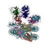
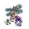
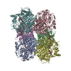
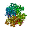
| Title | Cryo-EM structure of human MLL1-ubNCP complex (4.0 angstrom) | ||||||
 Components Components |
| ||||||
 Keywords Keywords | TRANSCRIPTION/DNA /  histone modification / nucleosome / MLL / TRANSCRIPTION / TRANSCRIPTION-DNA complex histone modification / nucleosome / MLL / TRANSCRIPTION / TRANSCRIPTION-DNA complex | ||||||
| Function / homology |  Function and homology information Function and homology informationprotein-cysteine methyltransferase activity / response to potassium ion / [histone H3]-lysine4 N-methyltransferase / histone H3K4 monomethyltransferase activity / unmethylated CpG binding / histone H3K4 trimethyltransferase activity / negative regulation of DNA methylation-dependent heterochromatin formation / regulation of short-term neuronal synaptic plasticity / MLL3/4 complex / Set1C/COMPASS complex ...protein-cysteine methyltransferase activity / response to potassium ion / [histone H3]-lysine4 N-methyltransferase / histone H3K4 monomethyltransferase activity / unmethylated CpG binding / histone H3K4 trimethyltransferase activity / negative regulation of DNA methylation-dependent heterochromatin formation / regulation of short-term neuronal synaptic plasticity / MLL3/4 complex / Set1C/COMPASS complex / MLL1/2 complex / ATAC complex / definitive hemopoiesis / NSL complex / histone H3K4 methyltransferase activity / T-helper 2 cell differentiation / : / Formation of the ternary complex, and subsequently, the 43S complex / Cardiogenesis / embryonic hemopoiesis / exploration behavior / anterior/posterior pattern specification / Ribosomal scanning and start codon recognition / histone methyltransferase complex / Translation initiation complex formation / regulation of tubulin deacetylation / : / regulation of cell division / Formation of WDR5-containing histone-modifying complexes / SARS-CoV-1 modulates host translation machinery / minor groove of adenine-thymine-rich DNA binding / Peptide chain elongation / Selenocysteine synthesis / membrane depolarization / MLL1 complex / Formation of a pool of free 40S subunits / Eukaryotic Translation Termination / regulation of embryonic development / Response of EIF2AK4 (GCN2) to amino acid deficiency / SRP-dependent cotranslational protein targeting to membrane / hemopoiesis / Viral mRNA Translation / Nonsense Mediated Decay (NMD) independent of the Exon Junction Complex (EJC) / GTP hydrolysis and joining of the 60S ribosomal subunit / histone acetyltransferase complex / L13a-mediated translational silencing of Ceruloplasmin expression / Major pathway of rRNA processing in the nucleolus and cytosol / Nonsense Mediated Decay (NMD) enhanced by the Exon Junction Complex (EJC) / negative regulation of fibroblast proliferation / homeostasis of number of cells within a tissue / Maturation of protein E / Maturation of protein E / positive regulation of gluconeogenesis / ER Quality Control Compartment (ERQC) / spleen development / Myoclonic epilepsy of Lafora / FLT3 signaling by CBL mutants / Prevention of phagosomal-lysosomal fusion / transcription initiation-coupled chromatin remodeling / IRAK2 mediated activation of TAK1 complex / Alpha-protein kinase 1 signaling pathway / Glycogen synthesis / IRAK1 recruits IKK complex / IRAK1 recruits IKK complex upon TLR7/8 or 9 stimulation / Membrane binding and targetting of GAG proteins / Constitutive Signaling by NOTCH1 HD Domain Mutants / NOTCH2 Activation and Transmission of Signal to the Nucleus / Endosomal Sorting Complex Required For Transport (ESCRT) / IRAK2 mediated activation of TAK1 complex upon TLR7/8 or 9 stimulation / Regulation of FZD by ubiquitination / PTK6 Regulates RTKs and Their Effectors AKT1 and DOK1 / Negative regulation of FLT3 / TICAM1,TRAF6-dependent induction of TAK1 complex / TICAM1-dependent activation of IRF3/IRF7 / cellular response to transforming growth factor beta stimulus / methylated histone binding / APC/C:Cdc20 mediated degradation of Cyclin B / Downregulation of ERBB4 signaling / p75NTR recruits signalling complexes / TRAF6 mediated IRF7 activation in TLR7/8 or 9 signaling / APC-Cdc20 mediated degradation of Nek2A / Transferases; Transferring one-carbon groups; Methyltransferases / PINK1-PRKN Mediated Mitophagy / TRAF6-mediated induction of TAK1 complex within TLR4 complex / InlA-mediated entry of Listeria monocytogenes into host cells / Pexophagy / Regulation of innate immune responses to cytosolic DNA / VLDLR internalisation and degradation / Downregulation of ERBB2:ERBB3 signaling / NRIF signals cell death from the nucleus / Activated NOTCH1 Transmits Signal to the Nucleus / Translesion synthesis by REV1 / NF-kB is activated and signals survival / Regulation of PTEN localization / Translesion synthesis by POLK / Regulation of BACH1 activity / small-subunit processome / Synthesis of active ubiquitin: roles of E1 and E2 enzymes / Translesion synthesis by POLI / Gap-filling DNA repair synthesis and ligation in GG-NERSimilarity search - Function | ||||||
| Biological species | Xenopus laevis (African clawed frog) Homo sapiens (human) Homo sapiens (human)synthetic construct (others) | ||||||
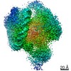
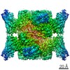
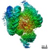
| Method | ELECTRON MICROSCOPY / single particle reconstruction / cryo EM / Resolution: 4 Å | ||||||
 Authors Authors | Huang, J. / Xue, H. / Yao, T. | ||||||
| Funding support |  China, 1items China, 1items
| ||||||
 Citation Citation | Journal: Nature / Year: 2019 Title: Structural basis of nucleosome recognition and modification by MLL methyltransferases. Authors: Han Xue / Tonghui Yao / Mi Cao / Guanjun Zhu / Yan Li / Guiyong Yuan / Yong Chen / Ming Lei / Jing Huang / Abstract: Methyltransferases of the mixed-lineage leukaemia (MLL) family-which include MLL1, MLL2, MLL3, MLL4, SET1A and SET1B-implement methylation of histone H3 on lysine 4 (H3K4), and have critical and ...Methyltransferases of the mixed-lineage leukaemia (MLL) family-which include MLL1, MLL2, MLL3, MLL4, SET1A and SET1B-implement methylation of histone H3 on lysine 4 (H3K4), and have critical and distinct roles in the regulation of transcription in haematopoiesis, adipogenesis and development. The C-terminal catalytic SET (Su(var.)3-9, enhancer of zeste and trithorax) domains of MLL proteins are associated with a common set of regulatory factors (WDR5, RBBP5, ASH2L and DPY30) to achieve specific activities. Current knowledge of the regulation of MLL activity is limited to the catalysis of histone H3 peptides, and how H3K4 methyl marks are deposited on nucleosomes is poorly understood. H3K4 methylation is stimulated by mono-ubiquitination of histone H2B on lysine 120 (H2BK120ub1), a prevalent histone H2B mark that disrupts chromatin compaction and favours open chromatin structures, but the underlying mechanism remains unknown. Here we report cryo-electron microscopy structures of human MLL1 and MLL3 catalytic modules associated with nucleosome core particles that contain H2BK120ub1 or unmodified H2BK120. These structures demonstrate that the MLL1 and MLL3 complexes both make extensive contacts with the histone-fold and DNA regions of the nucleosome; this allows ease of access to the histone H3 tail, which is essential for the efficient methylation of H3K4. The H2B-conjugated ubiquitin binds directly to RBBP5, orienting the association between MLL1 or MLL3 and the nucleosome. The MLL1 and MLL3 complexes display different structural organizations at the interface between the WDR5, RBBP5 and MLL1 (or the corresponding MLL3) subunits, which accounts for the opposite roles of WDR5 in regulating the activity of the two enzymes. These findings transform our understanding of the structural basis for the regulation of MLL activity at the nucleosome level, and highlight the pivotal role of nucleosome regulation in histone-tail modification. | ||||||
| History |
|
- Structure visualization
| Movie |
Movie viewer |
|---|---|
| Structure viewer | Molecule: MolmilJmol/JSmol |
- Downloads & links
-Download
| PDBx/mmCIF format | 6kiv.cif.gz | 571.1 KB | Display | PDBx/mmCIF format |
|---|---|---|---|---|
| PDB format | pdb6kiv.ent.gz | 411.9 KB | Display | PDB format |
| PDBx/mmJSON format | 6kiv.json.gz | Tree view | PDBx/mmJSON format | |
| Others |  Other downloads Other downloads |
-Validation report
| Arichive directory | https://data.pdbj.org/pub/pdb/validation_reports/ki/6kivftp://data.pdbj.org/pub/pdb/validation_reports/ki/6kiv | HTTPS FTP |
|---|
-Related structure data
| Related structure data | 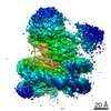 9999MC  0693C  0694C 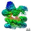 0695C 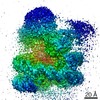 9998C  6kiuC 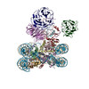 6kiwC 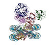 6kixC 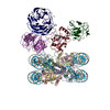 6kizC M: map data used to model this data C: citing same article ( |
|---|---|
| Similar structure data |
-Links
 PDBj
PDBj


- Assembly
| Deposited unit | 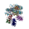
|
|---|---|
| 1 |
|
-Components
-Protein , 9 types, 13 molecules AEBFCGDHKNOTR
| #1: Protein | Mass: 15303.930 Da / Num. of mol.: 2 Source method: isolated from a genetically manipulated source Source: (gene. exp.) Xenopus laevis (African clawed frog) / Gene: XELAEV_18002543mg / Production host:  Escherichia coli (E. coli) / References: UniProt: A0A310TTQ1, UniProt: P84233*PLUS Escherichia coli (E. coli) / References: UniProt: A0A310TTQ1, UniProt: P84233*PLUS#2: Protein | Mass: 11263.231 Da / Num. of mol.: 2 Source method: isolated from a genetically manipulated source Source: (gene. exp.) Xenopus laevis (African clawed frog) / Production host: Escherichia coli (E. coli) / References: UniProt: P62799#3: Protein | Mass: 13978.241 Da / Num. of mol.: 2 Source method: isolated from a genetically manipulated source Source: (gene. exp.) Xenopus laevis (African clawed frog) / Gene: hist1h2aj, LOC494591, XELAEV_18003602mg / Production host: Escherichia coli (E. coli) / References: UniProt: Q6AZJ8, UniProt: P06897*PLUS#4: Protein | Mass: 13498.715 Da / Num. of mol.: 2 / Mutation: S29T/K117C Source method: isolated from a genetically manipulated source Source: (gene. exp.) Xenopus laevis (African clawed frog) / Production host: Escherichia coli (E. coli) / References: UniProt: P02281#7: Protein | | Mass: 24970.539 Da / Num. of mol.: 1 Source method: isolated from a genetically manipulated source Source: (gene. exp.) Homo sapiens (human) / Gene: KMT2A, ALL1, CXXC7, HRX, HTRX, MLL, MLL1, TRX1 / Production host: Escherichia coli (E. coli)References: UniProt: Q03164, histone-lysine N-methyltransferase#8: Protein | | Mass: 59223.477 Da / Num. of mol.: 1 Source method: isolated from a genetically manipulated source Source: (gene. exp.) Homo sapiens (human) / Gene: RBBP5, RBQ3 / Production host: Escherichia coli (E. coli) / References: UniProt: Q15291#9: Protein | | Mass: 8622.922 Da / Num. of mol.: 1 / Mutation: G76C Source method: isolated from a genetically manipulated source Source: (gene. exp.) Homo sapiens (human) / Gene: RPS27A, UBA80, UBCEP1 / Production host: Escherichia coli (E. coli) / References: UniProt: P62979#10: Protein | | Mass: 60288.758 Da / Num. of mol.: 1 Source method: isolated from a genetically manipulated source Source: (gene. exp.) Homo sapiens (human) / Gene: ASH2L, ASH2L1 / Production host: Escherichia coli (E. coli) / References: UniProt: Q9UBL3#11: Protein | | Mass: 36635.438 Da / Num. of mol.: 1 Source method: isolated from a genetically manipulated source Source: (gene. exp.) Homo sapiens (human) / Gene: WDR5, BIG3 / Production host: Escherichia coli (E. coli) / References: UniProt: P61964 |
|---|
-DNA chain , 2 types, 2 molecules IJ
| #5: DNA chain | Mass: 44521.367 Da / Num. of mol.: 1 / Source method: obtained synthetically / Source: (synth.) synthetic construct (others) |
|---|---|
| #6: DNA chain | Mass: 44992.648 Da / Num. of mol.: 1 / Source method: obtained synthetically / Source: (synth.) synthetic construct (others) |
-Non-polymers , 2 types, 2 molecules 

| #12: Chemical | ChemComp-SAH / S-Adenosyl-L-homocysteine Type: L-peptide linking / Mass: 384.411 Da / Num. of mol.: 1 / Source method: obtained synthetically / Formula: C14H20N6O5S Type: L-peptide linking / Mass: 384.411 Da / Num. of mol.: 1 / Source method: obtained synthetically / Formula: C14H20N6O5S |
|---|---|
| #13: Chemical | ChemComp-ZN /  Mass: 65.409 Da / Num. of mol.: 1 / Source method: obtained synthetically / Formula: Zn Mass: 65.409 Da / Num. of mol.: 1 / Source method: obtained synthetically / Formula: Zn |
-Details
| Has ligand of interest | N |
|---|
-Experimental details
-Experiment
| Experiment | Method: ELECTRON MICROSCOPY |
|---|---|
| EM experiment | Aggregation state: PARTICLE / 3D reconstruction method: single particle reconstruction |
- Sample preparation
| Component |
| ||||||||||||||||||||||||
|---|---|---|---|---|---|---|---|---|---|---|---|---|---|---|---|---|---|---|---|---|---|---|---|---|---|
| Source (natural) |
| ||||||||||||||||||||||||
| Source (recombinant) |
| ||||||||||||||||||||||||
| Buffer solution | pH: 7.5 | ||||||||||||||||||||||||
| Specimen | Embedding applied: NO / Shadowing applied: NO / Staining applied: NO / Vitrification applied: YES | ||||||||||||||||||||||||
| Vitrification | Cryogen name: ETHANE |
- Electron microscopy imaging
| Experimental equipment |  Model: Titan Krios / Image courtesy: FEI Company |
|---|---|
| Microscopy | Model: FEI TITAN KRIOS |
| Electron gun | Electron source: FIELD EMISSION GUN / Accelerating voltage: 300 kV / Illumination mode: FLOOD BEAM |
| Electron lens | Mode: BRIGHT FIELDBright-field microscopy |
| Image recording | Electron dose: 40 e/Å2 / Film or detector model: FEI FALCON III (4k x 4k) |
- Processing
| Software |
| ||||||||||||||||||||||||
|---|---|---|---|---|---|---|---|---|---|---|---|---|---|---|---|---|---|---|---|---|---|---|---|---|---|
| CTF correction | Type: PHASE FLIPPING ONLY | ||||||||||||||||||||||||
| Symmetry | Point symmetry: C1 (asymmetric) | ||||||||||||||||||||||||
| 3D reconstruction | Resolution: 4 Å / Resolution method: FSC 0.143 CUT-OFF / Num. of particles: 127898 / Symmetry type: POINT | ||||||||||||||||||||||||
| Refinement | Stereochemistry target values: GeoStd + Monomer Library | ||||||||||||||||||||||||
| Refine LS restraints |
|
