 Movie
Movie Controller


+ Open data

- Basic information
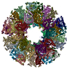
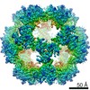
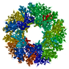
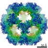
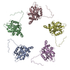
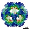
| Entry | Database: PDB / ID: 6h55 | ||||||
|---|---|---|---|---|---|---|---|
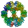
| Title | core of the human pyruvate dehydrogenase (E2) | ||||||
 Components Components | Dihydrolipoyllysine-residue acetyltransferase component of pyruvate dehydrogenase complex, mitochondrial Dihydrolipoyl transacetylase Dihydrolipoyl transacetylase | ||||||
 Keywords Keywords | OXIDOREDUCTASE / Pyruvate dehydrogenase / human | ||||||
| Function / homology |  Function and homology informationdihydrolipoyllysine-residue acetyltransferase / dihydrolipoyllysine-residue acetyltransferase activity / acetyl-CoA biosynthetic process from pyruvate / pyruvate dehydrogenase complex / : / Pyruvate metabolism / Glyoxylate metabolism and glycine degradation / Regulation of pyruvate dehydrogenase (PDH) complex / Signaling by Retinoic Acid / tricarboxylic acid cycle ...dihydrolipoyllysine-residue acetyltransferase / dihydrolipoyllysine-residue acetyltransferase activity / acetyl-CoA biosynthetic process from pyruvate / pyruvate dehydrogenase complex / : / Pyruvate metabolism / Glyoxylate metabolism and glycine degradation / Regulation of pyruvate dehydrogenase (PDH) complex / Signaling by Retinoic Acid / tricarboxylic acid cycle / glucose metabolic process / mitochondrial matrix / intracellular membrane-bounded organelle / mitochondrion / identical protein binding Function and homology informationdihydrolipoyllysine-residue acetyltransferase / dihydrolipoyllysine-residue acetyltransferase activity / acetyl-CoA biosynthetic process from pyruvate / pyruvate dehydrogenase complex / : / Pyruvate metabolism / Glyoxylate metabolism and glycine degradation / Regulation of pyruvate dehydrogenase (PDH) complex / Signaling by Retinoic Acid / tricarboxylic acid cycle ...dihydrolipoyllysine-residue acetyltransferase / dihydrolipoyllysine-residue acetyltransferase activity / acetyl-CoA biosynthetic process from pyruvate / pyruvate dehydrogenase complex / : / Pyruvate metabolism / Glyoxylate metabolism and glycine degradation / Regulation of pyruvate dehydrogenase (PDH) complex / Signaling by Retinoic Acid / tricarboxylic acid cycle / glucose metabolic process / mitochondrial matrix / intracellular membrane-bounded organelle / mitochondrion / identical protein bindingSimilarity search - Function | ||||||
| Biological species |  Homo sapiens (human) Homo sapiens (human) | ||||||
| Method | ELECTRON MICROSCOPY / single particle reconstruction / cryo EM / Resolution: 6 Å | ||||||
 Authors Authors | Haselbach, D. / Prajapati, S. / Tittmann, K. / Stark, H. | ||||||
| Funding support |  Germany, 1items Germany, 1items
| ||||||
 Citation Citation | Journal: Structure / Year: 2019 Title: Structural and Functional Analyses of the Human PDH Complex Suggest a "Division-of-Labor" Mechanism by Local E1 and E3 Clusters. Authors: Sabin Prajapati / David Haselbach / Sabine Wittig / Mulchand S Patel / Ashwin Chari / Carla Schmidt / Holger Stark / Kai Tittmann /  Abstract: The pseudo-atomic structural model of human pyruvate dehydrogenase complex (PDHc) core composed of full-length E2 and E3BP components, calculated from our cryoelectron microscopy-derived density maps ...The pseudo-atomic structural model of human pyruvate dehydrogenase complex (PDHc) core composed of full-length E2 and E3BP components, calculated from our cryoelectron microscopy-derived density maps at 6-Å resolution, is similar to those of prokaryotic E2 structures. The spatial organization of human PDHc components as evidenced by negative-staining electron microscopy and native mass spectrometry is not homogeneous, and entails the unanticipated formation of local clusters of E1:E2 and E3BP:E3 complexes. Such uneven, clustered organization translates into specific duties for E1-E2 clusters (oxidative decarboxylation and acetyl transfer) and E3BP-E3 clusters (regeneration of reduced lipoamide) corresponding to half-reactions of the PDHc catalytic cycle. The addition of substrate coenzyme A modulates the conformational landscape of PDHc, in particular of the lipoyl domains, extending the postulated multiple random coupling mechanism. The conformational and associated chemical landscapes of PDHc are thus not determined entirely stochastically, but are restrained and channeled through an asymmetric architecture and further modulated by substrate binding. | ||||||
| History |
|
- Structure visualization
| Movie |
Movie viewer |
|---|---|
| Structure viewer | Molecule: MolmilJmol/JSmol |
- Downloads & links
-Download
| PDBx/mmCIF format | 6h55.cif.gz | 50.3 KB | Display | PDBx/mmCIF format |
|---|---|---|---|---|
| PDB format | pdb6h55.ent.gz | 27.8 KB | Display | PDB format |
| PDBx/mmJSON format | 6h55.json.gz | Tree view | PDBx/mmJSON format | |
| Others |  Other downloads Other downloads |
-Validation report
| Arichive directory | https://data.pdbj.org/pub/pdb/validation_reports/h5/6h55ftp://data.pdbj.org/pub/pdb/validation_reports/h5/6h55 | HTTPS FTP |
|---|
-Related structure data
| Related structure data | 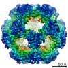 0138MC 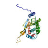 6h60C M: map data used to model this data C: citing same article ( |
|---|---|
| Similar structure data |

-Links
 PDBj
PDBj


- Assembly
| Deposited unit | 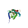
|
|---|---|
| 1 | x 60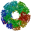
|
-Components
| #1: Protein | Dihydrolipoyl transacetylase / 70 kDa mitochondrial autoantigen of primary biliary cirrhosis / PBC / Dihydrolipoamide ...70 kDa mitochondrial autoantigen of primary biliary cirrhosis / PBC / Dihydrolipoamide acetyltransferase component of pyruvate dehydrogenase complex / M2 antigen complex 70 kDa subunit / Pyruvate dehydrogenase complex component E2 / PDCE2 Mass: 69069.398 Da / Num. of mol.: 1 Source method: isolated from a genetically manipulated source Source: (gene. exp.) Homo sapiens (human) / Gene: DLAT, DLTA / Production host:  Escherichia coli (E. coli) Escherichia coli (E. coli)References: UniProt: P10515, dihydrolipoyllysine-residue acetyltransferase |
|---|
-Experimental details
-Experiment
| Experiment | Method: ELECTRON MICROSCOPY |
|---|---|
| EM experiment | Aggregation state: PARTICLE / 3D reconstruction method: single particle reconstruction |
- Sample preparation
| Component | Name: single particle cryo EM-derived map of the full-length native human E2-E3BP core of the pyruvate dehydrogenase multienzyme complex Type: COMPLEX / Entity ID: all / Source: RECOMBINANT |
|---|---|
| Source (natural) | Organism: Homo sapiens (human) |
| Source (recombinant) | Organism: Escherichia coli (E. coli) |
| Buffer solution | pH: 8 |
| Specimen | Embedding applied: NO / Shadowing applied: NO / Staining applied: NO / Vitrification applied: YES |
| Specimen support | Grid type: Quantifoil R3.5/1 |
| Vitrification | Cryogen name: ETHANE |
- Electron microscopy imaging
| Experimental equipment |  Model: Titan Krios / Image courtesy: FEI Company |
|---|---|
| Microscopy | Model: FEI TITAN KRIOS |
| Electron gun | Electron source: FIELD EMISSION GUN / Accelerating voltage: 300 kV / Illumination mode: SPOT SCAN |
| Electron lens | Mode: BRIGHT FIELDBright-field microscopy / Cs: 0.01 mm |
| Image recording | Average exposure time: 1 sec. / Electron dose: 41 e/Å2 / Detector mode: INTEGRATING / Film or detector model: FEI FALCON II (4k x 4k) / Num. of grids imaged: 1 / Num. of real images: 2250 |
| EM imaging optics | Spherical aberration corrector: Microscope was modified with a Cs corrector with two hexapole elements |
| Image scans | Movie frames/image: 1 |
- Processing
| CTF correction | Type: PHASE FLIPPING AND AMPLITUDE CORRECTION |
|---|---|
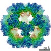
| Symmetry | Point symmetry: I (icosahedral) |
| 3D reconstruction | Resolution: 6 Å / Resolution method: FSC 0.143 CUT-OFF / Num. of particles: 29788 / Algorithm: FOURIER SPACE / Symmetry type: POINT |
