 Movie
Movie Controller


+ Open data

- Basic information
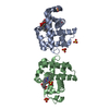
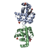
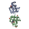
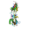
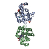
| Entry | Database: PDB / ID: 6cw1 | ||||||
|---|---|---|---|---|---|---|---|
| Title | Crystal structure of Neurexin-1 alpha ectodomain fragment, L2-L3 | ||||||
 Components Components | Neurexin-1 | ||||||
 Keywords Keywords | CELL ADHESION / LNS domain / Beta-Sandwich / Synapse | ||||||
| Function / homology |  Function and homology information Function and homology informationneuroligin family protein binding / cell projection / presynaptic membrane / cell adhesion / metal ion bindingSimilarity search - Function | ||||||
| Biological species |  Bos taurus (cattle) Bos taurus (cattle) | ||||||
| Method | X-RAY DIFFRACTION / SYNCHROTRON / MOLECULAR REPLACEMENT / molecular replacement / Resolution: 2.84 Å | ||||||
 Authors Authors | Misra, A. / Rudenko, G. | ||||||
| Funding support |  United States, 1items United States, 1items
| ||||||
 Citation Citation | Journal: J Mol Biol / Year: 2018 Title: Structural Plasticity of Neurexin 1α: Implications for its Role as Synaptic Organizer. Authors: Jianfang Liu / Anurag Misra / M V V V Sekhar Reddy / Mark Andrew White / Gang Ren / Gabby Rudenko / Abstract: α-Neurexins are synaptic organizing molecules implicated in neuropsychiatric disorders. They bind and arrange an array of different partners in the synaptic cleft. The extracellular region of ...α-Neurexins are synaptic organizing molecules implicated in neuropsychiatric disorders. They bind and arrange an array of different partners in the synaptic cleft. The extracellular region of neurexin 1α (n1α) contains six LNS domains (L1-L6) interspersed by three Egf-like repeats. N1α must encode highly evolved structure-function relationships in order to fit into the narrow confines of the synaptic cleft, and also recruit its large, membrane-bound partners. Internal molecular flexibility could provide a solution; however, it is challenging to delineate because currently no structural methods permit high-resolution structure determination of large, flexible, multi-domain protein molecules. To investigate the structural plasticity of n1α, in particular the conformation of domains that carry validated binding sites for different protein partners, we used a panel of structural techniques. Individual particle electron tomography revealed that the N-terminally and C-terminally tethered domains, L1 and L6, have a surprisingly limited range of conformational freedom with respect to the linear central core containing L2 through L5. A 2.8-Å crystal structure revealed an unexpected arrangement of the L2 and L3 domains. Small-angle X-ray scattering and electron tomography indicated that incorporation of the alternative splice insert SS6 relieves the restricted conformational freedom between L5 and L6, suggesting that SS6 may work as a molecular toggle. The architecture of n1α thus encodes a combination of rigid and flexibly tethered domains that are uniquely poised to work together to promote its organizing function in the synaptic cleft, and may permit allosterically regulated and/or concerted protein partner binding. | ||||||
| History |
|
- Structure visualization
| Structure viewer | Molecule: MolmilJmol/JSmol |
|---|
- Downloads & links
-Download
| PDBx/mmCIF format | 6cw1.cif.gz | 302.6 KB | Display | PDBx/mmCIF format |
|---|---|---|---|---|
| PDB format | pdb6cw1.ent.gz | 245.1 KB | Display | PDB format |
| PDBx/mmJSON format | 6cw1.json.gz | Tree view | PDBx/mmJSON format | |
| Others |  Other downloads Other downloads |
-Validation report
| Arichive directory | https://data.pdbj.org/pub/pdb/validation_reports/cw/6cw1ftp://data.pdbj.org/pub/pdb/validation_reports/cw/6cw1 | HTTPS FTP |
|---|
-Related structure data
| Related structure data |  7639C  7640C  7641C  7642C 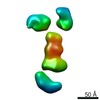 7643C 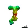 7644C 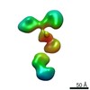 7645C 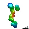 7646C 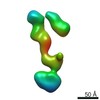 7647C 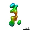 7648C 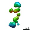 7649C  7650C 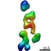 7651C  7652C  7653C  7654C  7655C  7656C  7657C  7658C  7659C 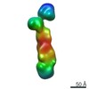 7660C 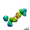 7661C 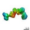 7662C 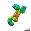 7663C 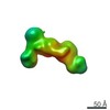 7664C 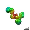 7665C 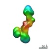 7666C 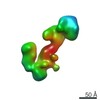 7667C 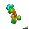 7668C 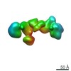 7669C 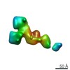 7670C 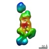 7671C 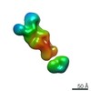 7672C 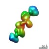 7673C 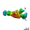 7674C 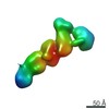 7675C 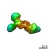 7676C 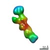 7677C 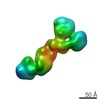 7678C  7679C 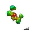 7680C 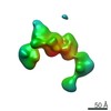 7681C 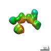 7682C 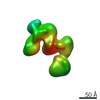 7683C 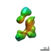 7684C 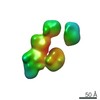 7685C 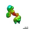 7686C 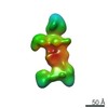 7687C 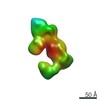 7688C 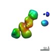 7689C 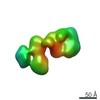 7690C 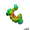 7691C 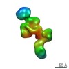 7692C 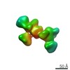 7693C 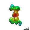 7694C 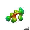 7695C 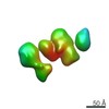 7696C 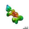 7697C 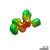 7698C 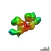 7699C 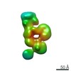 7700C 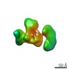 7701C 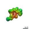 7702C  7703C 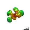 7704C 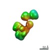 7705C  7706C  7707C  7708C  7709C 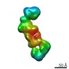 7710C 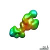 7711C 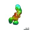 7712C 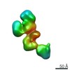 7713C 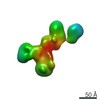 7714C 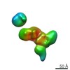 7715C 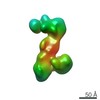 7716C  7717C 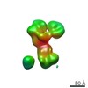 7718C 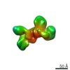 7719C 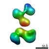 7720C 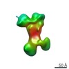 7721C 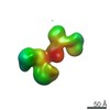 7722C 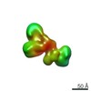 7723C 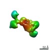 7724C 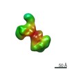 7725C 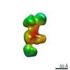 7726C 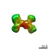 7727C 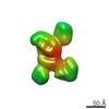 7728C 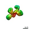 7729C 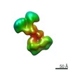 7730C 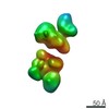 7731C 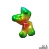 7732C 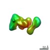 7733C 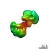 7734C  7735C  7736C  7737C  7738C 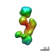 7739C 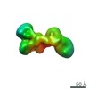 7740C 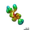 7741C 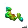 7742C 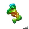 7743C  7744C 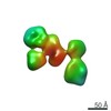 7745C  7746C 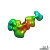 7747C 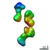 7748C 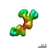 7749C 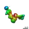 7750C 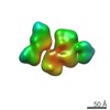 7751C 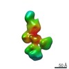 7752C 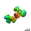 7753C 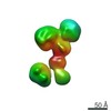 7754C 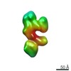 7755C 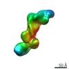 7756C  7757C  7758C  7759C  7760C 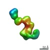 7761C 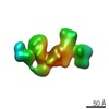 7762C 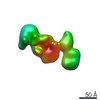 7763C 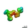 7764C 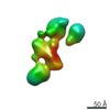 7765C 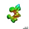 7766C 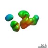 7767C 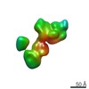 7768C  3qcwS S: Starting model for refinement C: citing same article ( |
|---|---|
| Similar structure data |


-Links
 PDBj
PDBj


- Assembly
| Deposited unit | 
| ||||||||||||||||||
|---|---|---|---|---|---|---|---|---|---|---|---|---|---|---|---|---|---|---|---|
| 1 | 
| ||||||||||||||||||
| 2 | 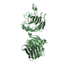
| ||||||||||||||||||
| Unit cell |
| ||||||||||||||||||
| Noncrystallographic symmetry (NCS) | NCS domain:
NCS domain segments: Component-ID: 0 / Ens-ID: 1 / Beg auth comp-ID: LYS / Beg label comp-ID: LYS / End auth comp-ID: THR / End label comp-ID: THR / Refine code: 0 / Auth seq-ID: 279 - 672 / Label seq-ID: 22 - 400
|
-Components
| #1: Protein | / Neurexin I-alpha / Neurexin-1-alpha Mass: 44517.121 Da / Num. of mol.: 2 / Fragment: Alpha fragment L2-L3, residues 258-674 Source method: isolated from a genetically manipulated source Source: (gene. exp.) Bos taurus (cattle) / Gene: NRXN1 / Plasmid: pGEX-KG / Production host:  Escherichia coli BL21(DE3) (bacteria) / Strain (production host): BL21(DE3) / References: UniProt: Q28146 Escherichia coli BL21(DE3) (bacteria) / Strain (production host): BL21(DE3) / References: UniProt: Q28146#2: Water | ChemComp-HOH / | Water Mass: 18.015 Da / Num. of mol.: 12 / Source method: isolated from a natural source / Formula: H2O Mass: 18.015 Da / Num. of mol.: 12 / Source method: isolated from a natural source / Formula: H2O |
|---|
-Experimental details
-Experiment
| Experiment | Method: X-RAY DIFFRACTION / Number of used crystals: 1 |
|---|
- Sample preparation
| Crystal | Density Matthews: 3.45 Å3/Da / Density % sol: 64.39 % / Mosaicity: 1.742 ° |
|---|---|
| Crystal grow | Temperature: 298 K / Method: vapor diffusion, hanging drop / pH: 8 / Details: 0.9 M NaCITRATE, 0.1 M TRIS PH 8.0, 5 MM CACL2 |
-Data collection
| Diffraction | Mean temperature: 100 K | ||||||||||||||||||||||||||||||||||||||||||||||||||||||||||||||||||||||||||||||||||||||||||||||||||||||||||||||
|---|---|---|---|---|---|---|---|---|---|---|---|---|---|---|---|---|---|---|---|---|---|---|---|---|---|---|---|---|---|---|---|---|---|---|---|---|---|---|---|---|---|---|---|---|---|---|---|---|---|---|---|---|---|---|---|---|---|---|---|---|---|---|---|---|---|---|---|---|---|---|---|---|---|---|---|---|---|---|---|---|---|---|---|---|---|---|---|---|---|---|---|---|---|---|---|---|---|---|---|---|---|---|---|---|---|---|---|---|---|---|---|
| Diffraction source | Source: SYNCHROTRON / Site: APS / Beamline: 21-ID-D / Wavelength: 1.10208 Å | ||||||||||||||||||||||||||||||||||||||||||||||||||||||||||||||||||||||||||||||||||||||||||||||||||||||||||||||
| Detector | Type: MARMOSAIC 300 mm CCD / Detector: CCD / Date: Mar 21, 2008 | ||||||||||||||||||||||||||||||||||||||||||||||||||||||||||||||||||||||||||||||||||||||||||||||||||||||||||||||
| Radiation | Protocol: SINGLE WAVELENGTH / Monochromatic (M) / Laue (L): M / Scattering type: x-ray | ||||||||||||||||||||||||||||||||||||||||||||||||||||||||||||||||||||||||||||||||||||||||||||||||||||||||||||||
| Radiation wavelength | Wavelength: 1.10208 Å / Relative weight: 1 | ||||||||||||||||||||||||||||||||||||||||||||||||||||||||||||||||||||||||||||||||||||||||||||||||||||||||||||||
| Reflection | Resolution: 2.84→50.01 Å / Num. obs: 28540 / % possible obs: 98.4 % / Redundancy: 3.4 % / Rmerge(I) obs: 0.091 / Rpim(I) all: 0.058 / Rrim(I) all: 0.108 / Χ2: 1.469 / Net I/σ(I): 15.2 | ||||||||||||||||||||||||||||||||||||||||||||||||||||||||||||||||||||||||||||||||||||||||||||||||||||||||||||||
| Reflection shell | Diffraction-ID: 1
|
-Phasing
| Phasing | Method: molecular replacement |
|---|
- Processing
| Software |
| |||||||||||||||||||||||||||||||||||||||||||||||||||||||||||||||||||||||||||||||||||||||||||||||||||||||||||||||||||||||||||||
|---|---|---|---|---|---|---|---|---|---|---|---|---|---|---|---|---|---|---|---|---|---|---|---|---|---|---|---|---|---|---|---|---|---|---|---|---|---|---|---|---|---|---|---|---|---|---|---|---|---|---|---|---|---|---|---|---|---|---|---|---|---|---|---|---|---|---|---|---|---|---|---|---|---|---|---|---|---|---|---|---|---|---|---|---|---|---|---|---|---|---|---|---|---|---|---|---|---|---|---|---|---|---|---|---|---|---|---|---|---|---|---|---|---|---|---|---|---|---|---|---|---|---|---|---|---|---|
| Refinement | Method to determine structure: MOLECULAR REPLACEMENT Starting model: 3QCW Resolution: 2.84→50.01 Å / Cor.coef. Fo:Fc: 0.923 / Cor.coef. Fo:Fc free: 0.889 / SU B: 35.561 / SU ML: 0.307 / SU R Cruickshank DPI: 0.7583 / Cross valid method: THROUGHOUT / σ(F): 0 / ESU R: 0.758 / ESU R Free: 0.341 Details: HYDROGENS HAVE BEEN ADDED IN THE RIDING POSITIONS U VALUES : WITH TLS ADDED
| |||||||||||||||||||||||||||||||||||||||||||||||||||||||||||||||||||||||||||||||||||||||||||||||||||||||||||||||||||||||||||||
| Solvent computation | Ion probe radii: 0.8 Å / Shrinkage radii: 0.8 Å / VDW probe radii: 1.2 Å | |||||||||||||||||||||||||||||||||||||||||||||||||||||||||||||||||||||||||||||||||||||||||||||||||||||||||||||||||||||||||||||
| Displacement parameters | Biso max: 138.21 Å2 / Biso mean: 52.259 Å2 / Biso min: 24.48 Å2
| |||||||||||||||||||||||||||||||||||||||||||||||||||||||||||||||||||||||||||||||||||||||||||||||||||||||||||||||||||||||||||||
| Refinement step | Cycle: final / Resolution: 2.84→50.01 Å
| |||||||||||||||||||||||||||||||||||||||||||||||||||||||||||||||||||||||||||||||||||||||||||||||||||||||||||||||||||||||||||||
| Refine LS restraints |
| |||||||||||||||||||||||||||||||||||||||||||||||||||||||||||||||||||||||||||||||||||||||||||||||||||||||||||||||||||||||||||||
| Refine LS restraints NCS | Ens-ID: 1 / Number: 22294 / Refine-ID: X-RAY DIFFRACTION / Type: interatomic distance / Rms dev position: 0.1 Å / Weight position: 0.05
| |||||||||||||||||||||||||||||||||||||||||||||||||||||||||||||||||||||||||||||||||||||||||||||||||||||||||||||||||||||||||||||
| LS refinement shell | Resolution: 2.837→2.911 Å / Rfactor Rfree error: 0 / Total num. of bins used: 20
| |||||||||||||||||||||||||||||||||||||||||||||||||||||||||||||||||||||||||||||||||||||||||||||||||||||||||||||||||||||||||||||
| Refinement TLS params. | Method: refined / Refine-ID: X-RAY DIFFRACTION
| |||||||||||||||||||||||||||||||||||||||||||||||||||||||||||||||||||||||||||||||||||||||||||||||||||||||||||||||||||||||||||||
| Refinement TLS group |
|
