 Movie
Movie Controller

[English] 日本語
 Yorodumi
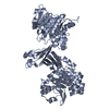
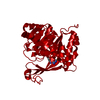
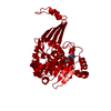
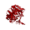
- PDB-6c67: Mycobacterium tuberculosis adenosine kinase bound to iodotubercidin -
+ Open data

- Basic information
| Entry | Database: PDB / ID: 6c67 | ||||||||||||
|---|---|---|---|---|---|---|---|---|---|---|---|---|---|
| Title | Mycobacterium tuberculosis adenosine kinase bound to iodotubercidin | ||||||||||||
 Components Components | Adenosine kinase | ||||||||||||
 Keywords Keywords | TRANSFERASE/TRANSFERASE INHIBITOR / Nucleoside analog / Complex / Inhibitor / Structural Genomics / PSI-2 / Protein Structure Initiative / TB Structural Genomics Consortium / TBSGC / TRANSFERASE-TRANSFERASE INHIBITOR complex | ||||||||||||
| Function / homology |  Function and homology informationadenosine kinase / adenosine kinase activity / dGTP binding / AMP salvage / purine ribonucleoside salvage / phosphorylation / GTP binding / magnesium ion binding / ATP binding / plasma membrane Function and homology informationadenosine kinase / adenosine kinase activity / dGTP binding / AMP salvage / purine ribonucleoside salvage / phosphorylation / GTP binding / magnesium ion binding / ATP binding / plasma membraneSimilarity search - Function | ||||||||||||
| Biological species |   Mycobacterium tuberculosis (bacteria) Mycobacterium tuberculosis (bacteria) | ||||||||||||
| Method | X-RAY DIFFRACTION / SYNCHROTRON / MOLECULAR REPLACEMENT / Resolution: 2.11 Å | ||||||||||||
 Authors Authors | Crespo, R.A. / TB Structural Genomics Consortium (TBSGC) | ||||||||||||
| Funding support |  United States, 3items United States, 3items
| ||||||||||||
 Citation Citation | Journal: J.Med.Chem. / Year: 2019 Title: Structure-Guided Drug Design of 6-Substituted Adenosine Analogues as Potent Inhibitors of Mycobacterium tuberculosis Adenosine Kinase. Authors: Crespo, R.A. / Dang, Q. / Zhou, N.E. / Guthrie, L.M. / Snavely, T.C. / Dong, W. / Loesch, K.A. / Suzuki, T. / You, L. / Wang, W. / O'Malley, T. / Parish, T. / Olsen, D.B. / Sacchettini, J.C. | ||||||||||||
| History |
|
- Structure visualization
| Structure viewer | Molecule: MolmilJmol/JSmol |
|---|
- Downloads & links
-Download
| PDBx/mmCIF format | 6c67.cif.gz | 235.9 KB | Display | PDBx/mmCIF format |
|---|---|---|---|---|
| PDB format | pdb6c67.ent.gz | 190.4 KB | Display | PDB format |
| PDBx/mmJSON format | 6c67.json.gz | Tree view | PDBx/mmJSON format | |
| Others |  Other downloads Other downloads |
-Validation report
| Arichive directory | https://data.pdbj.org/pub/pdb/validation_reports/c6/6c67ftp://data.pdbj.org/pub/pdb/validation_reports/c6/6c67 | HTTPS FTP |
|---|
-Related structure data
| Related structure data | 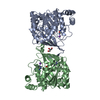 6c9nC 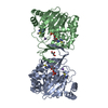 6c9pC  6c9qC 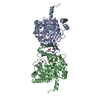 6c9rC 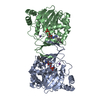 6c9sC 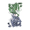 6c9vC 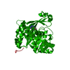 2pkmS S: Starting model for refinement C: citing same article ( |
|---|---|
| Similar structure data |


-Links
 PDBj
PDBj- Assembly
| Deposited unit | 
| ||||||||
|---|---|---|---|---|---|---|---|---|---|
| 1 |
| ||||||||
| Unit cell |
|
-Components
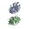
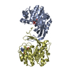
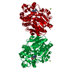
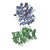
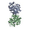
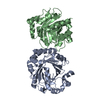
-Protein , 1 types, 2 molecules AB
| #1: Protein | / AK Mass: 34503.953 Da / Num. of mol.: 2 Source method: isolated from a genetically manipulated source Source: (gene. exp.) Mycobacterium tuberculosis (strain ATCC 25618 / H37Rv) (bacteria)Strain: ATCC 25618 / H37Rv / Gene: adoK, cbhK, Rv2202c, MTCY190.13c / Production host: Escherichia coli (E. coli) / References: UniProt: P9WID5, adenosine kinase |
|---|
-Non-polymers , 5 types, 268 molecules 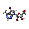


| #2: Chemical | ChemComp-5ID / (  Mass: 392.150 Da / Num. of mol.: 4 / Source method: obtained synthetically / Formula: C11H13IN4O4 Mass: 392.150 Da / Num. of mol.: 4 / Source method: obtained synthetically / Formula: C11H13IN4O4#3: Chemical |  Mass: 22.990 Da / Num. of mol.: 2 / Source method: obtained synthetically / Formula: Na Mass: 22.990 Da / Num. of mol.: 2 / Source method: obtained synthetically / Formula: Na#4: Chemical | ChemComp-GOL / | Glycerol Mass: 92.094 Da / Num. of mol.: 1 / Source method: obtained synthetically / Formula: C3H8O3 Mass: 92.094 Da / Num. of mol.: 1 / Source method: obtained synthetically / Formula: C3H8O3#5: Chemical | Sulfate Mass: 96.063 Da / Num. of mol.: 2 / Source method: obtained synthetically / Formula: SO4 Mass: 96.063 Da / Num. of mol.: 2 / Source method: obtained synthetically / Formula: SO4#6: Water | ChemComp-HOH / | WaterMass: 18.015 Da / Num. of mol.: 259 / Source method: isolated from a natural source / Formula: H2O |
|---|
-Experimental details
-Experiment
| Experiment | Method: X-RAY DIFFRACTION / Number of used crystals: 1 |
|---|
- Sample preparation
| Crystal | Density Matthews: 2.29 Å3/Da / Density % sol: 46.25 % |
|---|---|
| Crystal grow | Temperature: 290 K / Method: vapor diffusion, hanging drop Details: 0.1 M HEPES, pH 7.5, 2.0 M ammonium sulfate, 2.0% PEG400 |
-Data collection
| Diffraction | Mean temperature: 100 K |
|---|---|
| Diffraction source | Source: SYNCHROTRON / Site: APS / Beamline: 19-ID / Wavelength: 1 Å |
| Detector | Type: ADSC QUANTUM 315 / Detector: CCD / Date: Nov 25, 2013 |
| Radiation | Monochromator: double crystal Si(111) / Protocol: SINGLE WAVELENGTH / Monochromatic (M) / Laue (L): M / Scattering type: x-ray |
| Radiation wavelength | Wavelength: 1 Å / Relative weight: 1 |
| Reflection | Resolution: 2.105→34.69 Å / Num. obs: 34540 / % possible obs: 96.5 % / Redundancy: 7.3 % / Biso Wilson estimate: 40.73 Å2 / Rmerge(I) obs: 0.0793 / Net I/σ(I): 23.36 |
| Reflection shell | Resolution: 2.11→2.18 Å / Rmerge(I) obs: 0.2468 / Num. unique obs: 3485 / CC1/2: 0.985 / Rpim(I) all: 0.09564 / Rrim(I) all: 0.2648 |
- Processing
| Software |
| |||||||||||||||||||||||||||||||||||||||||||||||||||||||||||||||||||||||||||||||||||||||||||
|---|---|---|---|---|---|---|---|---|---|---|---|---|---|---|---|---|---|---|---|---|---|---|---|---|---|---|---|---|---|---|---|---|---|---|---|---|---|---|---|---|---|---|---|---|---|---|---|---|---|---|---|---|---|---|---|---|---|---|---|---|---|---|---|---|---|---|---|---|---|---|---|---|---|---|---|---|---|---|---|---|---|---|---|---|---|---|---|---|---|---|---|---|
| Refinement | Method to determine structure: MOLECULAR REPLACEMENT Starting model: PDB entry 2PKM Resolution: 2.11→34.69 Å / SU ML: 0.2 / Cross valid method: FREE R-VALUE / σ(F): 1.36 / Phase error: 26.01 / Stereochemistry target values: ML
| |||||||||||||||||||||||||||||||||||||||||||||||||||||||||||||||||||||||||||||||||||||||||||
| Solvent computation | Shrinkage radii: 0.9 Å / VDW probe radii: 1.11 Å / Solvent model: FLAT BULK SOLVENT MODEL | |||||||||||||||||||||||||||||||||||||||||||||||||||||||||||||||||||||||||||||||||||||||||||
| Refinement step | Cycle: LAST / Resolution: 2.11→34.69 Å
| |||||||||||||||||||||||||||||||||||||||||||||||||||||||||||||||||||||||||||||||||||||||||||
| Refine LS restraints |
| |||||||||||||||||||||||||||||||||||||||||||||||||||||||||||||||||||||||||||||||||||||||||||
| LS refinement shell |
|
