 Movie
Movie Controller

[English] 日本語
 Yorodumi
- PDB-5g6v: Crystal structure of the PCTAIRE1 kinase in complex with inhibitor -
+ Open data

- Basic information
| Entry | Database: PDB / ID: 5g6v | ||||||
|---|---|---|---|---|---|---|---|
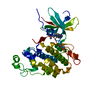
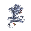
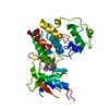
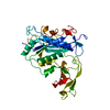
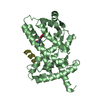
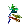
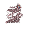
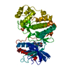
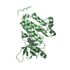
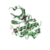
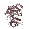
| Title | Crystal structure of the PCTAIRE1 kinase in complex with inhibitor | ||||||
 Components Components | CYCLIN-DEPENDENT KINASE 16 | ||||||
 Keywords Keywords | TRANSFERASE | ||||||
| Function / homology |  Function and homology information: / growth hormone secretion / regulation of insulin secretion involved in cellular response to glucose stimulus / exocytosis / cyclin-dependent protein kinase holoenzyme complex / cyclin-dependent kinase / cyclin-dependent protein serine/threonine kinase activity / positive regulation of autophagy / cytoplasmic side of plasma membrane / neuron projection development ...: / growth hormone secretion / regulation of insulin secretion involved in cellular response to glucose stimulus / exocytosis / cyclin-dependent protein kinase holoenzyme complex / cyclin-dependent kinase / cyclin-dependent protein serine/threonine kinase activity / positive regulation of autophagy / cytoplasmic side of plasma membrane / neuron projection development / microtubule cytoskeleton / synaptic vesicle / spermatogenesis / neuron projection / protein phosphorylation / protein serine kinase activity / protein serine/threonine kinase activity / ATP binding / nucleus / plasma membrane / cytosol / cytoplasm Function and homology information: / growth hormone secretion / regulation of insulin secretion involved in cellular response to glucose stimulus / exocytosis / cyclin-dependent protein kinase holoenzyme complex / cyclin-dependent kinase / cyclin-dependent protein serine/threonine kinase activity / positive regulation of autophagy / cytoplasmic side of plasma membrane / neuron projection development ...: / growth hormone secretion / regulation of insulin secretion involved in cellular response to glucose stimulus / exocytosis / cyclin-dependent protein kinase holoenzyme complex / cyclin-dependent kinase / cyclin-dependent protein serine/threonine kinase activity / positive regulation of autophagy / cytoplasmic side of plasma membrane / neuron projection development / microtubule cytoskeleton / synaptic vesicle / spermatogenesis / neuron projection / protein phosphorylation / protein serine kinase activity / protein serine/threonine kinase activity / ATP binding / nucleus / plasma membrane / cytosol / cytoplasmSimilarity search - Function | ||||||
| Biological species |  HOMO SAPIENS (human) HOMO SAPIENS (human) | ||||||
| Method | X-RAY DIFFRACTION / SYNCHROTRON / MOLECULAR REPLACEMENT / Resolution: 2.2 Å | ||||||
 Authors Authors | Dixon-Clarke, S.E. / Galan Bartual, S. / Elkins, J. / Savitsky, P. / Kopec, J. / Mackenzie, A. / Tallant, C. / Heroven, C. / Burgess-Brown, N. / von Delft, F. ...Dixon-Clarke, S.E. / Galan Bartual, S. / Elkins, J. / Savitsky, P. / Kopec, J. / Mackenzie, A. / Tallant, C. / Heroven, C. / Burgess-Brown, N. / von Delft, F. / Arrowsmith, C.H. / Edwards, A.M. / Bountra, C. / Bullock, A. | ||||||
 Citation Citation | Journal: Biochem.J. / Year: 2017 Title: Structure and inhibitor specificity of the PCTAIRE-family kinase CDK16. Authors: Dixon-Clarke, S.E. / Shehata, S.N. / Krojer, T. / Sharpe, T.D. / von Delft, F. / Sakamoto, K. / Bullock, A.N. | ||||||
| History |
|
- Structure visualization
| Structure viewer | Molecule: MolmilJmol/JSmol |
|---|
- Downloads & links
-Download
| PDBx/mmCIF format | 5g6v.cif.gz | 135.5 KB | Display | PDBx/mmCIF format |
|---|---|---|---|---|
| PDB format | pdb5g6v.ent.gz | 109.7 KB | Display | PDB format |
| PDBx/mmJSON format | 5g6v.json.gz | Tree view | PDBx/mmJSON format | |
| Others |  Other downloads Other downloads |
-Validation report
| Arichive directory | https://data.pdbj.org/pub/pdb/validation_reports/g6/5g6vftp://data.pdbj.org/pub/pdb/validation_reports/g6/5g6v | HTTPS FTP |
|---|
-Related structure data
-Links
 PDBj
PDBj
- Assembly
| Deposited unit | 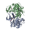
| |||||||||||||||||||||||||||||||||||||||||||||||||||||||||||||
|---|---|---|---|---|---|---|---|---|---|---|---|---|---|---|---|---|---|---|---|---|---|---|---|---|---|---|---|---|---|---|---|---|---|---|---|---|---|---|---|---|---|---|---|---|---|---|---|---|---|---|---|---|---|---|---|---|---|---|---|---|---|---|
| 1 | 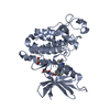
| |||||||||||||||||||||||||||||||||||||||||||||||||||||||||||||
| 2 | 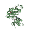
| |||||||||||||||||||||||||||||||||||||||||||||||||||||||||||||
| Unit cell |
| |||||||||||||||||||||||||||||||||||||||||||||||||||||||||||||
| Noncrystallographic symmetry (NCS) | NCS domain:
NCS domain segments: Ens-ID: 1 / End auth comp-ID: ALA / End label comp-ID: ALA / Refine code: 4
NCS oper:
|
-Components
| #1: Protein | / CDK16 / CELL DIVISION PROTEIN KINASE 16 / PCTAIRE-MOTIF PROTEIN KINASE 1 / SERINE/THREONINE-PROTEIN ...CDK16 / CELL DIVISION PROTEIN KINASE 16 / PCTAIRE-MOTIF PROTEIN KINASE 1 / SERINE/THREONINE-PROTEIN KINASE PCTAIRE-1 Mass: 37308.789 Da / Num. of mol.: 2 / Fragment: KINASE DOMAIN, RESIDUES 163-478 / Mutation: YES Source method: isolated from a genetically manipulated source Source: (gene. exp.) HOMO SAPIENS (human) / Production host:  ESCHERICHIA COLI (E. coli) / References: UniProt: Q00536, cyclin-dependent kinase ESCHERICHIA COLI (E. coli) / References: UniProt: Q00536, cyclin-dependent kinase#2: Chemical |   Mass: 553.587 Da / Num. of mol.: 2 / Source method: obtained synthetically / Formula: C30H28FN7O3 Mass: 553.587 Da / Num. of mol.: 2 / Source method: obtained synthetically / Formula: C30H28FN7O3#3: Chemical | ChemComp-EDO / Ethylene glycol  Mass: 62.068 Da / Num. of mol.: 4 / Source method: obtained synthetically / Formula: C2H6O2 Mass: 62.068 Da / Num. of mol.: 4 / Source method: obtained synthetically / Formula: C2H6O2#4: Water | ChemComp-HOH / | Water Mass: 18.015 Da / Num. of mol.: 262 / Source method: isolated from a natural source / Formula: H2O Mass: 18.015 Da / Num. of mol.: 262 / Source method: isolated from a natural source / Formula: H2OSequence details | ENGINEERED | |
|---|
-Experimental details
-Experiment
| Experiment | Method: X-RAY DIFFRACTION / Number of used crystals: 1 |
|---|
- Sample preparation
| Crystal | Density Matthews: 2.44 Å3/Da / Density % sol: 49.51 % / Description: NONE |
|---|---|
| Crystal grow | pH: 5.5 Details: 25% PEG MEDIUM SMEAR (PEG 2000, PEG 3350, PEG 4000, PEG 5000MME) AND 0.1 M CITRATE PH 5.5 |
-Data collection
| Diffraction | Mean temperature: 100 K |
|---|---|
| Diffraction source | Source: SYNCHROTRON / Site: Diamond  / Beamline: I02 / Wavelength: 0.91742 / Beamline: I02 / Wavelength: 0.91742 |
| Detector | Type: DECTRIS PILATUS / Detector: PIXEL / Date: Mar 6, 2015 |
| Radiation | Protocol: SINGLE WAVELENGTH / Monochromatic (M) / Laue (L): M / Scattering type: x-ray |
| Radiation wavelength | Wavelength: 0.91742 Å / Relative weight: 1 |
| Reflection | Resolution: 2.2→28.98 Å / Num. obs: 37175 / % possible obs: 98.3 % / Observed criterion σ(I): 2 / Redundancy: 5.2 % / Rmerge(I) obs: 0.09 / Net I/σ(I): 9.4 |
- Processing
| Software | Name: REFMAC / Version: 5.8.0155 / Classification: refinement | ||||||||||||||||||||||||||||||||||||||||||||||||||||||||||||||||||||||||||||||||||||||||||||||||||||||||||||||||||||||||||||||||||||||||||||||||||||||||||||||||||||||||||||||||||||||
|---|---|---|---|---|---|---|---|---|---|---|---|---|---|---|---|---|---|---|---|---|---|---|---|---|---|---|---|---|---|---|---|---|---|---|---|---|---|---|---|---|---|---|---|---|---|---|---|---|---|---|---|---|---|---|---|---|---|---|---|---|---|---|---|---|---|---|---|---|---|---|---|---|---|---|---|---|---|---|---|---|---|---|---|---|---|---|---|---|---|---|---|---|---|---|---|---|---|---|---|---|---|---|---|---|---|---|---|---|---|---|---|---|---|---|---|---|---|---|---|---|---|---|---|---|---|---|---|---|---|---|---|---|---|---|---|---|---|---|---|---|---|---|---|---|---|---|---|---|---|---|---|---|---|---|---|---|---|---|---|---|---|---|---|---|---|---|---|---|---|---|---|---|---|---|---|---|---|---|---|---|---|---|---|
| Refinement | Method to determine structure: MOLECULAR REPLACEMENT / Resolution: 2.2→86.94 Å / Cor.coef. Fo:Fc: 0.947 / Cor.coef. Fo:Fc free: 0.912 / SU B: 7.587 / SU ML: 0.187 / Cross valid method: THROUGHOUT / ESU R: 0.278 / ESU R Free: 0.234 / Stereochemistry target values: MAXIMUM LIKELIHOOD / Details: HYDROGENS HAVE BEEN ADDED IN THE RIDING POSITIONS.
| ||||||||||||||||||||||||||||||||||||||||||||||||||||||||||||||||||||||||||||||||||||||||||||||||||||||||||||||||||||||||||||||||||||||||||||||||||||||||||||||||||||||||||||||||||||||
| Solvent computation | Ion probe radii: 0.8 Å / Shrinkage radii: 0.8 Å / VDW probe radii: 1.2 Å / Solvent model: MASK | ||||||||||||||||||||||||||||||||||||||||||||||||||||||||||||||||||||||||||||||||||||||||||||||||||||||||||||||||||||||||||||||||||||||||||||||||||||||||||||||||||||||||||||||||||||||
| Displacement parameters | Biso mean: 40.421 Å2
| ||||||||||||||||||||||||||||||||||||||||||||||||||||||||||||||||||||||||||||||||||||||||||||||||||||||||||||||||||||||||||||||||||||||||||||||||||||||||||||||||||||||||||||||||||||||
| Refinement step | Cycle: LAST / Resolution: 2.2→86.94 Å
| ||||||||||||||||||||||||||||||||||||||||||||||||||||||||||||||||||||||||||||||||||||||||||||||||||||||||||||||||||||||||||||||||||||||||||||||||||||||||||||||||||||||||||||||||||||||
| Refine LS restraints |
|