 Movie
Movie Controller

[English] 日本語
 Yorodumi
- PDB-1p19: Hypoxanthine Phosphoribosyltransferase from Trypanosoma cruzi, in... -
+ Open data

- Basic information
| Entry | Database: PDB / ID: 1p19 | ||||||
|---|---|---|---|---|---|---|---|
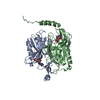
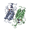
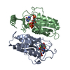
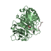
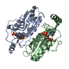
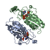
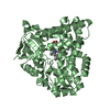
| Title | Hypoxanthine Phosphoribosyltransferase from Trypanosoma cruzi, in complex with the product IMP | ||||||
 Components Components | hypoxanthine phosphoribosyltransferase Hypoxanthine-guanine phosphoribosyltransferase Hypoxanthine-guanine phosphoribosyltransferase | ||||||
 Keywords Keywords | TRANSFERASE / GLYCOSYLTRANSFERASE / PHOSPHORIBOSYLTRANSFERASE / NUCLEOTIDE METABOLISM / PURINE SALVAGE / PRODUCT COMPLEX | ||||||
| Function / homology |  Function and homology informationhypoxanthine phosphoribosyltransferase / guanine phosphoribosyltransferase activity / hypoxanthine phosphoribosyltransferase activity / IMP salvage / purine ribonucleoside salvage / nucleotide binding / metal ion binding / cytoplasm Function and homology informationhypoxanthine phosphoribosyltransferase / guanine phosphoribosyltransferase activity / hypoxanthine phosphoribosyltransferase activity / IMP salvage / purine ribonucleoside salvage / nucleotide binding / metal ion binding / cytoplasmSimilarity search - Function | ||||||
| Biological species |  Trypanosoma cruzi (eukaryote) Trypanosoma cruzi (eukaryote) | ||||||
| Method | X-RAY DIFFRACTION / SYNCHROTRON / MOLECULAR REPLACEMENT / Resolution: 2.3 Å | ||||||
 Authors Authors | Medrano, F.J. / Eakin, A.E. / Craig III, S.P. | ||||||
 Citation Citation | Journal: J.Mol.Biol. / Year: 2004 Title: Interactions at the dimer interface influence the relative efficiencies for purine nucleotide synthesis and pyrophosphorolysis in a phosphoribosyltransferase. Authors: Canyuk, B. / Medrano, F.J. / Wenck, M.A. / Focia, P.J. / Eakin, A.E. / Craig III, S.P. | ||||||
| History |
|
- Structure visualization
| Structure viewer | Molecule: MolmilJmol/JSmol |
|---|
- Downloads & links
-Download
| PDBx/mmCIF format | 1p19.cif.gz | 181.2 KB | Display | PDBx/mmCIF format |
|---|---|---|---|---|
| PDB format | pdb1p19.ent.gz | 143.7 KB | Display | PDB format |
| PDBx/mmJSON format | 1p19.json.gz | Tree view | PDBx/mmJSON format | |
| Others |  Other downloads Other downloads |
-Validation report
| Arichive directory | https://data.pdbj.org/pub/pdb/validation_reports/p1/1p19ftp://data.pdbj.org/pub/pdb/validation_reports/p1/1p19 | HTTPS FTP |
|---|
-Related structure data

-Links
 PDBj
PDBj
- Assembly
| Deposited unit | 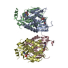
| ||||||||
|---|---|---|---|---|---|---|---|---|---|
| 1 | 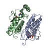
| ||||||||
| 2 | 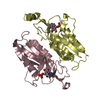
| ||||||||
| 3 | 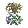
| ||||||||
| Unit cell |
| ||||||||
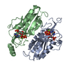
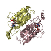
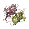
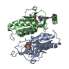
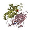
| Details | There are two copies of the biological dimer in the asymmetric unit |
-Components
| #1: Protein | Hypoxanthine-guanine phosphoribosyltransferase Mass: 25595.404 Da / Num. of mol.: 4 Source method: isolated from a genetically manipulated source Source: (gene. exp.) Trypanosoma cruzi (eukaryote) / Gene: HGPRT / Plasmid: pTcPRT / Production host:  Escherichia coli (E. coli) Escherichia coli (E. coli)References: UniProt: Q27796, UniProt: Q4DRC4*PLUS, hypoxanthine phosphoribosyltransferase#2: Chemical | ChemComp-IMP / Inosinic acid  Mass: 348.206 Da / Num. of mol.: 4 / Source method: obtained synthetically / Formula: C10H13N4O8P Mass: 348.206 Da / Num. of mol.: 4 / Source method: obtained synthetically / Formula: C10H13N4O8P#3: Water | ChemComp-HOH / | Water Mass: 18.015 Da / Num. of mol.: 780 / Source method: isolated from a natural source / Formula: H2O Mass: 18.015 Da / Num. of mol.: 780 / Source method: isolated from a natural source / Formula: H2O |
|---|
-Experimental details
-Experiment
| Experiment | Method: X-RAY DIFFRACTION / Number of used crystals: 1 |
|---|
- Sample preparation
| Crystal | Density Matthews: 2.43 Å3/Da / Density % sol: 49.35 % |
|---|---|
| Crystal grow | Temperature: 277 K / Method: vapor diffusion, hanging drop / pH: 4.4 Details: PEG 6000, Sodium Acetate, Ammonium Acetate, pH 4.4, VAPOR DIFFUSION, HANGING DROP, temperature 277K |
-Data collection
| Diffraction | Mean temperature: 100 K |
|---|---|
| Diffraction source | Source: SYNCHROTRON / Site: APS  / Beamline: 5ID-B / Wavelength: 1 Å / Beamline: 5ID-B / Wavelength: 1 Å |
| Detector | Type: MARRESEARCH / Detector: CCD / Date: Apr 4, 1999 |
| Radiation | Monochromator: Si III / Protocol: SINGLE WAVELENGTH / Monochromatic (M) / Laue (L): M / Scattering type: x-ray |
| Radiation wavelength | Wavelength: 1 Å / Relative weight: 1 |
| Reflection | Resolution: 2.3→20 Å / Num. all: 44180 / Num. obs: 44180 / % possible obs: 97.8 % / Observed criterion σ(F): -3 / Observed criterion σ(I): -3 / Redundancy: 4.3 % / Rmerge(I) obs: 0.065 / Net I/σ(I): 7.9 |
| Reflection shell | Resolution: 2.3→2.36 Å / Redundancy: 2.5 % / Rmerge(I) obs: 0.193 / Mean I/σ(I) obs: 3.9 / % possible all: 87.5 |
- Processing
| Software |
| ||||||||||||||||||||
|---|---|---|---|---|---|---|---|---|---|---|---|---|---|---|---|---|---|---|---|---|---|
| Refinement | Method to determine structure: MOLECULAR REPLACEMENT / Resolution: 2.3→20 Å / σ(F): 0 / Stereochemistry target values: Engh & Huber
| ||||||||||||||||||||
| Refinement step | Cycle: LAST / Resolution: 2.3→20 Å
|
