 Movie
Movie Controller

[English] 日本語
 Yorodumi
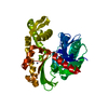
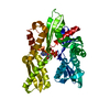
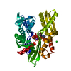
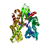
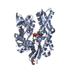
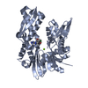
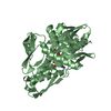
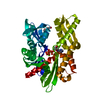
- PDB-1ba1: HEAT-SHOCK COGNATE 70KD PROTEIN 44KD ATPASE N-TERMINAL MUTANT WIT... -
+ Open data

- Basic information
| Entry | Database: PDB / ID: 1ba1 | ||||||
|---|---|---|---|---|---|---|---|
| Title | HEAT-SHOCK COGNATE 70KD PROTEIN 44KD ATPASE N-TERMINAL MUTANT WITH CYS 17 REPLACED BY LYS | ||||||
 Components Components | HEAT-SHOCK COGNATE 70KD PROTEIN | ||||||
 Keywords Keywords |  HYDROLASE / ACTING ON ACID ANHYDRIDES / ATP-BINDING / HEAT SHOCK HYDROLASE / ACTING ON ACID ANHYDRIDES / ATP-BINDING / HEAT SHOCK | ||||||
| Function / homology |  Function and homology information Function and homology informationRegulation of HSF1-mediated heat shock response / Attenuation phase / HSF1-dependent transactivation / Protein methylation / GABA synthesis, release, reuptake and degradation / PKR-mediated signaling / mRNA Splicing - Major Pathway / synaptic vesicle uncoating / HSP90 chaperone cycle for steroid hormone receptors (SHR) in the presence of ligand / chaperone-mediated autophagy translocation complex disassembly ...Regulation of HSF1-mediated heat shock response / Attenuation phase / HSF1-dependent transactivation / Protein methylation / GABA synthesis, release, reuptake and degradation / PKR-mediated signaling / mRNA Splicing - Major Pathway / synaptic vesicle uncoating / HSP90 chaperone cycle for steroid hormone receptors (SHR) in the presence of ligand / chaperone-mediated autophagy translocation complex disassembly / slow axonal transport / clathrin-uncoating ATPase activity / protein targeting to lysosome involved in chaperone-mediated autophagy / late endosomal microautophagy / AUF1 (hnRNP D0) binds and destabilizes mRNA / clathrin coat disassembly / Clathrin-mediated endocytosis / Prp19 complex / presynaptic cytosol / Neutrophil degranulation / postsynaptic cytosol / non-chaperonin molecular chaperone ATPase / chaperone cofactor-dependent protein refolding / autophagosome / protein folding chaperone / heat shock protein binding / RNA splicing / spliceosomal complex / ATP-dependent protein folding chaperone / terminal bouton / mRNA processing / melanosome / protein-macromolecule adaptor activity / protein refolding / lysosome / ribonucleoprotein complex / lysosomal membrane / negative regulation of DNA-templated transcription / dendrite / nucleolus / ATP hydrolysis activity / ATP binding / nucleus / plasma membrane / cytoplasmSimilarity search - Function | ||||||
| Biological species |  Bos taurus (cattle) Bos taurus (cattle) | ||||||
| Method | X-RAY DIFFRACTION / MOLECULAR REPLACEMENT / Resolution: 1.7 Å | ||||||
 Authors Authors | Wilbanks, S.M. / Mckay, D.B. | ||||||
 Citation Citation | Journal: Biochemistry / Year: 1998 Title: Structural replacement of active site monovalent cations by the epsilon-amino group of lysine in the ATPase fragment of bovine Hsc70. Authors: Wilbanks, S.M. / McKay, D.B. #1: Journal: J.Biol.Chem. / Year: 1994Title: Structural Basis of the 70-Kilodalton Heat Shock Cognate Protein ATP Hydrolytic Activity. II. Structure of the Active Site with Adp or ATP Bound to Wild Type and Mutant ATPase Fragment Authors: Flaherty, K.M. / Wilbanks, S.M. / Deluca-Flaherty, C. / Mckay, D.B. #2: Journal: Nature / Year: 1990Title: Three-Dimensional Structure of the ATPase Fragment of a 70K Heat-Shock Cognate Protein Authors: Flaherty, K.M. / Deluca-Flaherty, C. / Mckay, D.B. | ||||||
| History |
|
- Structure visualization
| Structure viewer | Molecule: MolmilJmol/JSmol |
|---|
- Downloads & links
-Download
| PDBx/mmCIF format | 1ba1.cif.gz | 93.8 KB | Display | PDBx/mmCIF format |
|---|---|---|---|---|
| PDB format | pdb1ba1.ent.gz | 71.8 KB | Display | PDB format |
| PDBx/mmJSON format | 1ba1.json.gz | Tree view | PDBx/mmJSON format | |
| Others |  Other downloads Other downloads |
-Validation report
| Arichive directory | https://data.pdbj.org/pub/pdb/validation_reports/ba/1ba1ftp://data.pdbj.org/pub/pdb/validation_reports/ba/1ba1 | HTTPS FTP |
|---|
-Related structure data
| Related structure data | 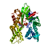 1ba0C 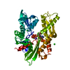 1hpmS S: Starting model for refinement C: citing same article ( |
|---|---|
| Similar structure data |


-Links
 PDBj
PDBj
- Assembly
| Deposited unit | 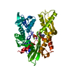
| ||||||||
|---|---|---|---|---|---|---|---|---|---|
| 1 |
| ||||||||
| Unit cell |
|
-Components
-Protein , 1 types, 1 molecules A
| #1: Protein | Mass: 42539.074 Da / Num. of mol.: 1 / Fragment: 44KD ATPASE N-TERMINAL FRAGMENT / Mutation: C17K Source method: isolated from a genetically manipulated source Source: (gene. exp.) Bos taurus (cattle) / Cell line: BL21 (DE3) / Cellular location: CYTOSOL / Organ: BRAIN / Plasmid: BL21 / Cell line (production host): BL21 (DE3) / Production host:  Escherichia coli (E. coli) / References: UniProt: P19120, EC: 3.6.1.3 Escherichia coli (E. coli) / References: UniProt: P19120, EC: 3.6.1.3 |
|---|
-Non-polymers , 5 types, 447 molecules 


| #2: Chemical | ChemComp-MG /  Mass: 24.305 Da / Num. of mol.: 1 / Source method: obtained synthetically / Formula: Mg Mass: 24.305 Da / Num. of mol.: 1 / Source method: obtained synthetically / Formula: Mg | ||||
|---|---|---|---|---|---|
| #3: Chemical | ChemComp-NA /  Mass: 22.990 Da / Num. of mol.: 1 / Source method: obtained synthetically / Formula: Na Mass: 22.990 Da / Num. of mol.: 1 / Source method: obtained synthetically / Formula: Na | ||||
| #4: Chemical | Chloride Mass: 35.453 Da / Num. of mol.: 2 / Source method: obtained synthetically / Formula: Cl Mass: 35.453 Da / Num. of mol.: 2 / Source method: obtained synthetically / Formula: Cl#5: Chemical | ChemComp-ADP / | Adenosine diphosphate Mass: 427.201 Da / Num. of mol.: 1 / Source method: obtained synthetically / Formula: C10H15N5O10P2 / Comment: ADP, energy-carrying molecule*YM Mass: 427.201 Da / Num. of mol.: 1 / Source method: obtained synthetically / Formula: C10H15N5O10P2 / Comment: ADP, energy-carrying molecule*YM#6: Water | ChemComp-HOH / | WaterMass: 18.015 Da / Num. of mol.: 442 / Source method: isolated from a natural source / Formula: H2O |
-Experimental details
-Experiment
| Experiment | Method: X-RAY DIFFRACTION / Number of used crystals: 1 |
|---|
- Sample preparation
| Crystal | Density Matthews: 2.5 Å3/Da / Density % sol: 50.73 % | ||||||||||||||||||||||||||||||||||||
|---|---|---|---|---|---|---|---|---|---|---|---|---|---|---|---|---|---|---|---|---|---|---|---|---|---|---|---|---|---|---|---|---|---|---|---|---|---|
| Crystal grow | pH: 9 Details: 20% PEG-8000 1.0M NACL 50MM CHES, PH 9 1MM MGATP, pH 9.0 | ||||||||||||||||||||||||||||||||||||
| Crystal grow | *PLUS Temperature: 4 ℃ / pH: 7 / Method: vapor diffusion, hanging drop / Details: used to seeding | ||||||||||||||||||||||||||||||||||||
| Components of the solutions | *PLUS
|
-Data collection
| Diffraction | Mean temperature: 100 K |
|---|---|
| Diffraction source | Source: ROTATING ANODE / Type: RIGAKU RUH2R / Wavelength: 1.5418 |
| Detector | Type: RIGAKU RAXIS / Detector: IMAGE PLATE / Date: Oct 1, 1995 |
| Radiation | Monochromator: GRAPHITE(002) / Monochromatic (M) / Laue (L): M / Scattering type: x-ray |
| Radiation wavelength | Wavelength: 1.5418 Å / Relative weight: 1 |
| Reflection | Resolution: 1.9→40 Å / Num. obs: 34106 / % possible obs: 99 % / Observed criterion σ(I): 3 / Redundancy: 3.2 % / Rmerge(I) obs: 1 / Rsym value: 0.056 / Net I/σ(I): 16 |
| Reflection shell | Resolution: 1.9→1.97 Å / Redundancy: 1.2 % / Rmerge(I) obs: 1 / Mean I/σ(I) obs: 12 / Rsym value: 0.133 / % possible all: 69.7 |
| Reflection | *PLUS Highest resolution: 1.7 Å / Num. obs: 45230 / % possible obs: 94.7 % / Num. measured all: 138386 / Rmerge(I) obs: 0.051 |
- Processing
| Software |
| ||||||||||||||||||||||||||||||||||||||||||||||||||||||||||||
|---|---|---|---|---|---|---|---|---|---|---|---|---|---|---|---|---|---|---|---|---|---|---|---|---|---|---|---|---|---|---|---|---|---|---|---|---|---|---|---|---|---|---|---|---|---|---|---|---|---|---|---|---|---|---|---|---|---|---|---|---|---|
| Refinement | Method to determine structure: MOLECULAR REPLACEMENT Starting model: PDB ENTRY 1HPM Resolution: 1.7→6 Å / Rfactor Rfree error: 0.6 / Data cutoff high absF: 100000 / Data cutoff low absF: 0.1 / Cross valid method: THROUGHOUT / σ(F): 2 / Details: SCALE ORTHORHOMBIC, LONGEST AXIS FIRST
| ||||||||||||||||||||||||||||||||||||||||||||||||||||||||||||
| Refine analyze | Luzzati coordinate error obs: 0.3 Å / Luzzati d res low obs: 40 Å | ||||||||||||||||||||||||||||||||||||||||||||||||||||||||||||
| Refinement step | Cycle: LAST / Resolution: 1.7→6 Å
| ||||||||||||||||||||||||||||||||||||||||||||||||||||||||||||
| Refine LS restraints |
| ||||||||||||||||||||||||||||||||||||||||||||||||||||||||||||
| LS refinement shell | Resolution: 1.7→1.76 Å / Rfactor Rfree error: 0.6 / Total num. of bins used: 10
|
