Movie
Movie Controller


+ Open data

- Basic information
| Entry | Database: EMDB / ID: EMD-7523 | |||||||||
|---|---|---|---|---|---|---|---|---|---|---|
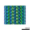
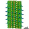
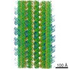
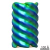
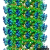
| Title | cryo-EM reconstruction of microtubule-bound 4R-tau | |||||||||
 Map data Map data | cryo-EM reconstruction of shortened tau construct (residues 197-400 of full-length tau) | |||||||||
 Sample Sample |
| |||||||||
| Biological species |   Homo sapiens (human) / Homo sapiens (human) /  Sus scrofa (pig) Sus scrofa (pig) | |||||||||
| Method | helical reconstruction / cryo EM / Resolution: 4.8 Å | |||||||||
 Authors Authors | Nogales E / Hejab NMA / Kellogg EH | |||||||||
 Citation Citation | Journal: Science / Year: 2018 Title: Near-atomic model of microtubule-tau interactions. Authors: Elizabeth H Kellogg / Nisreen M A Hejab / Simon Poepsel / Kenneth H Downing / Frank DiMaio / Eva Nogales /  Abstract: Tau is a developmentally regulated axonal protein that stabilizes and bundles microtubules (MTs). Its hyperphosphorylation is thought to cause detachment from MTs and subsequent aggregation into ...Tau is a developmentally regulated axonal protein that stabilizes and bundles microtubules (MTs). Its hyperphosphorylation is thought to cause detachment from MTs and subsequent aggregation into fibrils implicated in Alzheimer's disease. It is unclear which tau residues are crucial for tau-MT interactions, where tau binds on MTs, and how it stabilizes them. We used cryo-electron microscopy to visualize different tau constructs on MTs and computational approaches to generate atomic models of tau-tubulin interactions. The conserved tubulin-binding repeats within tau adopt similar extended structures along the crest of the protofilament, stabilizing the interface between tubulin dimers. Our structures explain the effect of phosphorylation on MT affinity and lead to a model of tau repeats binding in tandem along protofilaments, tethering together tubulin dimers and stabilizing polymerization interfaces. | |||||||||
| History |
|
- Structure visualization
| Movie |
 Movie viewer Movie viewer |
|---|---|
| Structure viewer | EM map: SurfViewMolmilJmol/JSmol |
| Supplemental images |

- Downloads & links
-EMDB archive
| Map data | emd_7523.map.gz | 481.8 MB | EMDB map data format | |
|---|---|---|---|---|
| Header (meta data) | emd-7523-v30.xmlemd-7523.xml | 16.2 KB 16.2 KB | Display Display | EMDB header |
| Images | 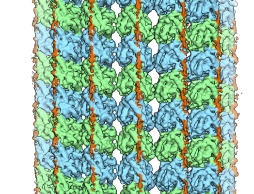 emd_7523.png emd_7523.png | 410.6 KB | ||
| Archive directory |  http://ftp.pdbj.org/pub/emdb/structures/EMD-7523ftp://ftp.pdbj.org/pub/emdb/structures/EMD-7523 http://ftp.pdbj.org/pub/emdb/structures/EMD-7523ftp://ftp.pdbj.org/pub/emdb/structures/EMD-7523 | HTTPS FTP |
-Related structure data
| Related structure data |  7520C  7522C 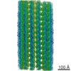 7769C 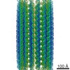 7771C 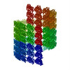 6cvjC 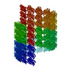 6cvnC C: citing same article ( |
|---|---|
| Similar structure data |

-Links
| EMDB pages | EMDB (EBI/PDBe) / EMDataResource |
|---|
-Map
| File | Download / File: emd_7523.map.gz / Format: CCP4 / Size: 512 MB / Type: IMAGE STORED AS FLOATING POINT NUMBER (4 BYTES) | ||||||||||||||||||||||||||||||||||||||||||||||||||||||||||||||||||||
|---|---|---|---|---|---|---|---|---|---|---|---|---|---|---|---|---|---|---|---|---|---|---|---|---|---|---|---|---|---|---|---|---|---|---|---|---|---|---|---|---|---|---|---|---|---|---|---|---|---|---|---|---|---|---|---|---|---|---|---|---|---|---|---|---|---|---|---|---|---|
| Annotation | cryo-EM reconstruction of shortened tau construct (residues 197-400 of full-length tau) | ||||||||||||||||||||||||||||||||||||||||||||||||||||||||||||||||||||
| Voxel size | X=Y=Z: 1.32 Å | ||||||||||||||||||||||||||||||||||||||||||||||||||||||||||||||||||||
| Density |
| ||||||||||||||||||||||||||||||||||||||||||||||||||||||||||||||||||||
| Symmetry | Space group: 1 | ||||||||||||||||||||||||||||||||||||||||||||||||||||||||||||||||||||
| Details | EMDB XML:
CCP4 map header:
| ||||||||||||||||||||||||||||||||||||||||||||||||||||||||||||||||||||
-Supplemental data
- Sample components
-Entire : Ternary complex of alpha-beta tubulin with shortened tau construct
| Entire | Name: Ternary complex of alpha-beta tubulin with shortened tau construct |
|---|---|
| Components |
|
-Supramolecule #1: Ternary complex of alpha-beta tubulin with shortened tau construct
| Supramolecule | Name: Ternary complex of alpha-beta tubulin with shortened tau construct type: complex / ID: 1 / Parent: 0 / Macromolecule list: all Details: Tau construct contains first four microtubule-targeting repeat motifs and portions of projection-domain and R' sequences |
|---|---|
| Source (natural) | Organism: Homo sapiens (human) |
-Macromolecule #1: 4Rtau
| Macromolecule | Name: 4Rtau / type: protein_or_peptide / ID: 1 / Enantiomer: LEVO |
|---|---|
| Source (natural) | Organism: Homo sapiens (human) |
| Recombinant expression | Organism:  Escherichia coli (E. coli) Escherichia coli (E. coli) |
| Sequence | String: YSSPGSPGTP GSRSRTPSLP TPPTREPKKV AVVRTPPKSP SSAKSRLQTA PVPMPDLKNV KSKIGSTENL KHQPGGGKVQ IINKKLDLSN VQSKCGSKDN IKHVPGGGSV QIVYKPVDLS KVTSKCGSLG NIHHKPGGGQ VEVKSEKLDF KDRVQSKIGS LDNITHVPGG ...String: YSSPGSPGTP GSRSRTPSLP TPPTREPKKV AVVRTPPKSP SSAKSRLQTA PVPMPDLKNV KSKIGSTENL KHQPGGGKVQ IINKKLDLSN VQSKCGSKDN IKHVPGGGSV QIVYKPVDLS KVTSKCGSLG NIHHKPGGGQ VEVKSEKLDF KDRVQSKIGS LDNITHVPGG GNKKIETHKL TFRENAKAKT DHGAEIVYKS PVVS |
-Macromolecule #2: alpha-tubulin
| Macromolecule | Name: alpha-tubulin / type: protein_or_peptide / ID: 2 / Enantiomer: LEVO |
|---|---|
| Source (natural) | Organism: Sus scrofa (pig) |
| Sequence | String: MRECISIHVG QAGVQIGNAC WELYCLEHGI QPDGQMPSDK TIGGGDDSFN TFFSETGAGK HVPRAVFVDL EPTVIDEVRT GTYRQLFHPE QLITGKEDAA NNYARGHYTI GKEIIDLVLD RIRKLADQCT GLQGFLVFHS FGGGTGSGFT SLLMERLSVD YGKKSKLEFS ...String: MRECISIHVG QAGVQIGNAC WELYCLEHGI QPDGQMPSDK TIGGGDDSFN TFFSETGAGK HVPRAVFVDL EPTVIDEVRT GTYRQLFHPE QLITGKEDAA NNYARGHYTI GKEIIDLVLD RIRKLADQCT GLQGFLVFHS FGGGTGSGFT SLLMERLSVD YGKKSKLEFS IYPAPQVSTA VVEPYNSILT THTTLEHSDC AFMVDNEAIY DICRRNLDIE RPTYTNLNRL ISQIVSSITA SLRFDGALNV DLTEFQTNLV PYPRIHFPLA TYAPVISAEK AYHEQLSVAE ITNACFEPAN QMVKCDPRHG KYMACCLLYR GDVVPKDVNA AIATIKTKRS IQFVDWCPTG FKVGINYQPP TVVPGGDLAK VQRAVCMLSN TTAIAEAWAR LDHKFDLMYA KRAFVHWYVG EGMEEGEFSE AREDMAALEK DYEEVGVDS |
-Macromolecule #3: beta-tubulin
| Macromolecule | Name: beta-tubulin / type: protein_or_peptide / ID: 3 / Enantiomer: LEVO |
|---|---|
| Source (natural) | Organism: Sus scrofa (pig) |
| Sequence | String: MREIVHIQAG QCGNQIGAKF WEVISDEHGI DPTGSYHGDS DLQLERINVY YNEAAGNKYV PRAILVDLEP GTMDSVRSGP FGQIFRPDNF VFGQSGAGNN WAKGHYTEGA ELVDSVLDVV RKESESCDCL QGFQLTHSLG GGTGSGMGTL LISKIREEYP DRIMNTFSVV ...String: MREIVHIQAG QCGNQIGAKF WEVISDEHGI DPTGSYHGDS DLQLERINVY YNEAAGNKYV PRAILVDLEP GTMDSVRSGP FGQIFRPDNF VFGQSGAGNN WAKGHYTEGA ELVDSVLDVV RKESESCDCL QGFQLTHSLG GGTGSGMGTL LISKIREEYP DRIMNTFSVV PSPKVSDTVV EPYNATLSVH QLVENTDETY CIDNEALYDI CFRTLKLTTP TYGDLNHLVS ATMSGVTTCL RFPGQLNADL RKLAVNMVPF PRLHFFMPGF APLTSRGSQQ YRALTVPELT QQMFDAKNMM AACDPRHGRY LTVAAVFRGR MSMKEVDEQM LNVQNKNSSY FVEWIPNNVK TAVCDIPPRG LKMSATFIGN STAIQELFKR ISEQFTAMFR RKAFLHWYTG EGMDEMEFTE AESNMNDLVS EYQQYQDATA D |
-Experimental details
-Structure determination
| Method | cryo EM |
|---|---|
 Processing Processing | helical reconstruction |
| Aggregation state | filament |
-Sample preparation
| Concentration | 0.5 mg/mL | ||||||||||||||||||
|---|---|---|---|---|---|---|---|---|---|---|---|---|---|---|---|---|---|---|---|
| Buffer | pH: 6.8 Component:
| ||||||||||||||||||
| Grid | Model: C-flat-1.2/1.3 4C / Material: COPPER / Mesh: 400 / Support film - Material: CARBON / Support film - topology: HOLEY / Pretreatment - Type: GLOW DISCHARGE / Pretreatment - Atmosphere: AIR / Details: unspecified | ||||||||||||||||||
| Vitrification | Cryogen name: ETHANE / Chamber humidity: 100 % / Chamber temperature: 310.15 K / Instrument: FEI VITROBOT MARK IV Details: blot force 10 pN, 6 second blot time. used C-flat 1.2/1.3 holey grids. First 2 uL of microtubules were adhered to grid for 30 seconds, followed by 2 4 uL washes with 30 second incubation for each wash.. | ||||||||||||||||||
| Details | tubulin concentration is 0.5 mg/mL, tau concentration is 1 mg/mL |
- Electron microscopy
| Microscope | FEI TITAN |
|---|---|
| Electron beam | Acceleration voltage: 300 kV / Electron source: FIELD EMISSION GUN |
| Electron optics | C2 aperture diameter: 100.0 µm / Illumination mode: FLOOD BEAM / Imaging mode: BRIGHT FIELDBright-field microscopy / Cs: 2.7 mm / Nominal defocus max: 2.5 µm / Nominal defocus min: 1.0 µm / Nominal magnification: 27500 |
| Sample stage | Specimen holder model: GATAN 626 SINGLE TILT LIQUID NITROGEN CRYO TRANSFER HOLDER Cooling holder cryogen: NITROGEN |
| Image recording | Film or detector model: GATAN K2 SUMMIT (4k x 4k) / Detector mode: COUNTING / Number grids imaged: 1 / Number real images: 422 / Average exposure time: 6.0 sec. / Average electron dose: 27.5 e/Å2 |
-Image processing
| Segment selection | Number selected: 36777 / Software - Name: Appion Details: manually selected helical segments, then filled in with overlapping boxes spaced 80 Angstrom apart. |
|---|---|
| CTF correction | Software - Name: CTFFIND (ver. 4) |
| Startup model | Type of model: EMDB MAP EMDB ID: Details: used a naked microtubule reconstruction as the initial model, low pass filtered to 20 Angstrom |
| Final angle assignment | Type: NOT APPLICABLE / Software - Name: FREALIGN (ver. 9.09) |
| Final reconstruction | Applied symmetry - Helical parameters - Δz: 8.6 Å Applied symmetry - Helical parameters - Δ&Phi: -25.76 ° Applied symmetry - Helical parameters - Axial symmetry: C1 (asymmetric) Algorithm: FOURIER SPACE / Resolution.type: BY AUTHOR / Resolution: 4.8 Å / Resolution method: FSC 0.143 CUT-OFF / Software - Name: FREALIGN (ver. 9.09) / Number images used: 24618 |
| Details | used motioncorr to correct for beam-induced motion |