National Institutes of Health/National Institute of General Medical Sciences (NIH/NIGMS)
R01GM120553
United States
Citation
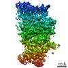
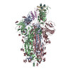
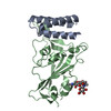
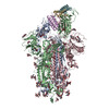
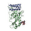
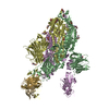
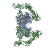
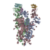
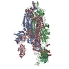
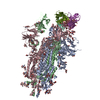
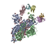
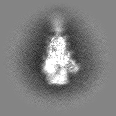
Journal: Science / Year: 2020 Title: De novo design of picomolar SARS-CoV-2 miniprotein inhibitors. Authors: Longxing Cao / Inna Goreshnik / Brian Coventry / James Brett Case / Lauren Miller / Lisa Kozodoy / Rita E Chen / Lauren Carter / Alexandra C Walls / Young-Jun Park / Eva-Maria Strauch / ...Authors: Longxing Cao / Inna Goreshnik / Brian Coventry / James Brett Case / Lauren Miller / Lisa Kozodoy / Rita E Chen / Lauren Carter / Alexandra C Walls / Young-Jun Park / Eva-Maria Strauch / Lance Stewart / Michael S Diamond / David Veesler / David Baker / Abstract: Targeting the interaction between the severe acute respiratory syndrome coronavirus 2 (SARS-CoV-2) spike protein and the human angiotensin-converting enzyme 2 (ACE2) receptor is a promising ...Targeting the interaction between the severe acute respiratory syndrome coronavirus 2 (SARS-CoV-2) spike protein and the human angiotensin-converting enzyme 2 (ACE2) receptor is a promising therapeutic strategy. We designed inhibitors using two de novo design approaches. Computer-generated scaffolds were either built around an ACE2 helix that interacts with the spike receptor binding domain (RBD) or docked against the RBD to identify new binding modes, and their amino acid sequences were designed to optimize target binding, folding, and stability. Ten designs bound the RBD, with affinities ranging from 100 picomolar to 10 nanomolar, and blocked SARS-CoV-2 infection of Vero E6 cells with median inhibitory concentration (IC) values between 24 picomolar and 35 nanomolar. The most potent, with new binding modes, are 56- and 64-residue proteins (IC ~ 0.16 nanograms per milliliter). Cryo-electron microscopy structures of these minibinders in complex with the SARS-CoV-2 spike ectodomain trimer with all three RBDs bound are nearly identical to the computational models. These hyperstable minibinders provide starting points for SARS-CoV-2 therapeutics.
History
Deposition
Sep 2, 2020
-
Header (metadata) release
Sep 23, 2020
-
Map release
Sep 23, 2020
-
Update
Dec 2, 2020
-
Current status
Dec 2, 2020
Processing site: RCSB / Status: Released
-
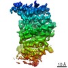
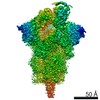
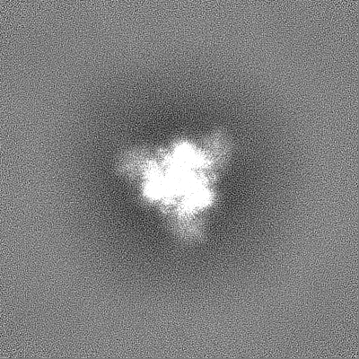
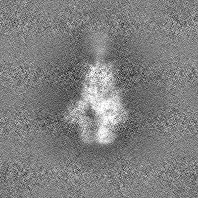
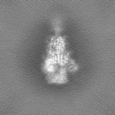
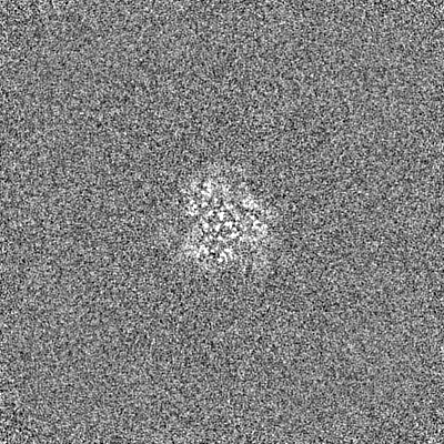
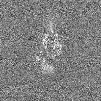
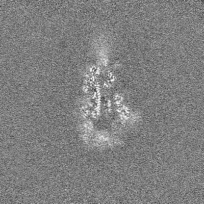
Structure visualization
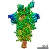
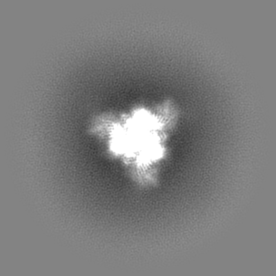
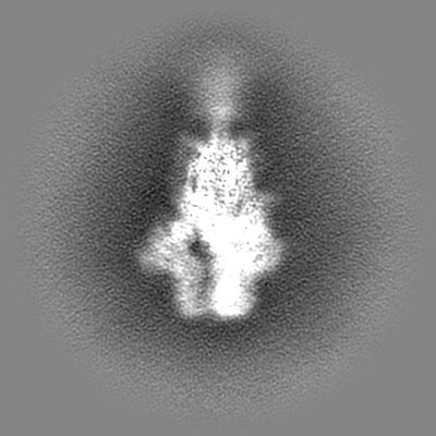
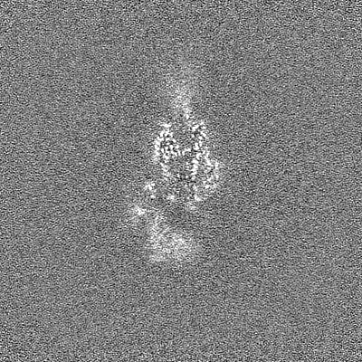
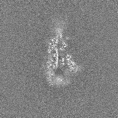
Movie
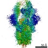
Surface view with section colored by density value
In the structure databanks used in Yorodumi, some data are registered as the other names, "COVID-19 virus" and "2019-nCoV". Here are the details of the virus and the list of structure data.
Jan 31, 2019. EMDB accession codes are about to change! (news from PDBe EMDB page)
EMDB accession codes are about to change! (news from PDBe EMDB page)
The allocation of 4 digits for EMDB accession codes will soon come to an end. Whilst these codes will remain in use, new EMDB accession codes will include an additional digit and will expand incrementally as the available range of codes is exhausted. The current 4-digit format prefixed with “EMD-” (i.e. EMD-XXXX) will advance to a 5-digit format (i.e. EMD-XXXXX), and so on. It is currently estimated that the 4-digit codes will be depleted around Spring 2019, at which point the 5-digit format will come into force.
The EM Navigator/Yorodumi systems omit the EMD- prefix.
Related info.:Q: What is EMD? / ID/Accession-code notation in Yorodumi/EM Navigator
Yorodumi is a browser for structure data from EMDB, PDB, SASBDB, etc.
This page is also the successor to EM Navigator detail page, and also detail information page/front-end page for Omokage search.
The word "yorodu" (or yorozu) is an old Japanese word meaning "ten thousand". "mi" (miru) is to see.
Related info.:EMDB / PDB / SASBDB / Comparison of 3 databanks / Yorodumi Search / Aug 31, 2016. New EM Navigator & Yorodumi / Yorodumi Papers / Jmol/JSmol / Function and homology information / Changes in new EM Navigator and Yorodumi
 Movie
Movie Controller


 Open data
 Basic information
 Map data
 Sample
 Function and homology information
 membrane fusion / positive regulation of viral entry into host cell / receptor-mediated virion attachment to host cell /


 Authors
 United States, 1 items
  Citation
 Structure visualization
 Downloads & links
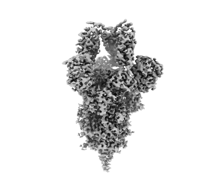 emd_22535.png
 http://ftp.pdbj.org/pub/emdb/structures/EMD-22535
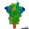


 Z
Z Y
Y X
X


 Sample components

 Processing
 Electron microscopy
