 Movie
Movie Controller


+ Open data

- Basic information
| Entry | Database: PDB / ID: 9ork | |||||||||
|---|---|---|---|---|---|---|---|---|---|---|
| Title | Antarctic Rhodopsin crystallized in LCP at pH 8.5 | |||||||||
 Components Components | Antarctic Rhodopsin | |||||||||
 Keywords Keywords | MEMBRANE PROTEIN / microbial rhodopsin / retinal-binding protein / inward proton pump | |||||||||
| Function / homology | DODECANE / HEXANE / RETINAL Function and homology information Function and homology information | |||||||||
| Biological species | unidentified (others) | |||||||||
| Method |  X-RAY DIFFRACTION / SYNCHROTRON / MOLECULAR REPLACEMENT / Resolution: 2.59 Å X-RAY DIFFRACTION / SYNCHROTRON / MOLECULAR REPLACEMENT / Resolution: 2.59 Å | |||||||||
 Authors Authors | Peng, S. / Besaw, J.E. / Ernst, O.P. | |||||||||
| Funding support |  Canada, 2items Canada, 2items
| |||||||||
 Citation Citation | Journal: Biophys.J. / Year: 2025 Title: Structural insights into an inward proton pumping rhodopsin. Authors: Besaw, J.E. / Peng, S. / Kuo, A. / Reichenwallner, J. / Miller, R.J.D. / Brown, L.S. / Ernst, O.P. | |||||||||
| History |
|
- Structure visualization
| Structure viewer | Molecule: MolmilJmol/JSmol |
|---|
- Downloads & links
-Download
| PDBx/mmCIF format | 9ork.cif.gz | 61.9 KB | Display | PDBx/mmCIF format |
|---|---|---|---|---|
| PDB format | pdb9ork.ent.gz | Display | PDB format | |
| PDBx/mmJSON format | 9ork.json.gz | Tree view | PDBx/mmJSON format | |
| Others |  Other downloads Other downloads |
-Validation report
| Summary document | 9ork_validation.pdf.gz | 616.4 KB | Display | wwPDB validaton report |
|---|---|---|---|---|
| Full document | 9ork_full_validation.pdf.gz | 618.5 KB | Display | |
| Data in XML | 9ork_validation.xml.gz | 11.4 KB | Display | |
| Data in CIF | 9ork_validation.cif.gz | 14.3 KB | Display | |
| Arichive directory | https://data.pdbj.org/pub/pdb/validation_reports/or/9orkftp://data.pdbj.org/pub/pdb/validation_reports/or/9ork | HTTPS FTP |
-Related structure data


-Links
 PDBj
PDBj


- Assembly
| Deposited unit | 
| ||||||||||||
|---|---|---|---|---|---|---|---|---|---|---|---|---|---|
| 1 | 
| ||||||||||||
| Unit cell |
|
-Components
| #1: Protein | Mass: 24856.141 Da / Num. of mol.: 1 Source method: isolated from a genetically manipulated source Source: (gene. exp.) unidentified (others) / Production host:  | ||||||||
|---|---|---|---|---|---|---|---|---|---|
| #2: Chemical | ChemComp-HEX / 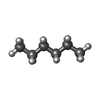  Mass: 86.175 Da / Num. of mol.: 1 / Source method: obtained synthetically / Formula: C6H14 Mass: 86.175 Da / Num. of mol.: 1 / Source method: obtained synthetically / Formula: C6H14 | ||||||||
| #3: Chemical | ChemComp-D12 /   Mass: 170.335 Da / Num. of mol.: 4 / Source method: obtained synthetically / Formula: C12H26 Mass: 170.335 Da / Num. of mol.: 4 / Source method: obtained synthetically / Formula: C12H26#4: Chemical | ChemComp-RET / |   Mass: 284.436 Da / Num. of mol.: 1 / Source method: obtained synthetically / Formula: C20H28O / Feature type: SUBJECT OF INVESTIGATION Mass: 284.436 Da / Num. of mol.: 1 / Source method: obtained synthetically / Formula: C20H28O / Feature type: SUBJECT OF INVESTIGATION#5: Water | ChemComp-HOH / |  Mass: 18.015 Da / Num. of mol.: 20 / Source method: isolated from a natural source / Formula: H2O Mass: 18.015 Da / Num. of mol.: 20 / Source method: isolated from a natural source / Formula: H2OHas ligand of interest | Y | Has protein modification | Y | |
-Experimental details
-Experiment
| Experiment | Method: X-RAY DIFFRACTION / Number of used crystals: 1 |
|---|
- Sample preparation
| Crystal | Density Matthews: 3.4 Å3/Da / Density % sol: 63.86 % |
|---|---|
| Crystal grow | Temperature: 279.15 K / Method: lipidic cubic phase / pH: 8.5 Details: 0.2 M NaCl, 20% w/v 2-methyl-2,4-pentanediol, 4% w/v glycerol PH range: 8.0-8.5 |
-Data collection
| Diffraction | Mean temperature: 100 K / Serial crystal experiment: N |
|---|---|
| Diffraction source | Source: SYNCHROTRON / Site: NSLS-II  / Beamline: 17-ID-1 / Wavelength: 0.92 Å / Beamline: 17-ID-1 / Wavelength: 0.92 Å |
| Detector | Type: DECTRIS EIGER X 9M / Detector: PIXEL / Date: Sep 1, 2020 |
| Radiation | Protocol: SINGLE WAVELENGTH / Monochromatic (M) / Laue (L): M / Scattering type: x-ray |
| Radiation wavelength | Wavelength: 0.92 Å / Relative weight: 1 |
| Reflection | Resolution: 2.59→28.91 Å / Num. obs: 10412 / % possible obs: 97.9 % / Redundancy: 5.2 % / Biso Wilson estimate: 52.47 Å2 / CC1/2: 0.999 / Net I/σ(I): 13.2 |
| Reflection shell | Resolution: 2.59→2.66 Å / Redundancy: 4.9 % / Mean I/σ(I) obs: 1.8 / Num. unique obs: 738 / CC1/2: 0.762 / % possible all: 93.8 |
- Processing
| Software |
| ||||||||||||||||||||||||||||||||||||||||||||||||||||||||
|---|---|---|---|---|---|---|---|---|---|---|---|---|---|---|---|---|---|---|---|---|---|---|---|---|---|---|---|---|---|---|---|---|---|---|---|---|---|---|---|---|---|---|---|---|---|---|---|---|---|---|---|---|---|---|---|---|---|
| Refinement | Method to determine structure: MOLECULAR REPLACEMENT / Resolution: 2.59→28.91 Å / SU ML: 0.2552 / Cross valid method: FREE R-VALUE / σ(F): 1.36 / Phase error: 23.5646 Stereochemistry target values: GeoStd + Monomer Library + CDL v1.2
| ||||||||||||||||||||||||||||||||||||||||||||||||||||||||
| Solvent computation | Shrinkage radii: 0.9 Å / VDW probe radii: 1.11 Å / Solvent model: FLAT BULK SOLVENT MODEL | ||||||||||||||||||||||||||||||||||||||||||||||||||||||||
| Displacement parameters | Biso mean: 55.63 Å2 | ||||||||||||||||||||||||||||||||||||||||||||||||||||||||
| Refinement step | Cycle: LAST / Resolution: 2.59→28.91 Å
| ||||||||||||||||||||||||||||||||||||||||||||||||||||||||
| Refine LS restraints |
| ||||||||||||||||||||||||||||||||||||||||||||||||||||||||
| LS refinement shell |
|
