 Movie
Movie Controller

[English] 日本語
 Yorodumi
- PDB-9e6u: Crystal structure of human Taspase1 in complex with ligand SMDC1041556 -
+ Open data

- Basic information
| Entry | Database: PDB / ID: 9e6u | ||||||
|---|---|---|---|---|---|---|---|
| Title | Crystal structure of human Taspase1 in complex with ligand SMDC1041556 | ||||||
 Components Components | Threonine aspartase 1 | ||||||
 Keywords Keywords | HYDROLASE/INHIBITOR / HYDROLASE-INHIBITOR complex | ||||||
| Function / homology |  Function and homology information Function and homology informationHydrolases; Acting on peptide bonds (peptidases); Threonine endopeptidases / Formation of WDR5-containing histone-modifying complexes / threonine-type endopeptidase activity / protein maturation / positive regulation of DNA-templated transcription / proteolysis / identical protein binding / cytoplasm / cytosol Similarity search - Function | ||||||
| Biological species |  Homo sapiens (human) Homo sapiens (human) | ||||||
| Method |  X-RAY DIFFRACTION / SYNCHROTRON / MOLECULAR REPLACEMENT / Resolution: 2.488 Å X-RAY DIFFRACTION / SYNCHROTRON / MOLECULAR REPLACEMENT / Resolution: 2.488 Å | ||||||
 Authors Authors | Doamekpor, S.K. / Hamilton, K. / Choi, P.H. / Tong, L. | ||||||
| Funding support |  United States, 1items United States, 1items
| ||||||
 Citation Citation | Journal: To Be Published Title: Design and mechanism of potent inhibitors for the Mixed Lineage Leukemia (MLL) activating protease, Taspase-1 Authors: Doamekpor, S.K. / Hamilton, K. / Choi, P.H. / Tong, L. / Delker, S. / Waterson, A. / Neitz, J. / Sambucetti, L. / Arkin, M. | ||||||
| History |
|
- Structure visualization
| Structure viewer | Molecule: MolmilJmol/JSmol |
|---|
- Downloads & links
-Download
| PDBx/mmCIF format | 9e6u.cif.gz | 130.1 KB | Display | PDBx/mmCIF format |
|---|---|---|---|---|
| PDB format | pdb9e6u.ent.gz | 97.1 KB | Display | PDB format |
| PDBx/mmJSON format | 9e6u.json.gz | Tree view | PDBx/mmJSON format | |
| Others |  Other downloads Other downloads |
-Validation report
| Summary document | 9e6u_validation.pdf.gz | 1020.9 KB | Display | wwPDB validaton report |
|---|---|---|---|---|
| Full document | 9e6u_full_validation.pdf.gz | 1 MB | Display | |
| Data in XML | 9e6u_validation.xml.gz | 28.5 KB | Display | |
| Data in CIF | 9e6u_validation.cif.gz | 35.9 KB | Display | |
| Arichive directory | https://data.pdbj.org/pub/pdb/validation_reports/e6/9e6uftp://data.pdbj.org/pub/pdb/validation_reports/e6/9e6u | HTTPS FTP |
-Related structure data
| Related structure data | 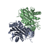 2a8jS S: Starting model for refinement |
|---|---|
| Similar structure data |
-Links
 PDBj
PDBj


- Assembly
| Deposited unit | 
| ||||||||
|---|---|---|---|---|---|---|---|---|---|
| 1 |
| ||||||||
| Unit cell |
|
-Components
| #1: Protein | Mass: 41825.266 Da / Num. of mol.: 2 Source method: isolated from a genetically manipulated source Source: (gene. exp.) Homo sapiens (human) / Gene: TASP1, C20orf13 / Production host:  References: UniProt: Q9H6P5, Hydrolases; Acting on peptide bonds (peptidases); Threonine endopeptidases #2: Chemical |   Mass: 22.990 Da / Num. of mol.: 2 / Source method: obtained synthetically / Formula: Na Mass: 22.990 Da / Num. of mol.: 2 / Source method: obtained synthetically / Formula: Na#3: Chemical | Mass: 479.449 Da / Num. of mol.: 2 / Source method: obtained synthetically / Formula: C18H21F4N5O4S / Feature type: SUBJECT OF INVESTIGATION Has ligand of interest | Y | Has protein modification | Y | |
|---|
-Experimental details
-Experiment
| Experiment | Method: X-RAY DIFFRACTION / Number of used crystals: 1 |
|---|
- Sample preparation
| Crystal | Density Matthews: 1.79 Å3/Da / Density % sol: 31.38 % |
|---|---|
| Crystal grow | Temperature: 293 K / Method: vapor diffusion, sitting drop / pH: 6.5 / Details: 20% MPD, 0.1 M MES, pH 6.5 |
-Data collection
| Diffraction | Mean temperature: 100 K / Serial crystal experiment: N | ||||||||||||||||||||||||||||||||||||||||||||||||||||||||||||||||||||||||||||||||||||||||||||||||||||
|---|---|---|---|---|---|---|---|---|---|---|---|---|---|---|---|---|---|---|---|---|---|---|---|---|---|---|---|---|---|---|---|---|---|---|---|---|---|---|---|---|---|---|---|---|---|---|---|---|---|---|---|---|---|---|---|---|---|---|---|---|---|---|---|---|---|---|---|---|---|---|---|---|---|---|---|---|---|---|---|---|---|---|---|---|---|---|---|---|---|---|---|---|---|---|---|---|---|---|---|---|---|
| Diffraction source | Source: SYNCHROTRON / Site: APS / Beamline: 24-ID-C / Wavelength: 0.9791 Å | ||||||||||||||||||||||||||||||||||||||||||||||||||||||||||||||||||||||||||||||||||||||||||||||||||||
| Detector | Type: DECTRIS PILATUS3 S 6M / Detector: PIXEL / Date: Feb 17, 2018 | ||||||||||||||||||||||||||||||||||||||||||||||||||||||||||||||||||||||||||||||||||||||||||||||||||||
| Radiation | Protocol: SINGLE WAVELENGTH / Monochromatic (M) / Laue (L): M / Scattering type: x-ray | ||||||||||||||||||||||||||||||||||||||||||||||||||||||||||||||||||||||||||||||||||||||||||||||||||||
| Radiation wavelength | Wavelength: 0.9791 Å / Relative weight: 1 | ||||||||||||||||||||||||||||||||||||||||||||||||||||||||||||||||||||||||||||||||||||||||||||||||||||
| Reflection | Resolution: 2.488→70.059 Å / Num. obs: 21399 / % possible obs: 96.4 % / Redundancy: 2.708 % / Biso Wilson estimate: 59.71 Å2 / CC1/2: 0.998 / Rmerge(I) obs: 0.055 / Rrim(I) all: 0.068 / Χ2: 1.096 / Net I/σ(I): 11.16 | ||||||||||||||||||||||||||||||||||||||||||||||||||||||||||||||||||||||||||||||||||||||||||||||||||||
| Reflection shell | Diffraction-ID: 1
|
- Processing
| Software |
| ||||||||||||||||||||||||||||||||||||||||||||||||||||||||||||||||||||||||||||||||||||||||||
|---|---|---|---|---|---|---|---|---|---|---|---|---|---|---|---|---|---|---|---|---|---|---|---|---|---|---|---|---|---|---|---|---|---|---|---|---|---|---|---|---|---|---|---|---|---|---|---|---|---|---|---|---|---|---|---|---|---|---|---|---|---|---|---|---|---|---|---|---|---|---|---|---|---|---|---|---|---|---|---|---|---|---|---|---|---|---|---|---|---|---|---|
| Refinement | Method to determine structure: MOLECULAR REPLACEMENT Starting model: PDB entry 2A8J Resolution: 2.488→70.059 Å / SU ML: 0.4 / Cross valid method: THROUGHOUT / σ(F): 1.34 / Phase error: 34.63
| ||||||||||||||||||||||||||||||||||||||||||||||||||||||||||||||||||||||||||||||||||||||||||
| Solvent computation | Shrinkage radii: 0.9 Å / VDW probe radii: 1.11 Å | ||||||||||||||||||||||||||||||||||||||||||||||||||||||||||||||||||||||||||||||||||||||||||
| Displacement parameters | Biso max: 132.37 Å2 / Biso mean: 72.3998 Å2 / Biso min: 39.82 Å2 | ||||||||||||||||||||||||||||||||||||||||||||||||||||||||||||||||||||||||||||||||||||||||||
| Refinement step | Cycle: final / Resolution: 2.488→70.059 Å
| ||||||||||||||||||||||||||||||||||||||||||||||||||||||||||||||||||||||||||||||||||||||||||
| Refine LS restraints |
| ||||||||||||||||||||||||||||||||||||||||||||||||||||||||||||||||||||||||||||||||||||||||||
| LS refinement shell | Refine-ID: X-RAY DIFFRACTION / Rfactor Rfree error: 0
|
