 Movie
Movie Controller

[English] 日本語
 Yorodumi
- PDB-7dvn: Crystal structure of a MarR family protein in complex with a lipi... -
+ Open data

- Basic information
| Entry | Database: PDB / ID: 7dvn | ||||||
|---|---|---|---|---|---|---|---|
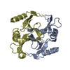
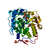
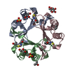
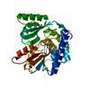
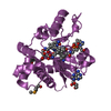
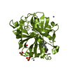
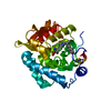
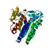
| Title | Crystal structure of a MarR family protein in complex with a lipid-like effector molecule from the psychrophilic bacterium Paenisporosarcina sp. TG-14 | ||||||
 Components Components | MarR family transcriptional regulator | ||||||
 Keywords Keywords | DNA BINDING PROTEIN / Transcription regulator DNA-binding Complex Lipid-like effector MarR family | ||||||
| Function / homology | PALMITIC ACID Function and homology information Function and homology information | ||||||
| Biological species |  Paenisporosarcina sp. TG-14 (bacteria) Paenisporosarcina sp. TG-14 (bacteria) | ||||||
| Method |  X-RAY DIFFRACTION / SYNCHROTRON / SAD / Resolution: 1.6 Å X-RAY DIFFRACTION / SYNCHROTRON / SAD / Resolution: 1.6 Å | ||||||
 Authors Authors | Lee, C.W. / Hwang, J. / Do, H. / Lee, J.H. | ||||||
| Funding support |  Korea, Republic Of, 1items Korea, Republic Of, 1items
| ||||||
 Citation Citation | Journal: Iucrj / Year: 2021 Title: Crystal structure of a MarR family protein from the psychrophilic bacterium Paenisporosarcina sp. TG-14 in complex with a lipid-like molecule. Authors: Hwang, J. / Park, S.H. / Lee, C.W. / Do, H. / Shin, S.C. / Kim, H.W. / Lee, S.G. / Park, H.H. / Kwon, S. / Lee, J.H. | ||||||
| History |
|
- Structure visualization
| Structure viewer | Molecule: MolmilJmol/JSmol |
|---|
- Downloads & links
-Download
| PDBx/mmCIF format | 7dvn.cif.gz | 46.2 KB | Display | PDBx/mmCIF format |
|---|---|---|---|---|
| PDB format | pdb7dvn.ent.gz | 30.2 KB | Display | PDB format |
| PDBx/mmJSON format | 7dvn.json.gz | Tree view | PDBx/mmJSON format | |
| Others |  Other downloads Other downloads |
-Validation report
| Summary document | 7dvn_validation.pdf.gz | 607.1 KB | Display | wwPDB validaton report |
|---|---|---|---|---|
| Full document | 7dvn_full_validation.pdf.gz | 607.7 KB | Display | |
| Data in XML | 7dvn_validation.xml.gz | 8.9 KB | Display | |
| Data in CIF | 7dvn_validation.cif.gz | 12.2 KB | Display | |
| Arichive directory | https://data.pdbj.org/pub/pdb/validation_reports/dv/7dvnftp://data.pdbj.org/pub/pdb/validation_reports/dv/7dvn | HTTPS FTP |
-Related structure data
| Similar structure data |
|---|


-Links
 PDBj
PDBj


- Assembly
| Deposited unit | 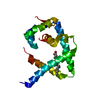
| ||||||||||||
|---|---|---|---|---|---|---|---|---|---|---|---|---|---|
| 1 | 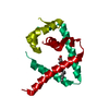
| ||||||||||||
| Unit cell |
| ||||||||||||
| Components on special symmetry positions |
|
-Components
| #1: Protein | Mass: 16937.818 Da / Num. of mol.: 1 Source method: isolated from a genetically manipulated source Details: complex with palmitic acid / Source: (gene. exp.) Paenisporosarcina sp. TG-14 (bacteria) / Gene: E2636_00495 / Production host: |
|---|---|
| #2: Chemical | ChemComp-PLM /   Mass: 256.424 Da / Num. of mol.: 1 / Source method: obtained synthetically / Formula: C16H32O2 / Feature type: SUBJECT OF INVESTIGATION Mass: 256.424 Da / Num. of mol.: 1 / Source method: obtained synthetically / Formula: C16H32O2 / Feature type: SUBJECT OF INVESTIGATION |
| #3: Water | ChemComp-HOH /  Mass: 18.015 Da / Num. of mol.: 143 / Source method: isolated from a natural source / Formula: H2O Mass: 18.015 Da / Num. of mol.: 143 / Source method: isolated from a natural source / Formula: H2O |
| Has ligand of interest | Y |
-Experimental details
-Experiment
| Experiment | Method: X-RAY DIFFRACTION / Number of used crystals: 1 |
|---|
- Sample preparation
| Crystal | Density Matthews: 2.86 Å3/Da / Density % sol: 57.02 % |
|---|---|
| Crystal grow | Temperature: 296 K / Method: vapor diffusion, hanging drop / pH: 7 / Details: 1.6M ammonium citrate tribasic |
-Data collection
| Diffraction | Mean temperature: 95 K / Serial crystal experiment: N |
|---|---|
| Diffraction source | Source: SYNCHROTRON / Site: PAL/PLS / Beamline: 5C (4A) / Wavelength: 0.9794 Å |
| Detector | Type: DECTRIS EIGER2 X 9M / Detector: PIXEL / Date: Nov 21, 2018 |
| Radiation | Protocol: SINGLE WAVELENGTH / Monochromatic (M) / Laue (L): M / Scattering type: x-ray |
| Radiation wavelength | Wavelength: 0.9794 Å / Relative weight: 1 |
| Reflection | Resolution: 1.6→50 Å / Num. obs: 26268 / % possible obs: 98 % / Redundancy: 26.4 % / Biso Wilson estimate: 23.63 Å2 / Rmerge(I) obs: 0.074 / Net I/σ(I): 82.4 |
| Reflection shell | Resolution: 1.6→1.63 Å / Rmerge(I) obs: 0.422 / Num. unique obs: 1300 |
- Processing
| Software |
| ||||||||||||||||||||||||||||||||||||||||||||||||||||||||||||||||||||||
|---|---|---|---|---|---|---|---|---|---|---|---|---|---|---|---|---|---|---|---|---|---|---|---|---|---|---|---|---|---|---|---|---|---|---|---|---|---|---|---|---|---|---|---|---|---|---|---|---|---|---|---|---|---|---|---|---|---|---|---|---|---|---|---|---|---|---|---|---|---|---|---|
| Refinement | Method to determine structure: SAD / Resolution: 1.6→32.34 Å / SU ML: 0.1657 / Cross valid method: FREE R-VALUE / σ(F): 1.37 / Phase error: 23.1222 / Stereochemistry target values: CDL v1.2
| ||||||||||||||||||||||||||||||||||||||||||||||||||||||||||||||||||||||
| Solvent computation | Shrinkage radii: 0.9 Å / VDW probe radii: 1.11 Å / Solvent model: FLAT BULK SOLVENT MODEL | ||||||||||||||||||||||||||||||||||||||||||||||||||||||||||||||||||||||
| Displacement parameters | Biso mean: 28.78 Å2 | ||||||||||||||||||||||||||||||||||||||||||||||||||||||||||||||||||||||
| Refinement step | Cycle: LAST / Resolution: 1.6→32.34 Å
| ||||||||||||||||||||||||||||||||||||||||||||||||||||||||||||||||||||||
| Refine LS restraints |
| ||||||||||||||||||||||||||||||||||||||||||||||||||||||||||||||||||||||
| LS refinement shell |
|
